Epidemiology
Over the past four decades, melanoma has shown a consistent increase in incidence in white populations, becoming one of the most frequent cancers in light skin populations. The incidence rate of this disease varies widely in relation to race, occurring much less commonly in black, Asian, or Hispanic populations. Although white and non-white populations have similar risks of developing plantar melanoma, non-white populations have a greater risk of developing noncutaneous melanomas (e.g., mucosal). Melanoma is now regarded as the fifth most common cancer in men and the sixth most common cancer in women in the United States. The highest recorded incidence of melanoma worldwide is in Australia and New Zealand, with an incidence of 27.3–55.8/100,000 for males and 23.4–41.1/100,000 for females. In the United States the incidence rate is 19.4/100,000 for men and 14.4/100,000 for women. In European countries the incidence rates are lower, being highest in Switzerland and Scandinavian countries. Moreover, they show a north to south gradient, with the highest rates in northern countries. This is probably related to both the increased protection against the sun’s rays of the pigmented skin of southern Europeans and the different pattern of sun exposure (chronic in southern Europeans, intermittent in northern Europeans). In many countries, early detection of primarily thin melanomas and improved survival rates have been observed, especially in young females. However, in most countries the incidence of thick melanomas remains constant or continues to increase, especially in the older age group. Melanoma occurs approximately 1.5 times more often in men than women, and prognosis is slightly better for women, considering other prognostic factors. The anatomic distribution of melanoma is sex-dependent, occurring more often on the trunk in males and on the arms and legs in women. Melanoma can occur at any age but is rare before the age of puberty. The median age at the time of diagnosis ranges between 45 and 55 years, with an increase in incidence after the age of 25 until 50 years.
Risk Factors
Current considerations in the epidemiology of melanoma have shown that melanoma results from an unidentified interaction between mutations in various genes and constitutional and/or inherited factors combined with environmental factors, mainly solar ultraviolet (UV) radiations. Exposure to UV is the most important environmental factor predisposing to melanoma in susceptible populations. It is reported that people incurring severe burns in childhood appear to be at higher risk for the development of melanoma in later age.
Iatrogenic exposures, such as immunosuppressive agents and nonsteroidal antiinflammatory drugs, have been studied as additional environmental factors. Well-known host risk factors, such as melanocytic nevi on the skin, skin type, family history, and genetic susceptibility have been validated as risk factors in large-scale association studies. Patients with more than 100 nevi or giant nevi (>20 cm) have an increased risk of melanoma. In addition, genome-wide association studies have revealed genetic loci that underlie the genetic susceptibility of melanoma, some of which are related to known risk factors. Mutations or epigenetic silencing of the cyclin-dependent kinase gene ( CDK ) are linked to 20%–40% of familial melanoma. Other genes involved in familial melanoma carcinogenesis include CDK4 , xeroderma pigmentosum genes and melanocortin 1 receptor gene ( MC1R ). Activating mutations in the BRAF gene have been discovered in over two-thirds of melanomas. Melanomas on skin without chronic sun-induced damage had frequent mutations in BRAF ; in contrast, acral and mucosal melanomas as well as skin melanomas with chronic sun damage are frequently characterized by KIT mutations. These mutations are responsible for cancer cell behavior through mechanisms that are still undefined.
Clinical Diagnosis
Melanoma usually appears as an irregularly pigmented skin lesion with an irregular border and a tendency to grow or change over time. Melanomas arise mostly de novo, just occasionally within a congenital or acquired nevus. In the past, they were often diagnosed in advanced stages as large, ulcerated or vegetating lesions. The need to detect melanomas at an early stage of disease led to the development of the ABCDE criteria, an acronym for asymmetry, border irregularity, color alterations, diameter >6 mm, and evolution. Not only are these simple criteria intended to allow both patients and nondermatologist healthcare professionals to differentiate common melanocytic lesions from suspicious pigmented lesions, they are also a useful tool for the dermatoncologist in guiding the decision to perform a biopsy. The sensitivity of clinical diagnosis by physicians with experience in differential diagnosis of suspected cutaneous lesions is about 70%. Dermoscopy, also known as epiluminescence microscopy, improves the diagnostic accuracy of melanocytic lesions. Dermoscopy, when adopted by experts, increases the sensitivity of clinical diagnosis up to 89%, reducing the number of negative biopsies. Since the diagnostic protocol of suspicious lesions includes a complete excision with safety margins, the problem of unnecessary scarring is significant. The real challenge in this case is to have a properly formulated diagnosis before acquiring a biopsy. Reflectance confocal microscopy is a currently available noninvasive technique that can be a reliable bridge between dermoscopy and histopathology. In high-risk patients (e.g., with atypical mole syndrome), a follow-up examination with digital dermoscopy and total-body microphotography is also helpful in detecting early changes in lesions or in discovering new atypical lesions. In patients with a previous melanoma, the risk of harboring a second melanoma is significant and implies scheduled examinations of the skin at regular intervals for detecting second tumors or skin metastasis.
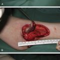
Biopsy
In the presence of a skin lesion with a clinical suspicion of melanoma, the patient is referred for an excisional biopsy. The biopsy of a suspected lesion is a simple procedure, practicable under local anesthesia on an outpatient basis. The procedure consists in complete removal of the lesion with about 1–2 mm of surrounding healthy skin up to the subcutaneous fat. During the procedure care should be taken to avoid causing trauma or injury to the specimen, which could compromise a correct histopathological analysis. The choice of incision lines of the biopsy must take into account the further excision of the skin involved, preferring longitudinal incisions in the biopsy of the limbs while trying to minimize as much as possible the need of skin grafts. Excisional biopsy allows an accurate histopathological diagnosis, providing all the histological parameters necessary to formulate a prognosis and to plan the best therapeutic strategy. However, excision of the whole lesion may not be indicated in some cases: for example, in the presence of a large lesion, or in the case of proximity of the lesion to difficult areas (subungual) or important structures (eye, nose, and ear). In these circumstances, punch or incisional biopsy should be considered. In the incisional or so‐called “punch” biopsy, only a full-thickness portion or a pill of the most suspected portion of the lesion is removed. Superficial biopsies, “shave biopsy,” and any type of laser or diathermocoagulation should be avoided, as they do not allow an accurate pathological staging and can lead to a significant delay in diagnosis of any potentially malignant lesions.
Histology
The histological diagnosis of cutaneous melanoma is not always easy and it would be desirable for an expert pathologist in the diagnosis of pigmented lesions of the skin to perform the analysis. In doubtful cases, morphology may indicate the use of immunohistochemical surveys (such as HMB‐45, Ki‐67, p16) and fluorescence in situ hybridization (FISH) techniques, the reading of which requires experienced operators.
Histologically, invasive cutaneous melanomas are divided into four major subtypes. These are based on growth pattern and location, and are categorized as:
- •
superficial spreading
- •
nodular
- •
lentigo maligna, or
- •
acral lentiginous.
A fifth subtype is also defined: desmoplastic melanoma. In general, the histologic subtype of melanoma is not a major factor in determining prognosis, but some histologic subtypes are more likely to be detected at an advanced stage, thus indirectly affecting prognosis.
Melanoma originates from the proliferation of melanocytes in the basal layers of the epidermis, which tend to expand radially and later invade deep into the dermis. Superficial spreading melanoma is the most common (70%), can occur in any location, and is characterized by a radial growth associated in the later stages to a pattern of vertical proliferation. Nodular melanomas, occurring in about 15%, are characterized by an exception to this pattern, with an exclusive vertical growth not accompanied by radial growth. Because of its invasiveness, it is usually diagnosed at a more advanced stage of invasion. Lentigo maligna accounts for about 10% of melanomas and is characterized by onset in old age, in chronically sun-exposed skin sites with a history of slow growth. These lesions are characterized by a long phase of intraepithelial growth and for this reason are considered a lesion with a better prognosis, though always remaining linked to the degree of invasion. Acral lentiginous melanoma is the less common type (5%); it is classified mainly for its site of origin (subnail bed and the palmar and plantar surfaces) rather than for histological features, which are similar to melanoma of the mucous membranes. Diagnosis is often delayed, especially for subungual forms often confused with posttraumatic hematoma. Among the rarer variants, desmoplastic melanoma has nontypical melanocytic cells and is characterized by a significant deposition of collagen, which hinders differential diagnosis with other fibrohistiocytic tumors or schwannomas.
The vertical growth phase is the tumorigenic phase in which the melanoma acquires the ability to metastasize. Morphologically, it is characterized by the presence of an expansive nodule larger than the intraepidermal component and/or by the presence of mitotic figures in the invasive component. In 1969, Clark formulated a histological classification with a prognostic value for melanoma based on the level of invasion in the anatomical layers of the skin, highlighting for the first time the prognostic significance of the degree of invasion ( Table 8.1 ). However, the Clark system has proven to be a source of significant variability among pathologists, especially in stages III and IV, thereby losing its prognostic relevance in more recent years. In 1970, Breslow described a more objective method of classification, based on the measurement in millimeters of the vertical thickness of the tumor. The Breslow thickness is measured from the granular layer or, if the lesion is ulcerated, from the bottom of ulceration, up to the point of maximum infiltration. This classification system has been shown to be more reproducible among pathologists, showing an excellent correlation with mortality. Prognosis tends to become progressively worse in logarithmic function, up to 8 mm, where it reaches a plateau. Among histological features assessed for their prognostic value, the presence of ulceration and mitosis are considered the most important, correlating in a totally independent way to the prognosis. Ulceration is defined as the absence of intact epithelium overlying the melanoma. Ulceration represents a phenotypic marker of aggressive tumor biology and predicts a greater propensity for invasion and metastasis.
| Level I | Confined to the epidermis |
|---|---|
| Level II | Invade but not fill or expand the papillary (superficial) dermis |
| Level III | Fill and expand the papillary dermis with extension to the papillary–reticular dermal interface |
| Level IV | Infiltrate into the reticular dermis |
| Level V | Infiltrate into the subcutaneous fat |
Staging
To improve the outcomes of patients with melanoma, treatment based on accurate staging and patient stratification into stage groups is fundamental. The most widely used staging system for melanoma is based on the American Joint Committee on Cancer (AJCC), eighth edition ( Tables 8.2 and 8.3 ). Today, 50%–70% of melanomas are diagnosed in an early stage (stage IA), with a very low risk of recurrence and distant spread. Therefore, it is important to identify which staging investigations are indicated in the different classes of risk. A meta-analysis performed comparing the usefulness of ultrasound, CT scan, PET, and PET–CT in the staging of patients with melanoma concluded that ultrasound is superior to the other methods in detecting metastasis to the lymph nodes, while PET–CT is superior in detecting distant metastases. It is difficult to draw clear guidelines for examinations that should be used for the staging of patients with melanoma. A general indication is to modulate the examinations in relationship with the risk and/or the presence of metastases. No examination staging should be performed in patients with melanoma in situ. For asymptomatic patients with T1 melanomas, clinical examination alone seems adequate for staging and follow-up. Careful skin and lymph node examination excludes other concomitant lesions and is considered sufficient for skin and lymph node metastasis. However, even in this lower-risk group, ultrasonography of the drainage basin (or basins) of the primary melanoma is advocated by some for its simplicity, high efficiency, low cost, and good tolerability, especially when the examination is performed by an experienced operator. In case of suspicious lymph node on ultrasound investigation, fine needle biopsy, combined with cytology, allows diagnostic confirmation in the vast majority of cases. Very controversial is the role of chest X-ray and abdominal ultrasound in the presence of intermediate risk (T2–T4a), while a certain consensus seems to emerge on the indication for total body CT scan or PET–CT in melanomas at high risk for distant metastasis (T4) and/or in the presence of lymph node metastases.
| T Category | Criteria/Thickness | Criteria/Ulceration Status |
|---|---|---|
| TX | Primary tumor thickness cannot be assessed (e.g., diagnosis by curettage) | Not applicable |
| T0 | No evidence of primary tumor (e.g., unknown primary or completely regressed melanoma) | Not applicable |
| Tis | Melanoma in situ | Not applicable |
| T1 | ≤1.0 mm | Unknown or unspecified |
| T1a | <0.8 mm | Without ulceration |
| T1b | <0.8 mm | With ulceration |
| T1b | 0.8–1.0 mm | With or without ulceration |
| T2 | >1.0–2.0 mm | Unknown or unspecified |
| T2a | >1.0–2.0 mm | Without ulceration |
| T2b | >1.0–2.0 mm | With ulceration |
| T3 | >2.0-4.0 mm | Unknown or unspecified |
| T3a | >2.0–4.0 mm | Without ulceration |
| T3b | >2.0–4.0 mm | With ulceration |
| T4 | >4.0 mm | Unknown or unspecified |
| T4a | >4.0 mm | Without ulceration |
| T4b | >4.0 mm | With ulceration |
| N Category | Number of Tumor-Involved Regional Lymph Nodes | Presence of In Transit, Satellite, and/or Microsatellite Metastases |
|---|---|---|
| NX | No regional metastases detected | No |
| N0 | One tumor-involved node or in transit, satellite, and/or microsatellite metastases with no tumor-involved nodes | One tumor-involved node or in transit, satellite, and/or microsatellite metastases with no tumor-involved nodes |
| N1a | One clinically occult (i.e., detected by SLN biopsy) | No |
| N1b | One clinically detected | No |
| N1c | No regional lymph node disease | Yes |
| N2 | Two or three tumor-involved nodes or in transit, satellite, and/or microsatellite metastases with one tumor-involved node | Two or three tumor-involved nodes or in transit, satellite, and/or microsatellite metastases with one tumor-involved node |
| N2a | Two or three clinically occult (i.e., detected by SLN biopsy) | No |
| N2b | Two or three, at least one of which was clinically detected | No |
| N2c | One clinically occult or clinically detected | Yes |
| N3 | Four or more tumor-involved nodes or in transit, satellite, and/or microsatellite metastases with two or more tumor-involved nodes, or any number of matted nodes without or with in transit, satellite, and/or microsatellite metastases | Four or more tumor-involved nodes or in transit, satellite, and/or microsatellite metastases with two or more tumor-involved nodes, or any number of matted nodes without or with in transit, satellite, and/or microsatellite metastases |
| N3a | Four or more clinically occult (i.e., detected by SLN biopsy) | No |
| N3b | Four or more, at least one of which was clinically detected, or presence of any number of matted nodes | No |
| N3c | Two or more clinically occult or clinically detected and/or presence of any number of matted nodes | Yes |
| M Category | Criteria | LDH Level |
|---|---|---|
| cM0 | No evidence of distant metastasis | Not applicable |
| cM1 | Evidence of distant metastasis | Any |
| cM1a | Distant metastasis to skin, soft tissue including muscle, and/or nonregional lymph node | Not recorded or unspecified |
| cM1a(0) | Distant metastasis to skin, soft tissue including muscle, and/or nonregional lymph node | Not elevated |
| cM1a(1) | Distant metastasis to skin, soft tissue including muscle, and/or nonregional lymph node | Elevated |
| cM1b | Distant metastasis to lung with or without M1a sites of disease | Not recorded or unspecified |
| cM1b(0) | Distant metastasis to lung with or without M1a sites of disease | Not elevated |
| cM1b(1) | Distant metastasis to lung with or without M1a sites of disease | Elevated |
| cM1c | Distant metastasis to non-CNS visceral sites with or without M1a or M1b sites of disease | Not recorded or unspecified |
| cM1c(0) | Distant metastasis to non-CNS visceral sites with or without M1a or M1b sites of disease | Not elevated |
| cM1c(1) | Distant metastasis to non-CNS visceral sites with or without M1a or M1b sites of disease | Elevated |
| cM1d | Distant metastasis to CNS with or without M1a, M1b, or M1c sites of disease | Not recorded or unspecified |
| cM1d(0) | Distant metastasis to CNS with or without M1a, M1b, or M1c sites of disease | Not elevated |
| cM1d(1) | Distant metastasis to CNS with or without M1a, M1b, or M1c sites of disease | Elevated |
| pM1 | Evidence of distant metastasis, microscopically proven | Any |
| pM1a | Distant metastasis to skin, soft tissue including muscle, and/or nonregional lymph node, microscopically proven | Not recorded or unspecified |
| pM1a(0) | Distant metastasis to skin, soft tissue including muscle, and/or nonregional lymph node, microscopically proven | Not elevated |
| pM1a(1) | Distant metastasis to skin, soft tissue including muscle, and/or nonregional lymph node, microscopically proven | Elevated |
| pM1b | Distant metastasis to lung with or without M1a sites of disease, microscopically proven | Not recorded or unspecified |
| pM1b(0) | Distant metastasis to lung with or without M1a sites of disease, microscopically proven | Not elevated |
| pM1b(1) | Distant metastasis to lung with or without M1a sites of disease, microscopically proven | Elevated |
| pM1c | Distant metastasis to non-CNS visceral sites with or without M1a or M1b sites of disease, microscopically proven | Not recorded or unspecified |
| pM1c(0) | Distant metastasis to non-CNS visceral sites with or without M1a or M1b sites of disease, microscopically proven | Not elevated |
| pM1c(1) | Distant metastasis to non-CNS visceral sites with or without M1a or M1b sites of disease, microscopically proven | Elevated |
| pM1d | Distant metastasis to CNS with or without M1a, M1b, or M1c sites of disease, microscopically proven | Not recorded or unspecified |
| pM1d(0) | Distant metastasis to CNS with or without M1a, M1b, or M1c sites of disease, microscopically proven | Not elevated |
| pM1d(1) | Distant metastasis to CNS with or without M1a, M1b, or M1c sites of disease, microscopically proven | Elevated |
Related posts:


Stay updated, free articles. Join our Telegram channel

Full access? Get Clinical Tree


