, Shimin Chang2, Jian Lin3 and Dajiang Song1
(1)
Department of Orthopedic Surgery, Changzheng Hospital Second Military Medical University, Shanghai, China
(2)
Department of Orthopedic Surgery, Yangpu Hospital Tongji University School of Medicine, Shanghai, China
(3)
Department of Microsurgery, Xinhu Hospital Shanghai Jiao Tong University, Shanghai, China
The medial supramalleolar perforator flap has proved an excellent option for the coverage of soft tissue defects in the lower third of the leg, ankle, and foot [1–6].
29.1 Vascular Anatomy
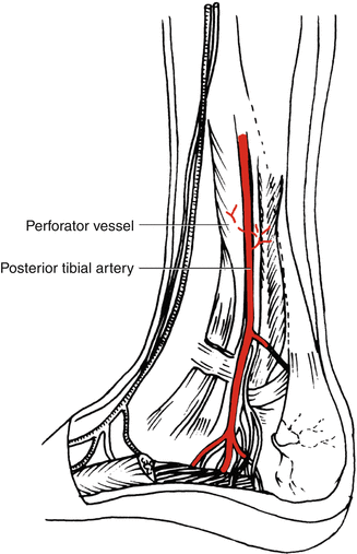
In the medial aspect of the leg, some perforators from the posterior tibial artery are located in the medial aspect of the lower leg and the medial plantar region. Perforators from the posterior tibial artery emerge at periodic intervals between the soleus and flexor digitorum longus muscles. The medial supramalleolar perforator, located 5 cm above the medial malleolus, supplies the medial supramalleolar perforator flap (Fig. 29.1).


Fig. 29.1
Vascular anatomy of the medial supramalleolar perforator flap
29.2 Illustrative Case
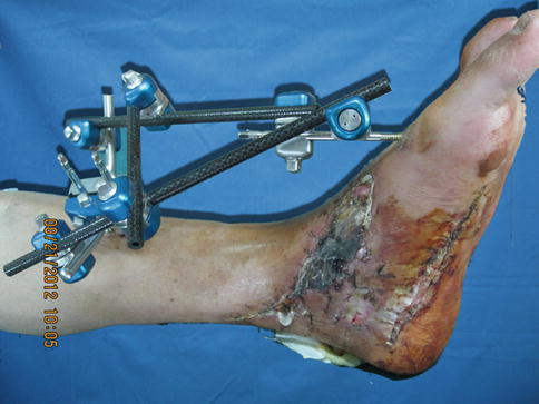
A 56-year-old man sustained a traffic accident injury on his left foot. The resulting wound measured 13 × 4 cm (Fig. 29.2).


Fig. 29.2
Preoperative view
Flap Design
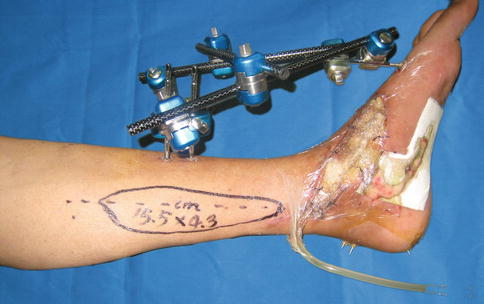
According to the location of the defect, perforators of the posterior tibial vessels were preoperatively selected and marked with a handheld Doppler device. All perforators proximal to the defect were marked, with special attention to the dominant vessel (Fig. 29.3). The flap was designed based on the location of the pivot point of the major perforator. A line was drawn from the malleolus to the medial tibial condyle. The flap was then outlined and centered on this line according to the size of the tissue defect (Fig. 29.4).


Fig. 29.3
Flap design
Stay updated, free articles. Join our Telegram channel

Full access? Get Clinical Tree


