An understanding of the pathophysiology, diagnosis, and management of leg ulcers is very important to health care providers as these occur in a significant number of patients. Approximately 1% to 3% of the population, or up to 9 million people in the United States are affected.1 The annual cost of leg ulcers is proposed to be $8 to $10 billion per year, with an estimated loss of 2 million workdays per year.2 The majority of leg ulcers are seen in middle-aged to elderly patients, and there is a female:male predilection of 2:1. The three most common types of leg ulcers are venous, arterial, and neuropathic.
Ulcers caused by venous insufficiency are the most common type of leg ulcerations, accounting for 70% to 80%. They are sometimes called stasis ulcers. About 10% to 20% of leg ulcerations have a mixed venous and arterial etiology. Leg ulcers caused by chronic venous insufficiency lead to significant morbidity and can have a long-term negative impact on an individual’s quality of life. Diagnosis can be difficult, and management is often expensive and labor-intensive.
Venous ulcers most commonly arise secondary to varicose veins or postphlebitic syndrome. They may also be seen in patients with a history of a deep vein thrombosis (DVT), obesity, or previous leg injury or surgery. When a patient with normal venous return stands or walks, the calf muscle acts in concert with veins and associated valves to empty the venous system and reduce its pressure.3 Venous hypertension develops when the valves become incompetent. This leads to tissue hypoxia and ultimately to skin destruction and breakdown. In addition, wound healing processes are compromised and autolytic processes take action. The result is loss of the epidermis and dermis and the formation of an ulcer.
Most commonly, patients complain of a heavy or swollen feeling in the affected leg. Pain ranges from mild with a superficial ulceration to severe with a deep ulceration. Patients may describe limitation of movement of the affected extremity, depending on the location of the ulcer. In addition, patients with venous stasis and dermatitis may have significant pruritus of the skin surrounding an ulcer.
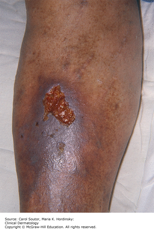
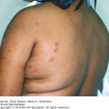
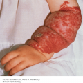
Most patients with venous ulceration have some degree of nonpitting or pitting edema. Varicosities may be visible, and there is often hyperpigmentation from hemosiderin deposition over the shin. Typically, venous ulcers occur over or proximal to the medial malleolus, but they may occur anywhere below the knee. They can be single or multiple, small or large, shallow or deep. They are usually well marginated with sloped borders, but can present with irregular shapes (Figure 29-1). Often, there is fibrinoid material and/or granulation tissue at the base. The surrounding skin may have an inflamed, eczematous appearance. These ulcers can sometimes have copious drainage.
There are no specific laboratory findings that point toward a diagnosis of venous ulceration. However, a complete blood count (CBC), erythrocyte sedimentation count (ESR), and blood glucose can help to diagnose an underlying hematologic, inflammatory, or diabetic condition. A culture will likely yield mixed flora, and may not be relevant unless the wound appears clinically infected. A venous Doppler ultrasound can help to locate venous occlusion or incompetent perforating veins.
The key diagnostic findings of venous ulcers are well-circumscribed ulcerations usually over the shin or medial malleolus, on a backdrop of hyperpigmentation, varicosities, and lower extremity edema. Pedal pulses are usually present. Fibrinoid material or granulation tissue is often observed at the base of the ulcer.
See Table 29-1 for the differential diagnosis of leg ulcers.
Differential diagnosis of leg ulcers.
| Type of Ulcer | Risk Factors | History and Physical | Notes |
|---|---|---|---|
| Venous | Deep venous thrombosis (DVT), varicose veins, previous lower extremity surgery or injury, and obesity | Lower extremity edema, varicosities, hyperpigmentation, and dermatitis, ulcer over shin or medial malleolus. Pulses usually palpable | Compression is the key to treatment |
| Arterial | Peripheral arterial disease, smoking, hyperlipidemia, hypertension, and diabetes | Intermittent claudication, painful ulcer, shiny skin, eschar may be present at base of ulcer, ulcer over distal lower extremities. Pulses usually not palpable | Low threshold for referral to vascular surgery. Do not use compression |
| Neuropathic | Diabetes, spinal cord injury or disease, alcohol abuse, and leprosy | Ulcer over pressure points of plantar foot, surrounded by callus, foot deformities, insensate lower extremities, deep | Prevention is critical as a significant number of these ulcers can lead to amputation |
| Inflammatory | Vasculitis and systemic lupus erythematosus | May have signs and symptoms of systemic inflammatory disease | Systemic workup is appropriate |
| Infectious | Diabetes and obesity | Significant exudate, foul odor, pain, and warmth of surrounding skin | Culture prior to starting treatment with antibiotics |
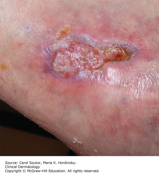
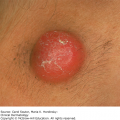
| Pyoderma gangrenosum | Inflammatory bowel disease, arthritis, and myeloproliferative disorder | Irregularly shaped ulcer with undermined edges (Figure 29-2) | Diagnosis of exclusion |
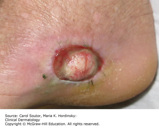
| Malignancy | History of ionizing radiation | Nonhealing ulcer (Figure 29-3) | Must be considered if standard therapy fails |
One must always know the cause of an ulcer before designing a treatment plan. In treating venous ulcers, the primary goal is to reverse venous hypertension so that there is an environment amenable to wound healing.4 The most effective way to accomplish this is with compression, the gold standard for the treatment of venous leg ulcers. Compression reverses venous hypertension, has positive effects on microcirculation, reduces deep venous reflux, reduces lower leg edema, and allows for improved oxygenation of the skin. There are two categories of compression products available: inelastic compression products, which are used for reduction of edema and healing of ulcers, and elastic compression products, which are used for maintenance to prevent ulcer recurrence. The most widely used inelastic products are Unna boots or Profore boots. These are occlusive wraps that are applied as an ace wrap would be applied in the office and removed 1 week later. They may also be applied at the patient’s residence by a trained home health professional. An important companion to the use of these leg wraps is frequent elevation of the legs. Elastic compression is achieved with products such as TEDS or Jobst stockings, which can be worn on a regular basis for maintenance, once a venous ulcer has healed. Compression should always be a component of treating a venous ulcer, but one must rule out the possibility of arterial insufficiency prior to applying a compressive dressing to a patient.
Related posts:
Stay updated, free articles. Join our Telegram channel

Full access? Get Clinical Tree


