Refinement of the wide, ptotic, under protected tip is one of the most difficult challenges in cosmetic nasal surgery yet also among the most common. Although excisional techniques can produce reductions in lobular width, long-term contour alterations are unpredictable and subject to stigmatic tip deformity. Preservation of natural tip support is a fundamental requirement of a successful rhinoplasty. The traditional lateral crural steal is a useful technique for tip refinement, but, when combined with a sturdy septal extension graft, the modified lateral crural steal (lateral crural tensioning) becomes a more potent and versatile rhinoplasty technique that can improve tip contour without jeopardizing function or structural stability.
Key points
- •
Excisional rhinoplasty techniques, such as the cephalic trim maneuver, often alter nasal tip size at the expense of structural stability.
- •
Effective refinement of the wide nasal tip does not mandate aggressive excision of the cephalic margin.
- •
The septal extension graft (SEG) creates a sturdy and stationary platform to allow precise positioning and suspension of the tip cartilage complex.
- •
The lateral crural steal (LCS) borrows from the overly long lateral crura to elongate the foreshortened medial crura to correct the alar cartilage length imbalance typical of the wide and underprojected nasal tip.
- •
In addition to cosmetic benefits of the traditional LCS, lateral crural tensioning (LCT) improves lower nasal sidewall tone and increases the threshold for dynamic nasal valve collapse by preserving the lateral crus and the nasal scroll and by stretching and tensioning the lateral crus.
Background
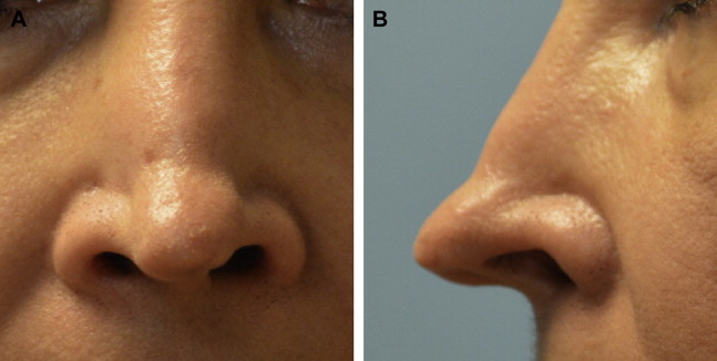
Refining the overly wide nasal tip is among the most common, yet also among the most difficult, challenges in cosmetic rhinoplasty. Until recently, surgical strategies to reduce tip width have been largely dependent on cartilage excision for alterations in lobular size and shape. Despite the immediate and discernable reduction in nasal tip size, aggressive cartilage excision often fails to enhance tip contour in a controlled and predictable manner. As a consequence, aggressive excision-based techniques are increasingly recognized as haphazard, unpredictable, and disproportionately prone to undesirable postoperative contour deformities. The outcome is frequently a nasal tip that is both unattractive and dysfunctional and one that usually deteriorates significantly over time ( Fig. 1 ).
In response to the unacceptably high morbidity of aggressive excisional rhinoplasty techniques, most accomplished rhinoplasty surgeons have adopted strategies that preserve tip cartilage and/or augment skeletal tip support, thereby improving long-term contour stability and airway patency. Although this trend is rapidly spreading among rhinoplasty enthusiasts, the number of failed rhinoplasty outcomes stemming from cartilage over-resection seems to be growing rapidly, suggesting that aggressive excisional techniques are still practiced widely even today. Nonetheless, there are now safe and effective alternatives to excisional rhinoplasty in which little if any tip cartilage excision is required. These techniques seek to preserve the existing tip cartilage and to alter tip contour via suture techniques, cartilage repositioning, and/or augmentation grafting to achieve an elegant and stable tip contour. And because the overly wide nasal tip is perhaps the most common morphology prompting cosmetic tip surgery, mastery of nonexcisional/structurally based rhinoplasty techniques is essential for the contemporary rhinoplasty surgeon.
The lateral crural steal (LSC) is the pejorative name given to an effective and tissue-conservative technique of nasal tip refinement. Resurrected in the contemporary rhinoplasty literature by Kridel and colleagues in 1989, the traditional LCS achieves several cosmetic improvements with one comparatively simple surgical maneuver: relocation of the domal apices. Moreover, unlike excisional rhinoplasty techniques, the traditional LCS is not contingent on aggressive cartilage excision to achieve tip refinement. Instead, the LCS uses redistribution and/or repositioning of the existing skeletal elements to derive a more attractive, stable, and functional tip configuration. Although a modest amount of cartilage must be excised from the nasal dome when performing an aggressive LCS, cartilage removal is confined to the medial-most aspect of the lateral crus in an area of comparatively minimal structural consequence, thereby preserving virtually all of the naturally derived skeletal support. And, when the traditional LCS is used in combination with a SEG, the LCS/SEG combination—herein referred to as LCT—becomes a far more potent and versatile surgical workhorse for tip refinement. With skillful execution, LCT not only achieves contour elegance with reliable long-term contour stability but also serves to protect or improve nasal valve patency.
The overly wide nasal tip is perhaps the most common tip malformation encountered in cosmetic nasal surgery. Although excess tip width may occur in isolation, it more commonly occurs in combination with inadequate tip projection and/or tip ptosis (ie, inadequate tip rotation). Historically, treatment of the wide, underprojected, and ptotic nasal tip—herein referred to as the compound tip deformity (CTD)—has been directed at volume reduction of the nasal tip cartilages. However, the CTD stems from more than just oversized tip cartilages, and volumetric reduction alone seldom achieves a satisfactory tip contour. Optimal refinement of the CTD necessitates correction of each anatomic malformation contributing to the unsightly tip morphology, not just volume reduction. For the CTD, excessive rounding of the nasal domes, excessive divergence of the nasal domes, and a length imbalance between the medial and lateral crura must all be corrected to achieve an elegant and natural-appearing tip contour. Rounding of the domal arches and excessive separation of the domal apices have both long been recognized as a major source of excessive lobular width, even when alar cartilage length is normal. And when the transverse (vertical) height of the lateral crura is also excessive, additional lateral crural deformity, characterized by increased convexity of the entire crural span, exacerbates the CTD by creating a wide supratip and/or an unsightly polly beak fullness of the supratip ( Fig. 2 ). A less commonly recognized abnormality of the CTD, however, is the length imbalance between the medial and lateral crura created by medial displacement of the domal apices (ie, the tip defining points [TDPs]). Length discrepancies between the medial and lateral crura and their effects on positioning of the TDPs have been previously described by Adamson and colleagues in their delineation of the M-Arch model of tip dynamics. In the healthy and attractive nasal tip, longitudinal stiffness of the lateral crura thrusts the tip anteriorly and inferiorly. This is counterbalanced by the opposing anterior and superior thrust of the medial crura to create both equilibrium and stability within the lower lateral cartilage (LLC) arch. The equilibrium is further stabilized by the surrounding soft tissues. In the CTD, however, these relationships are anomalous. Although the overall length of the widened LLC arch is often normal or near normal, in the CTD, the nasal domes (and thus the TDPs) are skewed medially, resulting in abnormally long lateral crura and disproportionately short medial crura ( Fig. 3 ). Overly long lateral crura bow outward and exaggerate the downward tip displacement creating a ptotic tip configuration and excessive width in the tip and supratip. In a review of 500 consecutive cases of nasal tip ptosis, Foda found inferiorly oriented alar cartilages were the main cause of tip ptosis in 85% of patients presenting with a drooping tip. The CTD is also frequently exacerbated by pronounced convex cupping (ie, bulbosity) of the lateral crura, both longitudinally and transversely, which not only adds to lobular width but also dramatically increases supratip fullness ( Fig. 4 ). Ironically, although bulbous cupping of the lateral crura increases crural stiffness, and therefore enhances lower nasal sidewall support, bulbosity also creates a highly objectionable cosmetic deformity that frequently prompts over-resection of the lateral crura and subsequent destabilization of the tip architecture. The anatomic counterpart to overly long lateral crura is overly short medial crura. Medial displacement of the domal breakpoint results in medial crura that are abnormally short and stubby, exacerbating the CTD with inadequate projection of the nasal tip (see Fig. 3 ). Moreover, inadequate tip projection is compounded by secondary splaying of the alar base, which further exacerbates the unsightly width deformity. Perhaps the most extreme example of alar cartilage maldistribution is the unilateral cleft-lip nasal deformity. In the unilateral cleft-lip nose, a severe ipsilateral length disparity between the foreshortened medial crus and the elongated lateral crus results from lateral, inferior, and posterior displacement of the ipsilateral alar base. This developmental deformity is best corrected by repositioning the ectopic alar base and redistributing the malformed LLC with a unilateral LCS-type domal repositioning.
Background
Refining the overly wide nasal tip is among the most common, yet also among the most difficult, challenges in cosmetic rhinoplasty. Until recently, surgical strategies to reduce tip width have been largely dependent on cartilage excision for alterations in lobular size and shape. Despite the immediate and discernable reduction in nasal tip size, aggressive cartilage excision often fails to enhance tip contour in a controlled and predictable manner. As a consequence, aggressive excision-based techniques are increasingly recognized as haphazard, unpredictable, and disproportionately prone to undesirable postoperative contour deformities. The outcome is frequently a nasal tip that is both unattractive and dysfunctional and one that usually deteriorates significantly over time ( Fig. 1 ).

In response to the unacceptably high morbidity of aggressive excisional rhinoplasty techniques, most accomplished rhinoplasty surgeons have adopted strategies that preserve tip cartilage and/or augment skeletal tip support, thereby improving long-term contour stability and airway patency. Although this trend is rapidly spreading among rhinoplasty enthusiasts, the number of failed rhinoplasty outcomes stemming from cartilage over-resection seems to be growing rapidly, suggesting that aggressive excisional techniques are still practiced widely even today. Nonetheless, there are now safe and effective alternatives to excisional rhinoplasty in which little if any tip cartilage excision is required. These techniques seek to preserve the existing tip cartilage and to alter tip contour via suture techniques, cartilage repositioning, and/or augmentation grafting to achieve an elegant and stable tip contour. And because the overly wide nasal tip is perhaps the most common morphology prompting cosmetic tip surgery, mastery of nonexcisional/structurally based rhinoplasty techniques is essential for the contemporary rhinoplasty surgeon.
The lateral crural steal (LSC) is the pejorative name given to an effective and tissue-conservative technique of nasal tip refinement. Resurrected in the contemporary rhinoplasty literature by Kridel and colleagues in 1989, the traditional LCS achieves several cosmetic improvements with one comparatively simple surgical maneuver: relocation of the domal apices. Moreover, unlike excisional rhinoplasty techniques, the traditional LCS is not contingent on aggressive cartilage excision to achieve tip refinement. Instead, the LCS uses redistribution and/or repositioning of the existing skeletal elements to derive a more attractive, stable, and functional tip configuration. Although a modest amount of cartilage must be excised from the nasal dome when performing an aggressive LCS, cartilage removal is confined to the medial-most aspect of the lateral crus in an area of comparatively minimal structural consequence, thereby preserving virtually all of the naturally derived skeletal support. And, when the traditional LCS is used in combination with a SEG, the LCS/SEG combination—herein referred to as LCT—becomes a far more potent and versatile surgical workhorse for tip refinement. With skillful execution, LCT not only achieves contour elegance with reliable long-term contour stability but also serves to protect or improve nasal valve patency.
The overly wide nasal tip is perhaps the most common tip malformation encountered in cosmetic nasal surgery. Although excess tip width may occur in isolation, it more commonly occurs in combination with inadequate tip projection and/or tip ptosis (ie, inadequate tip rotation). Historically, treatment of the wide, underprojected, and ptotic nasal tip—herein referred to as the compound tip deformity (CTD)—has been directed at volume reduction of the nasal tip cartilages. However, the CTD stems from more than just oversized tip cartilages, and volumetric reduction alone seldom achieves a satisfactory tip contour. Optimal refinement of the CTD necessitates correction of each anatomic malformation contributing to the unsightly tip morphology, not just volume reduction. For the CTD, excessive rounding of the nasal domes, excessive divergence of the nasal domes, and a length imbalance between the medial and lateral crura must all be corrected to achieve an elegant and natural-appearing tip contour. Rounding of the domal arches and excessive separation of the domal apices have both long been recognized as a major source of excessive lobular width, even when alar cartilage length is normal. And when the transverse (vertical) height of the lateral crura is also excessive, additional lateral crural deformity, characterized by increased convexity of the entire crural span, exacerbates the CTD by creating a wide supratip and/or an unsightly polly beak fullness of the supratip ( Fig. 2 ). A less commonly recognized abnormality of the CTD, however, is the length imbalance between the medial and lateral crura created by medial displacement of the domal apices (ie, the tip defining points [TDPs]). Length discrepancies between the medial and lateral crura and their effects on positioning of the TDPs have been previously described by Adamson and colleagues in their delineation of the M-Arch model of tip dynamics. In the healthy and attractive nasal tip, longitudinal stiffness of the lateral crura thrusts the tip anteriorly and inferiorly. This is counterbalanced by the opposing anterior and superior thrust of the medial crura to create both equilibrium and stability within the lower lateral cartilage (LLC) arch. The equilibrium is further stabilized by the surrounding soft tissues. In the CTD, however, these relationships are anomalous. Although the overall length of the widened LLC arch is often normal or near normal, in the CTD, the nasal domes (and thus the TDPs) are skewed medially, resulting in abnormally long lateral crura and disproportionately short medial crura ( Fig. 3 ). Overly long lateral crura bow outward and exaggerate the downward tip displacement creating a ptotic tip configuration and excessive width in the tip and supratip. In a review of 500 consecutive cases of nasal tip ptosis, Foda found inferiorly oriented alar cartilages were the main cause of tip ptosis in 85% of patients presenting with a drooping tip. The CTD is also frequently exacerbated by pronounced convex cupping (ie, bulbosity) of the lateral crura, both longitudinally and transversely, which not only adds to lobular width but also dramatically increases supratip fullness ( Fig. 4 ). Ironically, although bulbous cupping of the lateral crura increases crural stiffness, and therefore enhances lower nasal sidewall support, bulbosity also creates a highly objectionable cosmetic deformity that frequently prompts over-resection of the lateral crura and subsequent destabilization of the tip architecture. The anatomic counterpart to overly long lateral crura is overly short medial crura. Medial displacement of the domal breakpoint results in medial crura that are abnormally short and stubby, exacerbating the CTD with inadequate projection of the nasal tip (see Fig. 3 ). Moreover, inadequate tip projection is compounded by secondary splaying of the alar base, which further exacerbates the unsightly width deformity. Perhaps the most extreme example of alar cartilage maldistribution is the unilateral cleft-lip nasal deformity. In the unilateral cleft-lip nose, a severe ipsilateral length disparity between the foreshortened medial crus and the elongated lateral crus results from lateral, inferior, and posterior displacement of the ipsilateral alar base. This developmental deformity is best corrected by repositioning the ectopic alar base and redistributing the malformed LLC with a unilateral LCS-type domal repositioning.

The cephalic trim
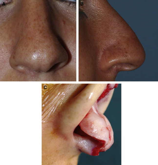
Historically, a variety of surgical techniques have been advocated for refinement of the overly wide nasal tip. Perhaps the least effective technique for tip refinement is the cephalic trim maneuver. The cephalic trim maneuver seeks to simultaneously narrow, refine, and rotate the ptotic and overly wide nasal tip simply by resecting the cephalic margin of both lateral crural cartilages. In theory, precise and judicious trimming of the cephalic margin strategically weakens the lateral crura leading to a refined and slightly rotated nasal tip, but only if the volume and location of the excised crural cartilage correspond perfectly to the required distribution and degree of structural weakening. In reality, determining how much cartilage can be safely excised without triggering secondary crural deformities is virtually impossible, and over-resections are commonplace. Because the average lateral crus measures only approximately 12 mm in (vertical) width, even the generally accepted residual crural width of 6.0 mm preserves only approximately half of the original crural height. Furthermore, because lateral crural thickness averages only 0.7 mm, resecting half of the crural height often results in a narrow and flimsy crural remnant that is incapable of supporting either the nasal tip or the lower nasal sidewall. Because LLC stiffness is a primary component of tip contour and support, an overaggressive cephalic resection can destabilize the tip architecture with disastrous consequences. The eventual result is often severe distortion of the nasal tip leading to lobular pinching, alar retraction, bossae formation, asymmetry, excessive tip rotation, unwanted loss of tip projection, and/or symptomatic nasal valve collapse. Patients with naturally weak tip cartilage are at disproportionally high risk for morbidity after the cephalic trim maneuver because the tip is already at or near the threshold for collapse, and these patients often develop unsightly tip deformities despite comparatively modest cephalic resections. Moreover, tip width does not correlate with cartilage stiffness, and overly pliable, weak tip cartilages are often encountered in ultrawide bulbous noses. Ironically, the combination of weak tip cartilage and a comparatively severe cosmetic deformity often prompts overzealous treatment and subsequent tip deformity. Similarly, over-resection of the cephalic margin is also more likely to distort the tip architecture in noses with extremes of skin thickness. In the thin-skinned nose, shrink-wrap contracture is often forceful and sustained, leading to a higher incidence of bossae, buckling, and alar retraction. However, the morbidity of over-resection is also exacerbated by ultrathick skin that adds additional weight to the frail and surgically dilapidated tip framework ( Fig. 5 ). Ironically, severe crural over-resection may not become immediately evident in the thick-skinned nose because postoperative swelling—which is typically more severe and longer lasting in thick nasal skin—may conceal the initial tip deformity for many months. However, as the swelling subsides and the surgically weakened tip framework is subjected to the sustained and potent forces of fibrosis combined with the repetitive inward sidewall flexion generated during nasal inspiration, stigmatic tip deformities and/or functional impairment eventually become evident. Finally, even when the cephalic trim fails to initially exceed the threshold for skeletal collapse, age or disease-related deterioration in crural stiffness may also lead to eventual tip deformities, particularly because many surgeons fail to account for future losses in cartilage strength when planning crural resections. Although tip suturing techniques are now commonly used in combination with the cephalic trim for tip refinement, the inappropriate or overzealous use of tip sutures can themselves cause postsurgical tip deformities, and aggressive resections of the cephalic margin usually serve to increase the likelihood of such problems. Owing to the synergistic and destabilizing triad of (1) surgically compromised structural support, (2) chronic deformational forces of wound healing, and (3) age-related losses in cartilage strength, the adverse effects of crural over-resection frequently worsen for decades, making the cephalic trim a risky undertaking associated with considerable long-term morbidity in susceptible patients. And when crural over-resection is combined with over-resection of the anterior nasal septum, which undergirds and supports the tip complex, virtually all of the adverse consequences of the cephalic trim are intensified.
Overly long lateral crura—a frequently neglected deformity
Treating the constellation of LLC deformities that characterizes the CTD—in particular, the overly wide tip cartilages and the abnormalities of domal shape and spacing—has improved greatly in the past 3 decades. Structural techniques that enhance skeletal support for improved contour stability and nondestructive suture-based techniques that reshape and reposition malformed or malpositioned tip cartilages have transformed the quality and long-term predictability of tip rhinoplasty. Over this same time period, however, comparatively little attention has been directed at another important anatomic deformity common to the CTD: the crural length disparity that results from malposition of the nasal domes. Despite the adverse impact on tip aesthetics, maldistribution of tip cartilage is a critical aberration of tip architecture that can dramatically affect lobular contour, supratip contour, tip support, sidewall aesthetics, and nasal valve function, yet one that is often overlooked, undertreated, and/or mismanaged. And although placement of a columellar strut graft or an SEG enhances central tip support by augmenting medial crural length, such techniques alone fail to treat the corresponding excess in lateral crural length that results from malposition of the apical fold. The persistent excess length of the lateral crura coupled with their caudally directed forces of tip displacement may explain the failure of columellar strut grafts to consistently maintain tip projection. Similarly, the unrecognized crural length surplus may also explain the continued use of overaggressive cephalic resections in a misguided and ill-fated attempt to eliminate unwanted supratip fullness. And when over-resection of the oversized lateral crus does occur, a flail segment usually ensues because the excess crural length remains unreconciled. Although some surgeons have advocated lateral crural overlap (LCO) techniques, in which the lateral crura are divided vertically, overlapped by several millimeters to reduce crural length, and then reattached with mattress sutures, vertical sectioning of the alar cartilage, although effective at truncating crural length, reduces tip projection and potentially destabilizes the lateral crural span – both consequences that can be avoided entirely with the use of the LCS. Moreover, in a 500-patient (consecutive) series comparing the standard LCS and the LCO technique for treatment of the ptotic and underprojected nasal tip, the LCS was deemed preferable because tip projection and rotation were both increased simultaneously. This confirmed findings of previous work in which the LCS was preferred over the LCO for simultaneous increases in tip projection and tip rotation. Regardless of the preferred treatment method, excessive lateral crural length is an often-ignored yet fundamental anomaly of the CTD that has a profound impact on form and function of the nasal base; and failure to shorten the overly long lateral crus while maintaining the structural integrity of the lateral crural span inevitably taints an otherwise satisfactory surgical outcome.
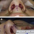
Sidewall tension—the unrecognized benefit of the lateral crural steal
Unlike the cephalic trim technique, which sacrifices natural skeletal support and ignores the crural length discrepancy, thereby converting a wide and overly prominent lateral crural span into a collapsed and flail segment vulnerable to distortion from scar contracture, the traditional LCS restores balanced and aesthetically pleasing crural proportions by lengthening the undersized medial crura at the expense of the overly long lateral crura ( Fig. 6 ). The redistribution of tip cartilage is accomplished without excising large segments of the cephalic margin or vertically dividing the LLC but simply by relocating the natural domal fold (or apex) that establishes the breakpoint between the medial and lateral crus and which delineates the TDP. Relocating the domal fold and creating a neodome results in several simultaneous functional and cosmetic benefits. First, as the relocated nasal domes are approximated in the midline, tip width is substantially reduced. Spacing of the neodomes and acuity of the domal angles can also be independently adjusted with tip sutures to fine-tune lobular width according to variations in skin thickness and cosmetic preferences. Second, neodomal approximation also simultaneously increases both tip rotation and tip projection as the length imbalance between the medial and lateral crura is eliminated. Thus, with a single nondestructive maneuver, the traditional LCS addresses all 3 major cosmetic abnomalities of the CTD—excessive lobular width, tip ptosis, and inadequate tip projection. And because each neodome is constructed independently, modest preexisting asymmetries in domal arch projection and/or tip rotation can be offset with differential dome positioning. Finally, when the LCS is used for aggressive increases in tip projection, a secondary reduction in nasal base width often occurs as an additional cosmetic benefit of alar cartilage redistribution.
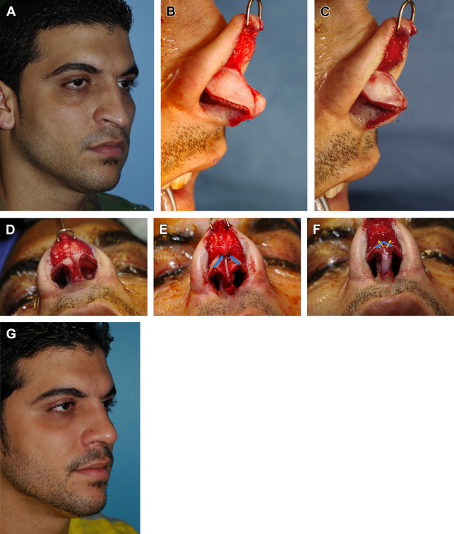
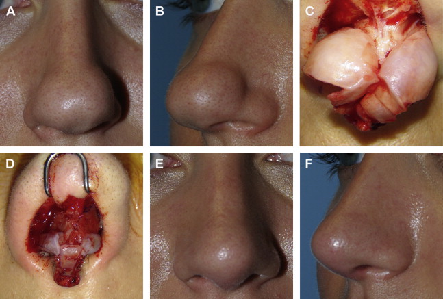
One of the most important but unrecognized benefits of an aggressive LCS involves secondary improvements in nasal tip dynamics. As the neodomes are suture approximated in the midline, longitudinal tensioning forces are generated that stretch and tighten both lateral crura. Unlike many other contemporary tip refinement strategies that rely on bulky structural grafts, such as the lateral crural strut graft (LCSG) or the crural batten graft, to contour and support the lax lower nasal sidewall (with or without cephalic resection), the LCT approach to tip refinement exploits the tensioning forces generated from tip refinement to increase crural rigidity and subsequently to strengthen and contour the lower nasal sidewall. And because lateral crural augmentation grafts are frequently obviated, limited graft materials are conserved, and the additional weight and mass effect of structural grafts can be avoided. Because LCT also shortens and tightens the lateral crura without the need for cephalic resection, the entire nasal scroll and its sizable contribution to sidewall support are also preserved. And because the nasal scroll lies at the epicenter of the internal nasal valve—a dynamic flow-regulating apparatus that is sensitive to even minor reductions in cross-sectional area resulting from bulky sidewall grafts or crural over-resection—the nondestructive LCT maneuver is far less likely to disrupt nasal airflow. Moreover, additional cosmetic enhancements are also derived from LCT. Because the lateral crura are tethered laterally at the piriform aperture, tensioning forces created by LCT also stretch and flatten the lateral crura with a noticeable reduction in crural convexity and bulbosity, particularly in patients with weak tip cartilage. The result of this sidewall tensioning effect is a more slender and elegant supra-tip contour, accompanied by a concomitant increase in resting sidewall tone and a corresponding increase in the threshold for dynamic nasal valve collapse. Hence, unlike most other contemporary strategies for treating the CTD, the LCT approach also simultaneously enhances nasal valve physiology by (1) preserving virtually all of the existing natural skeletal support, (2) eliminating laxity derived from excess lateral crural length, and (3) increasing lower nasal sidewall tone with tensioning forces—all without the use of lateral crural augmentation grafts. In previously operated noses presenting with concave sagging of the lower nasal sidewall from lateral crural over-resection, the tensioning forces generated by LCT also serve to lift and tighten flail crural segments, thereby minimizing unsightly sidewall pinching while dramatically enlarging internal nasal valve dimensions. Similarly, sidewall tensioning can also be used to prevent and/or minimize alar retraction. In primary rhinoplasty, stretching and tightening of the lateral crura with LCT not only flattens the crura but also creates a guitar string effect that opposes upward displacement from scar contracture. And because sidewall tensioning generally obviates a traditional cephalic trim, preservation of the full vertical height of the lateral crus further buttresses the alar rim against vertical scar contracture. In the over-resected tip presenting with iatrogenic alar retraction, sidewall tensioning, combined with lysis of cephalic adhesions and unfurling of the contractured internal lining, can stabilize the repositioned crural remnant against recurrent retraction ( Fig. 7 ), particularly if the crural remnant is also further supported with modified alar rim grafts. Although severe alar retraction may require more-aggressive techniques to stabilize the alar rim, such as the LCSG, with or without lateral crural repositioning, aggressive sidewall tensioning alone is sufficient in a large percentage of cases. However, the use of LCT does not preclude the combined use of the LCSG for the treatment of severe alar retraction, and because the mechanisms of alar rim stabilization are compatible with LCT, the combined use of LCT and LCSG is likely to be more effective, albeit with a greater risk of internal nasal valve impingement from LCSG bulk. In summary, LCT mimics the natural dynamics of an attractive and fully functional nasal sidewall by stiffening the existing crural cartilage and raising the threshold for dynamic internal valve collapse, all while maintaining a thin, lightweight, and flexible nasal sidewall—a particularly useful benefit when treating the long and ultraslender nose where LCSGs may compromise internal valve patency, efface the supra-alar crease, and/or partially restrict mimetic movement. LCT also expands the already potent cosmetic benefits of the traditional LCS by flattening the entire lateral crus to eliminate unsightly fullness of the supratip. Hence, by reallocating and reshaping the LLC using almost entirely reversible suture techniques, LCT can custom-contour the CTD while enhancing or preserving airway function and reducing the dependence on large structural grafts.