, Shimin Chang2, Jian Lin3 and Dajiang Song1
(1)
Department of Orthopedic Surgery, Changzheng Hospital Second Military Medical University, Shanghai, China
(2)
Department of Orthopedic Surgery, Yangpu Hospital Tongji University School of Medicine, Shanghai, China
(3)
Department of Microsurgery, Xinhu Hospital Shanghai Jiao Tong University, Shanghai, China
The lateral arm flap was first described by Song et al. in 1982 [1].
These indications have been well described by Katsaros et al. [2], Rivet et al. [3], and Scheker et al. [4] for reconstruction of the extremities and the head and neck.
8.1 Vascular Anatomy
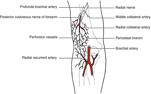
The lateral upper arm flap is supplied by septocutaneous branches of the posterior radial collateral artery (PRCA), which develops from the profunda brachii artery. The perforators of the flap run within the lateral intermuscular septum, which separates the brachialis from the triceps muscle (Fig. 8.1).


Fig. 8.1
Vascular anatomy of the lateral upper arm flap
In close proximity to the radial nerve, the vascular pedicle spirals around the humerus, and proximal to the lateral intermuscular septum, it divides into the small anterior and the stronger posterior radial collateral artery (PRCA). Whereas the small anterior radial collateral artery runs together with the radial nerve, the PRCA is the main nutrient artery of the flap, giving off the septocutaneous branches. After having traversed the septum at its base, the PRCA anastomoses with the interosseous recurrent artery, on which the flap can be perfused in a retrograde fashion.
The posterior cutaneous nerve of the arm (PCNA), which accompanies the PRCA, can be used to create sensate flaps [1, 5, 6].
Harvesting a cortical segment of the humerus is technically possible, but only to a size of 10 × 1 cm, leaving a muscle cuff on either side of the septum to include periosteal vessels of the PRCA [7].
8.2 Illustrative Case
A 33-year-old male underwent a traumatic severe crush injury of the left index finger. Emergency debridement was performed in another hospital. The patient refused to undergo toe pulp flap transplantation. So a free-arm flap was selected.
Flap Design
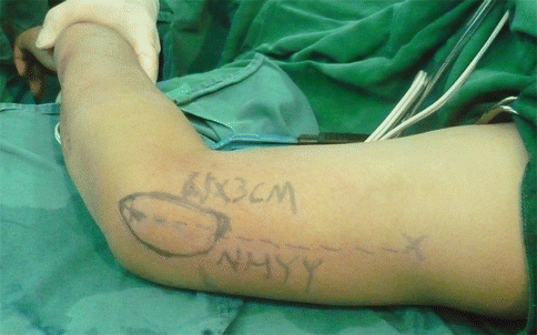
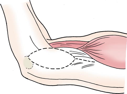
Draw a line from the insertion of the deltoid muscle to the lateral epicondyle of the humerus, corresponding to the lateral intermuscular septum (LIM), where the dominant pedicle and venae comitantes are found (Fig. 8.2). The perforator was identified preoperatively by using an 8 MHz handheld Doppler (Fig. 8.3).


Fig. 8.2
Harvest a lateral arm perforator flap for finger coverage. (The case is offered by Professor Songlin Xie and Dr. Xiangwu Deng, Hand Surgical Centre, Nanhua Hospital, Nanhua University, Hengyang, China)

Fig. 8.3
Schematic drawing of the flap design
Flap Elevation
Flap elevation began from the posterior side and moved toward the anterior side through the supracutaneous tissue plane. Once the perforator was identified, the lateral intermuscular septum was divided. The intermuscular septum and a tiny ellipse of deep fascia cuff were preserved around the radial collateral vessel and its skin perforator on the skin flap. The lateral cutaneous nerve of the arm was identified and contained in the flap. The entire flap was isolated (Fig. 8.4


Stay updated, free articles. Join our Telegram channel

Full access? Get Clinical Tree


