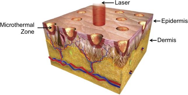
The demand for facial rejuvenation and cosmetic procedures is rising among all ethnicities and skin types. The authors present a review of lasers and how to select a laser based on skin type and the treatment goals of laser resurfacing: skin laxity, dyschromia, hair removal, keloid, and hypertrophic scarring. In addition, they discuss preprocedural and postprocedural considerations, potential complications, and their management to maximize patient outcomes and minimize risk.
Key points
- •
Ethnic skin presents a unique challenge for laser skin rejuvenation because of higher density of larger melanosomes, thicker collagen bundles, and increased fibroblast responses.
- •
Lasers may be safely used in patients with dark skin tones by choosing fractional technologies with longer wavelengths, lower fluences, and longer pulse durations.
- •
The risks of laser therapy include scarring, postinflammatory hyperpigmentation, and hypopigmentation.
- •
Developing careful treatment plans based on patient goals and maintaining careful attention to preprocedural and postprocedural management strategies can minimize the risk of complications.
- •
In the hands of an experienced laser surgeon, laser resurfacing in dark skin types may improve the appearance of fine wrinkles and even skin tone, texture, and pigmentation.
Introduction
In the last decade, there has been an increase in the use of lasers for facial skin rejuvenation. Owing to improved technologies, patients are able to confront dermatologic concerns in an office-based setting with outpatient procedures. Conditions such as photoaging, acne vulgaris, and dyschromia can be treated with laser therapy, with improved risk profiles and decreased recovery times. Although the demand for facial rejuvenation and cosmetic procedures continues to increase among all ethnic populations and skin types, not all patients and skin types are the same and there is no one-size-fits-all treatment algorithm. In addition, the complications of therapy vary between skin types, and careful attention must be paid to these reaction patterns and specific treatment options.
Skin types and colors are divided into 6 phototypes, Fitzpatrick skin types I through VI, with I being the fairest and VI being the darkest ( Table 1 ). Within a single ethnicity, there may be variable phototypes, and it is important to tailor the treatment to the patient. The number of melanocytes is consistent throughout all ethnicities. Melanocytes derive from neural crest cells and transfer melanosomes, which contain melanin, into keratinocytes. The color of skin depends on the density, size, and activity of melanosomes, as darker skin has a higher density of larger melanosomes. In addition, darker skin types, Fitzpatrick types V and VI, have thicker and more compact skin layers with thicker collagen bundles, which increase the epidermal barrier and reduce skin sensitivity ( Fig. 1 ). This barrier delays skin damage from the environment and ultraviolet radiation and aging in darker phototypes when compared with lighter skin types. Due to these histologic differences, dark skin is at increased risk for injury due to incidental laser absorption by melanin, problems with postinflammatory hyperpigmentation, and decrease in melanin production leading to hypopigmentation.
| Fitzpatrick Skin Type | Skin Characteristics | Sun Exposure |
|---|---|---|
| I | Pale white skin; blonde or red hair; blue eyes; freckles | Burns easily, never tans |
| II | White fair skin; blonde or red hair; blue, green, hazel eyes | Burns easily, tans minimally with difficulty |
| III | Cream white skin; any hair or eye color | Burns moderately, tans moderately and uniformly |
| IV | Moderate brown skin, Mediterranean | Burns minimally, tans moderately and easily |
| V | Dark brown skin, Middle Eastern | Rarely burns, tans profusely |
| VI | Deeply pigmented dark brown to black | Never burns, tans profusely |

Although there are many types of lasers, the fundamental principle is the same: all lasers treat the skin by targeting a specific chromophore. The main chromophores of the skin are hemoglobin, melanin, and water. In general, resurfacing lasers are designed at specific wavelengths that use water as a chromophore to cause targeted thermal damage in the dermis to promote new collagen formation and skin tightening. Other targetable chromophores include melanin, which has a broad, but gradually decreasing, absorption coefficient from 250 to 1200 nm. The selection of a laser with a longer wavelength can allow for targeting of deep melanin or tattoo pigmentation in darker skin types.
Other variables important to lasers include the thermal relaxation time, pulse duration, and energy fluence ( Table 2 ). The thermal relaxation time is the time required for a tissue to cool to half the temperature to which it was heated. Heating the tissue for time longer than the thermal relaxation time can cause thermal damage to surrounding tissue. In dark-skinned individuals, it is important to select a pulse duration longer than the thermal relaxation time of the epidermis but shorter than the target chromophore to avoid epidermal blistering, crusting, pigmentation changes, and scarring. The fluence is the joules per square centimeter of energy emitted from the laser handpiece. The laser fluence may need to be decreased to protect the epidermis to safely treat patients with darker skin types compared with those with lighter skin types. Other helpful strategies in safely treating patient of color include longer wavelengths, longer pulse durations, and skin cooling before, during, and/or after the procedure to avoid overheating the epidermis.
| Variable | Function | Example |
|---|---|---|
| Chromophore | Laser target molecule, unique absorption spectrum and peak absorption wavelength | Hemoglobin, melanin, water |
| Wavelength | Property of light measured in nanometers that influences how chromophores are targeted | Hemoglobin (variable absorption from 300 nm to infrared) Melanin (gradually decreasing absorption from 250 to 1200 nm) Water (1000 to 1 mm) |
| Thermal relaxation time | Time required for tissue to cool to half the temperature to which it was heated | Melanosome (250 ns) Vessels (2–10 ms) Hair follicles (100 ms) |
| Pulse duration | Time to heat tissue to target tissue; choose pulse duration less than or equal to thermal relaxation time of target chromophore to avoid damage to surrounding tissue | Pulse duration 10 to 100 ns to target melanosome |
| Energy fluence | Joules per square centimeter of energy emitted by a pulsed laser device | 25 J/cm 2 used by a 1064-nm Nd:YAG for laser hair removal; highest tolerated fluences are 100 J/cm 2 (skin types IV, V) and 50 J/cm 2 (skin type VI) |
Introduction
In the last decade, there has been an increase in the use of lasers for facial skin rejuvenation. Owing to improved technologies, patients are able to confront dermatologic concerns in an office-based setting with outpatient procedures. Conditions such as photoaging, acne vulgaris, and dyschromia can be treated with laser therapy, with improved risk profiles and decreased recovery times. Although the demand for facial rejuvenation and cosmetic procedures continues to increase among all ethnic populations and skin types, not all patients and skin types are the same and there is no one-size-fits-all treatment algorithm. In addition, the complications of therapy vary between skin types, and careful attention must be paid to these reaction patterns and specific treatment options.
Skin types and colors are divided into 6 phototypes, Fitzpatrick skin types I through VI, with I being the fairest and VI being the darkest ( Table 1 ). Within a single ethnicity, there may be variable phototypes, and it is important to tailor the treatment to the patient. The number of melanocytes is consistent throughout all ethnicities. Melanocytes derive from neural crest cells and transfer melanosomes, which contain melanin, into keratinocytes. The color of skin depends on the density, size, and activity of melanosomes, as darker skin has a higher density of larger melanosomes. In addition, darker skin types, Fitzpatrick types V and VI, have thicker and more compact skin layers with thicker collagen bundles, which increase the epidermal barrier and reduce skin sensitivity ( Fig. 1 ). This barrier delays skin damage from the environment and ultraviolet radiation and aging in darker phototypes when compared with lighter skin types. Due to these histologic differences, dark skin is at increased risk for injury due to incidental laser absorption by melanin, problems with postinflammatory hyperpigmentation, and decrease in melanin production leading to hypopigmentation.
| Fitzpatrick Skin Type | Skin Characteristics | Sun Exposure |
|---|---|---|
| I | Pale white skin; blonde or red hair; blue eyes; freckles | Burns easily, never tans |
| II | White fair skin; blonde or red hair; blue, green, hazel eyes | Burns easily, tans minimally with difficulty |
| III | Cream white skin; any hair or eye color | Burns moderately, tans moderately and uniformly |
| IV | Moderate brown skin, Mediterranean | Burns minimally, tans moderately and easily |
| V | Dark brown skin, Middle Eastern | Rarely burns, tans profusely |
| VI | Deeply pigmented dark brown to black | Never burns, tans profusely |
Although there are many types of lasers, the fundamental principle is the same: all lasers treat the skin by targeting a specific chromophore. The main chromophores of the skin are hemoglobin, melanin, and water. In general, resurfacing lasers are designed at specific wavelengths that use water as a chromophore to cause targeted thermal damage in the dermis to promote new collagen formation and skin tightening. Other targetable chromophores include melanin, which has a broad, but gradually decreasing, absorption coefficient from 250 to 1200 nm. The selection of a laser with a longer wavelength can allow for targeting of deep melanin or tattoo pigmentation in darker skin types.
Other variables important to lasers include the thermal relaxation time, pulse duration, and energy fluence ( Table 2 ). The thermal relaxation time is the time required for a tissue to cool to half the temperature to which it was heated. Heating the tissue for time longer than the thermal relaxation time can cause thermal damage to surrounding tissue. In dark-skinned individuals, it is important to select a pulse duration longer than the thermal relaxation time of the epidermis but shorter than the target chromophore to avoid epidermal blistering, crusting, pigmentation changes, and scarring. The fluence is the joules per square centimeter of energy emitted from the laser handpiece. The laser fluence may need to be decreased to protect the epidermis to safely treat patients with darker skin types compared with those with lighter skin types. Other helpful strategies in safely treating patient of color include longer wavelengths, longer pulse durations, and skin cooling before, during, and/or after the procedure to avoid overheating the epidermis.
| Variable | Function | Example |
|---|---|---|
| Chromophore | Laser target molecule, unique absorption spectrum and peak absorption wavelength | Hemoglobin, melanin, water |
| Wavelength | Property of light measured in nanometers that influences how chromophores are targeted | Hemoglobin (variable absorption from 300 nm to infrared) Melanin (gradually decreasing absorption from 250 to 1200 nm) Water (1000 to 1 mm) |
| Thermal relaxation time | Time required for tissue to cool to half the temperature to which it was heated | Melanosome (250 ns) Vessels (2–10 ms) Hair follicles (100 ms) |
| Pulse duration | Time to heat tissue to target tissue; choose pulse duration less than or equal to thermal relaxation time of target chromophore to avoid damage to surrounding tissue | Pulse duration 10 to 100 ns to target melanosome |
| Energy fluence | Joules per square centimeter of energy emitted by a pulsed laser device | 25 J/cm 2 used by a 1064-nm Nd:YAG for laser hair removal; highest tolerated fluences are 100 J/cm 2 (skin types IV, V) and 50 J/cm 2 (skin type VI) |
Stay updated, free articles. Join our Telegram channel

Full access? Get Clinical Tree


