Fig. 5.1
An infected, exposed tumor prosthesis with extensive skin necrosis
Multiple revised knees may have a large amount of bone loss, which would otherwise require massive allografts for arthroplasty.
Patients who are candidate for skin breakdown like with rheumatologic problems (rheumatoid arthritis, vasculitis, etc.) and dermatologic disorders or have skin problems due to a previous incident.
Patients using immunosuppressive agents like methotrexate have a tendency towards nonunion (who require gradual compression for solid union).
Recurrent infection of young, active patients.
Osteoporotic bone which will require a period of gradual compression for union.
Vascular problems which would render extensive exposures for a two-stage revision difficult or inapplicable.
Arthrodesis with an intramedullary nail or with other internal implants is not suggested if there is rigorous infection, especially for a Cierny–Mader host B or C patients.
Arthrodesis with external fixation is an alternative for patients with high risk of fat embolism which makes reaming contraindicated.
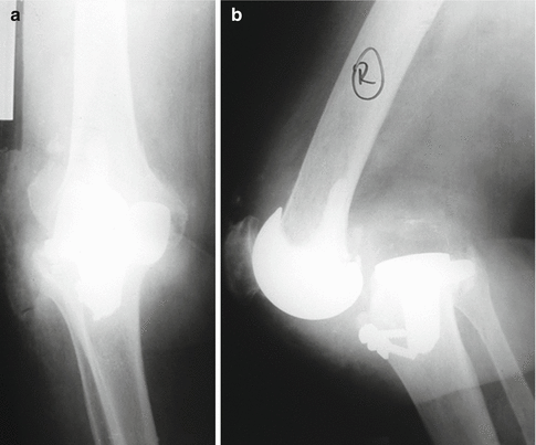
Unrepairable extensor mechanism problem with gross instability of the knee which is not possible to reconstruct (Fig. 5.2).


Fig. 5.2
A patient with a gross instability. (a) Anteroposterior and (b) lateral X-rays
5.3 Examination/Imaging
A patient has to be examined for the extent of the infection. Cardinal findings of infection have to be noted for postoperative follow-up.
The condition of the soft tissue is one of the most important issues. Debridement may cause soft tissue defects necessitating local or free flaps. Too much skin tension also may lead to skin necrosis. Thus, any soft tissue reconstruction has to be planned carefully preoperatively.
Plain anteroposterior and lateral X-rays, including an orthoroentgenogram, are required for preoperative planning. Femoral and tibial cuts have to be planned preoperatively, relative to current shortening. The limb has to be 1 cm shorter than the site with a mobile knee, but any shortening exceeding 1 cm requires support.
Most of the uncontrolled infections cause fistulae. Its tract has to be removed with debridement. Preoperative fistulography with radiopaque contrast will state its extent.
In case there is marked bone loss, a computerized tomography will display the amount and location of the defect.
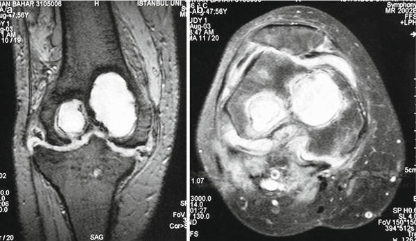
Widespread osteomyelitis will require an extended bone debridement, even resection. Magnetic resonance imaging is a sensitive method to state the extent of infection (Fig. 5.3).

Fig. 5.3
(a, b) Magnetic resonance scan reveals osteomyelitis sequela with huge bone defect
In case there is doubt for infected arthroplasty, an indium-labeled leukocyte bone scan is a sensitive and specific technique for diagnosis.
5.4 Surgical Anatomy
5.4.1 Structures at Risk
The preferred application direction of the Schanz screws is in the sagittal plane.
The anterior half of the femur is a safe zone and far from neurovascular structures.
For the tibia, care is required not to miss the medial wall, as the anterior edge of the tibia makes an acute angle with the lateral wall. It is safe to check every Schanz screw at the AP view.
The application of the fixator does not jeopardize neurovascular structures around the popliteal region, but during debridement and cuts, especially if posterior capsulotomy is being performed, care must be taken.
5.5 Positioning
Supine position with a sandbag under the ipsilateral buttock is required to overcome external rotation of the limb.
For initial debridement, knee flexion over 90° is required.
After debridement and resection, limb alignment should be checked in extension to 5° of flexion in anteroposterior and lateral planes with image guidance. If required, additional resection can be done.
The limb is supported with folded sheets in full length for external fixator application.
Fluoroscopy guidance is mandatory for this procedure. The device should be a C-arm and able to turn for lateral view.
A radiolucent operating table is required.
5.6 Exposures
There are various methods to access the knee joint. However, patients will have a previous knee replacement surgery incision, which may be used multipl times for debridements or revisions. Using it including deep planes facilitates skin healing and reduces the risk of skin necrosis.
If there is a fistula, incision should center it with fistulectomy.
Stay updated, free articles. Join our Telegram channel

Full access? Get Clinical Tree


