1 Intracranial Region
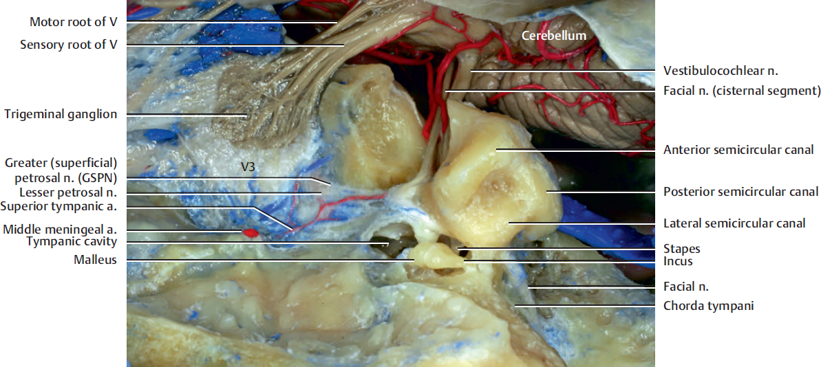
Fig. 1.1. Middle fossa from above. The tegmen tympani and the roof of the internal acoustic meatus have been opened.
The facial nerve contains motor fibers, which are responsible for facial expression, as well as other nerve fibers involved in sensation (auricular concha; see Fig. 13.3b), taste (anterior two-thirds of the tongue; see Fig. 1.3), and secretory function (lacrimal gland, submandibular gland, and sublingual gland; see Fig. 5.9a and Fig. 12.1).
The facial nerve emerges from the brainstem at the junction of the pons and medulla. The course of the facial nerve may be divided into intracranial, intratemporal, and extra-temporal segments. The intracranial segment, known as the pontine or cisternal segment, spans roughly 25 mm and connects the facial nerve from its origin to the entrance of the internal acoustic meatus. The intratemporal segment can be divided into four segments: meatal, labyrinthine, horizontal, and vertical. The facial nerve, accompanied by the vestibulocochlear nerve, enters the temporal bone through the internal acoustic meatus. Initially, there are two separate components, which are the motor root supplying the muscle of the face and the nervus intermedius that contains sensory fibers concerned with the perception of taste and parasympathetic (secretomotor) fibers to the lacrimal, submandibular, and sublingual glands. The two components merge within the meatus.
The greater (superficial) petrosal nerve (GSPN) is a branch of the facial nerve and carries parasympathetic fibers to the pterygopalatine fossa. It emerges within the facial canal of the temporal bone, close to the geniculate ganglion of the facial nerve. It then passes through the bone to appear on the floor of the middle cranial fossa and runs medially in a shallow groove to the foramen lacerum. Passing within the foramen lacerum, the greater petrosal nerve enters the pterygoid canal that lies at the base of the pterygoid process. On leaving the pterygoid canal, the nerve emerges into the pterygopalatine fossa and joins the pterygopalatine ganglion (see Fig. 10.15).
Stay updated, free articles. Join our Telegram channel

Full access? Get Clinical Tree


