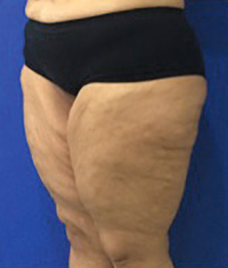
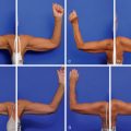
The Clinical Problem ( Fig. 38.1 )
The medial thigh is a problematic zone for body contouring both in the aging patient, who has skin laxity and lipodystrophy, and in the massive weight loss patient, with critical skin flaccidity in the medial thigh.
Patients with morbid obesity who have undergone massive weight loss (MWL) following a diet or bariatric surgery develop body dysmorphia due to skin laxity and dermal fat excesses. They suffer interference in their quality of life because of the difficulty of personal hygiene, ambulation, and physical activities, as well as skin infections, postural changes, low self-esteem, and changes to the body image.
To remove the dermal fat excesses and improve the thigh contour, plastic surgeons have performed thigh lift procedures, associated with or without liposuction. The medial thigh lift was first introduced by Lewis in 1957 for the treatment of extreme flaccidness of the medial thigh.
However, this traditional technique was related to the recurrence of ptosis, scar migration, and vulvar deformities. Therefore, in 1988, Lockwood proposed a technique of anchoring the dermal tissue from the distal medial thigh to the deep layer of the superficial perineal fascia (Colles fascia) to reduce scar migration, leading to a more stable and long-term outcome.
Surgical Preparation and Technique
Indications and Classification
Thigh lift techniques are used to treat thigh laxity, in particular its medial and proximal portion. Such techniques may be associated with liposuction to reduce the fat content of the entire thigh or more localized procedures, such as at the medial and lateral thigh or a region of the knee. Medial thigh patients can be divided in two categories: MWL patients and non-MWL patients.
Massive Weight Loss Patients
These are divided in two groups: deflated and nondeflated.
Deflated
In the first group (deflated), the thickness of fat tissue is lower, so a combination of surgeries is possible because the surgical time and bleeding are minor. Deflated patients usually present with skin flaccidity over the thigh, with minor or even without residual lipodystrophy. These patients are treated using an extended vertical medial skin excision, with or without liposuction.
Nondeflated
In the second group (nondeflated), the association of procedures at the same time is not recommended because the thicker adipose tissue is related to greater surgical manipulation, blood loss, impact on morbidity, and higher incidence of postoperative complications. Nondeflated patients generally present with both skin laxity and lipodystrophy. For these patients, liposuction is performed at the first stage, combined with a lower body lift, and followed by an extended vertical medial skin excision 4 to 6 months later.
Non–Massive Weight Loss Patients or Patients With an Aging Thigh
These patients usually present with lipodystrophy, with varying degrees of skin laxity. Those with only lipodystrophy without skin laxity require liposuction alone. Those with lipodystrophy and skin laxity confined to the upper third of the thigh are treated by liposuction and a horizontal medial skin excision at the level of the inguinal fold. According to the progression of skin flaccidity from the upper to the entire thigh, it can be associated with a vertical skin excision.
To correlate thigh deformities with the appropriate surgical treatment, some scales that rank the degree of thigh impairment and laxity have been developed. Here we highlight the Medial Thigh Classification and Treatment Scale ( Table 38.1 ).
| Type | Description | Treatment |
|---|---|---|
| I | Lipodystrophy with no sign of skin laxity | SAL, UAL alone |
| II | Lipodystrophy and skin laxity confined to the upper third of the thigh | SAL, UAL + horizontal medial thighplasty |
| III | Lipodystrophy and moderate skin laxity that extends to the middle third of the thigh | SAL, UAL + extended vertical medial thighplasty |
| IV | Lipodystrophy and moderate skin laxity that extends the length of the thigh | SAL, UAL + extended vertical medial thighplasty |
Preoperative Care
The critical point for successful intervention is the preoperative evaluation for surgical planning. The surgeon should clarify the limitations of the procedure, discuss possible complications, and emphasize the presence of extensive scarring, especially in patients for whom a vertical medial incision will be needed. A consent form must be signed. We evaluate the quality of the skin (MWL patients have depleted collagen fibers ), degree of elasticity, laxity, identification of lipodystrophy areas, and presence of previous scars. We also evaluate the vertical and horizontal skin excess by the pinch test and traction.
It is important that the patient undergo a psychological evaluation to check her or his expectations about the outcome. A nutritional screening must be performed, especially in MWL patients. Nutritional deficiencies such as iron, albumin, and vitamins depletion can be predictors of complications. Before surgery, antimycotic treatment should be performed in patients with fungal infections. Moreover, patients should stop smoking ; the prevention of venous thromboembolism is crucial and should be adopted.
Marking and Surgical Technique
In general, most of the techniques with skin excision involve skin markings at the proximal and medial thigh using the pinch test to predict the amount of skin to be removed, which is adjusted perioperatively to achieve closure without tension. The patient is marked in the standing position, anterior and posterior, with the knees apart. The femoral triangle is marked to avoid dissection into the lymph vessels. The location of fat deposits for liposuction is also marked.
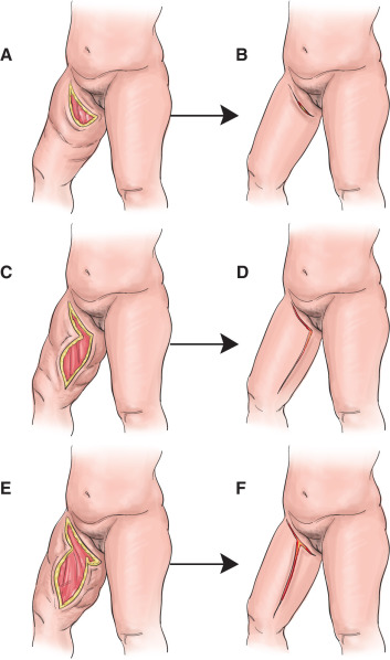
The degree of thigh laxity determines the location of the incisions and the need to add a vertical component. Usually, when skin laxity is located only at the upper third of the thigh, a horizontal incision at the level of the inguinal fold is enough ( Fig. 38.2A ). However, if skin laxity reaches the entire thigh, beyond its upper third, a vertical medial incision, inverted L –shaped incision (see Fig. 38.2B ), or T -shaped incision (see Fig. 38.2C ) may be needed, according to the amount of skin to be resected. In these cases, an attempt is made to hide the resultant scar so it is less visible in an anatomic position.
Related posts:


Stay updated, free articles. Join our Telegram channel

Full access? Get Clinical Tree


