The Clinical Problem
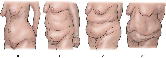
Contour deformities after post–bariatric surgery weight loss are varied and often complex. The Pittsburgh Rating Scale is a useful classification system that allows grading of 10 areas of the body following massive weight loss. Each area is scored on a scale of 0 to 3 and considered as part of the overall picture. Mild abdominal deformities call for standard measures, and increasing levels of deformity require more complex surgery and modifications to achieve adequate correction.
The grades of abdominal deformity are shown in Fig. 32.1 .
Synopsis
There has been an exponential increase in obese and overweight patients throughout the world, with rates in Europe and North America estimated to be approximately 20% to 30% of the adult population. One of the few means to achieve significant and maintained weight loss consistently is bariatric surgery. Evidence has shown that this brings a reduction in health-related complications, such as diabetes, psychosocial functioning, personal health perceptions, and health-related quality of life.
An inevitable consequence of significant weight loss is the persistence of large quantities of excess and inelastic skin and subcutaneous tissue. Body contouring surgery involves many areas and even more techniques. The best results are obtained by tailoring surgery to create individual packages for each patient. The abdomen is normally the first priority. Depending on the deformity, it can be treated in isolation or with a circumferential technique to address the buttocks and back.
The Aesthetic Problem
Patient Perspective
After massive weight loss, large areas of redundant skin and fat cause significant distress to patients. They often experience skin irritation, mycotic infection, and secondary self-image problems. The excess skin also affects activities of daily living and often leaves patients more embarrassed and self-conscious than the obesity itself.
Surgical Perspective
It is essential to ensure that patients are optimized before surgery and, in many cases, it is appropriate to liaise with their bariatric team. They need to have achieved weight stability for at least 1 year. A body mass index (BMI) of 30 or less is optimal. Folate, vitamin B 12 , and iron deficiencies must all be corrected. Patients need to be counseled that multiple, often staged procedures may be necessary to meet their goals, and their expectations have to be realistic. Each stage encompasses further risk, recovery time, and often expense.
Surgical Preparation and Technique
Surgical Options
The key deciding factors used in choosing the abdominoplasty technique are as follows:
- ▪
The amount of abdominal laxity
- ▪
Patient preference regarding the extent and location of scars
- ▪
Risk factors, such as previous abdominal incisions (e.g., Kocher’s incision) and keloid or hypertrophic scar
- ▪
Resistant fat pads—their quantity and location
Abdominoplasty Techniques
Traditional Abdominoplasty
The traditional abdominoplasty involves elevation of skin to the xiphisternum and costal margin laterally. The skin is then pulled taught by flexing the patient at the waist to reduce the tension, and a new opening is created for the umbilicus. This technique has several limitations, which restricts its use.
Large areas of undermining limit the blood supply to the skin and divide the retaining ligaments. This opens a large cavity with the potential for seroma and hematoma formation, both of which have a higher incidence in weight loss patients. Tensing the skin and dividing the retaining ligaments to enable closure gives a barrel-shaped abdomen, with loss of the definition of the rectus muscle and waist. The undermining inevitably compromises blood supply and prevents liposuction in zones 1 and 2.
Most weight loss patients will retain significant fat pads and laxity under the inframammary fold (IMF). Given the degree of skin laxity in these patients, most patents will have significant dog-ears laterally, which need a second stage for removal. In addition, the vertical laxity cannot be addressed.
Lipoabdominoplasty
This technique does not usually allow for tightening of the skin in the vertical dimension.
Fleur De Lis Abdominoplasty
This results in a vertical scar from the xiphisternum to the pubis, with a horizontal scar in the lower abdomen. Unfortunately, the patient is commonly left with a bulge in the xiphisternum as a result of a dog-ear created in this area.
Circumferential Abdominoplasty (Belt Lipectomy)
A standard abdominoplasty is extended round the lower torso to address laxity in the flanks, buttocks, and thighs. This allows resuspension of the lateral and anterior thighs, along with the traditional abdominoplasty improvements. It is an extensive procedure with a high risk of wound breakdown and seroma.
Two-Stage Procedure
A standard abdominoplasty alone will not address laxity and fat deposits above the rib cage. For some patients, a standard abdominoplasty followed by a reverse abdominoplasty at a second stage will allow the abdomen to be tightened without the need for a midline scar.
Corset Body Lift
This incorporates incisions from the fleur de lis and reverse abdominoplasty without undermining. It addresses laxity in the vertical and horizontal directions and the posterior waistline, as well as excising the folds of skin and fat above the rib cage. A corset body lift is performed with the patient in the supine position, without the need to turn the patient. It is therefore results in a 360-degree improvement of the trunk, with a low risk of skin complications. The technique is suitable for most massive weight loss patients but should be carried out in specialist unit.
Preoperative Markings ( Fig. 32.2 )
The patient is marked in the standing position. The following reference lines are drawn:
- ▪
IMF
- ▪
Midline
- ▪
Lower abdominal incision at the pubic hairline
Resection and Plication
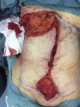
Vertical Resection ( Fig. 32.3 )
The extent of the vertical resection is best marked with the patient in the supine position on the operating table. The maximum resection is marked on each side by pinching the flank skin into the midline. The skin is tailor-tacked with staples in the correct position. A vertical line is drawn from the IMF to the pubis on each side of the staple line. When the staples are removed, the amount of skin resection can be seen. There should be the same amount of skin resection on each side, which can be checked with a ruler.
The skin markings are incised with a No. 10 blade, and dissection is commenced vertically down to the rectus fascia using the hand-held diathermy. The umbilicus is circumscribed and dissected free from the abdominal flap. The vertical skin flap is resected off the abdominal wall. Hemostasis is secured.
Abdominal Wall Plication and Vertical Closure
The abdominal wall is plicated in the midline using a barbed suture. The plication commences at the level of the umbilicus and proceeds proximally and distally. A second row of interrupted sutures supports the closure. This reduces the abdominal defect, thereby facilitating a tension-free closure. In addition, it allows the waist to be narrowed and the body contour improved.
Attention is then turned to closing the wound itself. A barbed suture is used to close Scarpa’s fascia and the dermal layer. Several interrupted deep dermal sutures reduce the tension on the skin edges during closure. The umbilical stalk is trimmed and defatted. It is then inset into a circular opening in the skin and secured with 5-0 Vicryl Rapide sutures. The repair is stopped short of the superior and inferior dog-ear. The sutures are left temporarily and protected.
Inframammary Fold Resection ( Fig. 32.4 )
The upper abdominal resection removes the dog-ear created by the midline closure and allows the lateral excess skin at the level of the IMF to be resected. It can be continued as far laterally as necessary. Tailor tacking is used to establish the amount of tissue resected. The length of the inferior resection margin should correspond to the length of the upper abdominal incision.