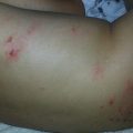
Fig. 17.1
Lymphocutaneous sporotrichosis. Erythematous nodule and ulceration present along the lymphatics of the upper extremity (Photo courtesy of Sylvia Hsu, MD)
Chromomycosis is caused by inoculation of a dematiaceous fungus from the soil into skin. Typical lesions of chromomycosis in children or adolescents are erythematous nodules or verrucous plaques, most often located on the upper extremity. This is of notable contrast to disease presentation in adults, which usually occurs on the lower extremity. In one study of chromomycosis in South American children, Cladophialophora carrionii was the most common cause, while most studies in adults have show Fonsecaea pedrosoi to be the most common cause [2, 3].
Eumycetoma is a chronic mycotic infection of the skin and soft tissue. Infection follows inoculation injury from a contaminated thorn or splinter. At least 30 species of molds are implicated; Pseudallescheria boydii is the most common causative species in the United States, while Madurella mycetomatis is the most common cause worldwide. Infection most often involves the feet or lower extremities, and is characterized by large verrucous nodules with abscesses, sinus tracts, and macroscopic grains (Fig. 17.2). Eumycetomas are usually confined to subcutaneous tissues, but can involve fascia, bone, and regional lymph nodes via contiguous dissemination. Fibrosis, deformity, and lymphedema eventually result if untreated [4].
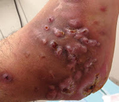
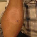
Fig. 17.2
Eumycetoma. Multiple coalescent verrucous nodules with sinus tract formation on the foot, resulting in deformity (Photo courtesy of Sylvia Hsu, MD)
Rhinosporidiosis is a non-contagious chronic granulomatous infection caused by Rhinosporidium seeberi, and is characterized by polyps which are sessile or pedunculated, and mainly affect the nasal mucosa and, less commonly, the conjunctival or ocular mucosa. Cutaneous lesions may occur due to spread from adjacent mucosa, direct inoculation, or hematogenous spread. Infection in adolescents and young adults is common [5, 6].
Specific Investigations
For diagnosis |
Fungal culture |
KOH |
Histopathology |
For treatment |
CBC, LFTs, lipid panel, metabolic panel (potassium, blood glucose), kidney function tests (BUN and creatinine), EKG |
Thyroid function tests (TFTs) |
Fungal culture is the most sensitive method for the diagnosis of sporotrichosis. Aspirate from a nodule or tissue from a punch biopsy should be inoculated onto Sabourad dextrose agar; growth occurs within 5 days at room temperature. Histopathology may be supportive, but is rarely diagnostic, given the difficulty of finding the sparse organisms, which are 3 μm or smaller in diameter. Nonspecific findings, including suppurative granulomas and asteroid bodies, are often present [7]. An enzyme immunoassay for serologic testing of S. schenkii has demonstrated up to 90 % sensitivity and 80 % specificity, but is not available widely [8].
In chromomycosis, KOH is highly sensitive in identifying the characteristic sclerotic or medlar bodies. Fungal culture on Sabourad or Mycosel agar is sensitive and specific. Histopathology often demonstrates granulomatous inflammation, pseudocarcinomatous hyperplasia, and pigmented yeast forms with single septations [9].
Eumycetoma can be diagnosed by the clinical findings in combination with black macroscopic grains, which are only found with fungal infections; yellow or white grains indicate fungal or bacterial infection. KOH demonstrates broad, septate, and branching hyphae. Culture should be performed but requires 6–8 weeks for growth. Histopathology is also helpful and demonstrates hyaline or pigmented hyphae in microscopic grains. Radiography, including X-ray, computed tomography, and magnetic resonance imaging should be considered in extensive cases, to exclude bony and soft tissue involvement [10].
In rhinosporidiosis, KOH or histopathology are diagnostic, demonstrating large sporangia 200 μm in diameter and filled with smaller endospores. Culture is not helpful, as R. seeberi is intractable to isolation or growth in microbiologic culture media [11].
Given the lengthy duration of systemic azole therapy required for theses diseases, baseline and periodic evaluation of CBC and LFTs is recommended. In the case of itraconazole, periodic evaluation of serum lipids is recommended, given the risk of hypertriglyceridemia. SSKI therapy requires monitoring of thyroid function tests, including TSH and FT4. Kidney function tests as well as potassium levels should be monitored during amphotericin B treatment; EKG is also recommended, given the risk of arrhythmia.
Clinical cure was obtained in almost 95 % of patients with cutaneous sporotrichosis, including children, treated with oral itraconazole. In another study, clinical response rate to itraconazole was over 80 %.
In a randomized non-blinded study, clinical cure rates were high and similar (above 89 %) for pediatric patients treated with either daily dosing or four times daily dosing of SSKI.
Medication | Dosing | Evidence level |
|---|---|---|
Amphotericin B deoxycholate followed by itraconazole | Amphotericin B: | E* |
0.7 mg/kg/day until improved | ||
Itraconazole: | ||
6–10 mg/kg/day up to maximum daily dose 400 mg × 12 months |
Initial treatment with intravenous amphotericin B followed by long-term therapy with itraconazole is reserved for disseminated sporotrichosis and based on case reports.
Complete clinical and mycologic remission was achieved in 85 % of the patients treated with itraconazole or 5-FU cream. Similar results were obtained for electrodesiccation or fulguration, but given the more invasive nature and potential for scarring, this is considered an alternative to itraconazole or 5-FU. Ajoene gel (isolated from alcoholic extracts of garlic) also demonstrated a high rate of efficacy, but may be difficult to obtain.
Low cure rates (31 %) were observed in a study of 51 cases, but cryosurgery and itraconazole produced the best results overall, sometimes in combination.
Medication | Dosing | Evidence level |
|---|---|---|
Ajoene 0.5 % gel | D* |
In one study, itraconazole was moderately efficacious, associated with improvement in 42 % of cases, although none showed mycologic or clinical remission. Voriconazole is the treatment of choice for eumycetoma caused by P. boydii. Posaconazole can be used as an equally effective alternative to itraconazole and voriconazole.
Radical surgical procedures should be avoided, but combined medical therapy and conservative excision have produced good results. Relapse rates after surgery alone are high (over 50 %), so antifungals should be administered for at least 6 months prior to surgery, and then in the post-surgical period to reduce recurrence. In a small study of oral ketoconazole, 5 of 13 patients were completely cured, and 4 improved following at least 6 months of treatment. Despite its historical role as the preferred agent for this disease, toxicity limits its use, and it is now only considered an alternative agent.
Medication | Dosing | Evidence level |
|---|---|---|
Surgery | E* |
Local surgical excision is the treatment of choice, but has been associated with 10 % recurrence rate. Concurrent medical treatment with dapsone has been used to decrease this risk.
Case reports have described long courses of dapsone being used as monotherapy or in combination with surgery. It has also been used in combination with surgery or other antimicrobials such as cycloserine and ketoconazole for disseminated disease.
Systemic Mycoses: Blastomycosis, Coccidioidomycosis, Paracoccidioidomycosis, Histoplasmosis
Clinical Features
Blastomycosis is caused by inhalation of the conidia of the dimorphic fungus Blastomyces dermatitidis. The lungs are the most common site of disease, and infection may be asymptomatic or severe. Cutaneous disease results from hematogenous spread from the lungs, and occurs in up to one-fifth of patients. Verrucous lesions with irregular borders and microabscesses, ulcerative plaques with elevated borders, subcutaneous nodules, and cold abscesses may be seen [28].
Coccidioidomycosis is caused by the dimorphic fungi, Coccidioides immitis, or Coccidioides posadasii, which are endemic to arid regions. Patients of African or Filipino ancestry or those with a history of immunosuppression are at increased risk of infection. Cutaneous lesions are either due to disseminated disease via hematogenous spread from a pulmonary nidus or, less commonly, primary infection. Organism-specific manifestations include nodules, pustular lesions, verrucous plaques, abscesses, and fistulae. Reactive cutaneous manifestations include erythema nodosum, erythema multiforme, an acute exanthem, Sweet’s syndrome, and interstitial granulomatous dermatitis [29, 30].
Paracoccidioidomycosis is a systemic mycotic disease caused by the dimorphic fungus Paracoccidioides brasiliensis. It is endemic in Central and South America, where it is widely present as a soil saprophyte. Exposure is often occupational, and the main portal of entry is inhalation. Acute or subacute disease is most often seen in children and adolescents: features include lymphadenopathy, hepatosplenomegaly, fever, and bone marrow dysfunction, but skin and pulmonary involvement are uncommon. In contrast, the chronic form of the disease involves the lungs, mucosa, skin, lymph nodes, and adrenal glands. Mucosal and skin findings simulate those of leishmaniasis. Painful ulcers with ragged borders and petechiae are seen most often in the mouth or larynx. Ulcerative or verrucous nodules or plaques are seen in the skin [31, 32].
Histoplasma capsulatum is a dimorphic intracellular fungus found worldwide. Cutaneous lesions are present in up to 15 % of patients with disseminated histoplasmosis (Figs. 17.3 and 17.4). A variety of manifestations are seen, including nodules, plaques, ulcers, pustules, abscesses, erythroderma, cellulitis and panniculitis, and purpura [33].
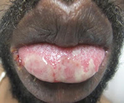
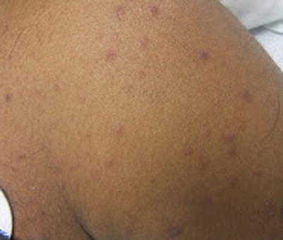
Fig. 17.3
Disseminated histoplasmosis. Erosive plaques of the oral mucosa in a patient with AIDS
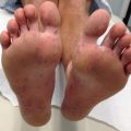
Fig. 17.4
Disseminated histoplasmosis. Violaceous papules distributed over the trunk and extremities in a patient with AIDS
Specific Investigations
For diagnosis |
KOH |
Fungal culture |
Histopathology |
Antigen detection (EIA) |
Serology (EIA, immunodiffusion, complement fixation) |
Skin testing (Coccidioidomycosis) |
Imaging (Computed tomography or x-ray, for paracoccidioidomycosis) |
For treatment |
Repeat serology to monitor treatment response (paracoccidioidomycosis) |
CBC, LFTs, lipid panel, metabolic panel (potassium, blood glucose), kidney function tests (BUN and creatinine), EKG |
For blastomycosis, KOH preparation has a diagnostic yield of less than 50 %, despite multiple specimens. Histopathology demonstrates suppurative granulomas, but yeast forms may be difficult to visualize. When identified, they are 8–15 μm in diameter, with refractile walls and single broad-based buds. Definitive diagnosis requires fungal culture, and B. dermatitidis grows within 1–4 weeks [34]. Antigen detection assays for blastomycosis demonstrate overall sensitivity of 90 %, but specificity is less than 80 % due to cross-reactive antigens in histoplasmosis, paracoccidioidomycosis, and penicilliosis. Sensitivity is higher in urine than in serum. Given the lower specificity, culture is still the gold standard [35].
Cutaneous coccidioidomycosis should be diagnosed either by direct visualization of the organism or by culture. Coccidioides spp will grow on routine media, but may take more than 1 week to isolate. Spherules of Coccidioides are large, up to 70 μm in diameter, and can be detected with KOH prep or in histologic sections. Despite their size, organisms are often sparse, so multiple level sections should be examined by the dermatopathologist [36]. Overall, serologic tests for the detection of IgG and IgM antibodies against Coccidioides are highly specific, but sensitivity is variable in early infection, as antibody production may not occur for weeks to months after illness onset. Immunodiffusion testing is the most specific serology available, while enzyme-linked immunoassay (EIA) has a sensitivity of 100 %. Thus, EIA should be used a screening test and immunodiffusion should be used for confirmation [37–39]. Complement fixation and tube precipitin-type assays are less accurate than these two newer methods. EIA for antigenuria is over 70 % sensitive, but also detects Histoplasma antigen. Skin testing for coccidioidomycosis is not recommended to diagnose current illness. Skin tests are positive for life, even in healthy patients with adequate prior treatment. In contrast, skin tests can be negative in infected patients with anergy. Thus, it is more useful as a prognostic test [40].
Paracoccidioidomycosis is diagnosed via direct microscopic visualization and/or by culturing P. brasiliensis from clinical specimens. KOH prep is positive in over 90 % of cases. Skin biopsy may be obtained in chronic disease, and demonstrates suppurative granulomas in most cases; P. brasiliensis is seen as a round or oval yeast 4–40 μm, with two or more narrow-necked budding cells (resembling a “pilot’s wheel” or “Mickey mouse head”). Quantitative immunodiffusion is the most useful serologic test for diagnosis and for monitoring response to therapy, given its high sensitivity and specificity—up to 97 and 100 %, respectively. Cultures are positive in up to 80 % of cases, but can take up to 30 days to grow. In addition to imaging of affected areas (computed tomography or X-ray) to evaluate lymphadenopathy and pulmonary lesions, all patients with suspected paracoccidioidomycosis should have specimens submitted for direct microscopy, culture, and serology [41].
In a study of HIV-infected patients with disseminated histoplasmosis, skin biopsy with special stains for fungi (gomori methenamine silver or PAS) allowed direct visualization of Histoplasma in over 86 % of cases. The most common histologic pattern is a diffuse infiltrate of macrophages parasitized by yeast 2–3 μm in size. However, organisms may also be extracellular [42, 43]. EIA antigen testing in disseminated histoplasmosis demonstrates a sensitivity ranging from 75 % to 100 %, with increased sensitivity in serum compared to urine, and in immunocompromised patients. However, false-positive tests can occur in patients with blastomycosis or coccidioidomycosis [44]. PCR is positive in over 70 % of culture-positive tissue samples [45]. Immunodiffusion and complement fixation methods detect anti-Histoplasma antibodies in 70 % of immunocompromised and 90 % of immunocompetent patients with disseminated infection, but are often negative in patients on tumor necrosis-alpha inhibitor therapy [46]. Blood cultures are positive in 65 % of patients with disseminated histoplasmosis; tissue cultures from skin biopsy specimens can also be submitted.
Blastomycosis
For patients with severe disseminated infection, amphotericin B should be used. Pooled retrospective data show that amphotericin B is up to 91 % effective for blastomycosis. Although liposomal amphotericin B does not have as much supportive data, it should be used when available, and is particularly preferred in cases with CNS involvement. Itraconazole may be used in patients with mild to moderate disease not involving the CNS. In an open study, 90 % of patients with blastomycosis demonstrated clinical response to treatment with 6 months of itraconazole, making this a first-line therapy for blastomycosis. Although ketoconazole has strong supportive trial data for its curative success in blastomycosis, its use can be associated with severe hepatotoxicity as well as infection relapse. Therefore it is not recommended for the treatment of any endemic mycosis, including blastomycosis.
Fluconazole was 65 % effective at doses under or equal to 400 mg/day, but was successful in 87 % of patients treated with doses above 400 mg/day for 6 months in open studies. Voriconazole has been successful in small series for the treatment of refractory blastomycosis with CNS involvement. Posaconazole has also been used.
Coccidioidomycosis
Medication | Dosing | Evidence level |
|---|---|---|
Itraconazole | For 12 months | C/A* |
Fluconazole | For 12 months | C/A* |
A randomized, controlled trial demonstrated 72 % response rate for itraconazole and 57 % response rate for fluconazole after 12 months of treatment. While the majority of patients included were adults, there were a few cases in children as young as 6 years old.
Case series support the use of posaconazole, which has shown up to 73 % efficacy in refractory infections. Voriconazole demonstrated similar results for the treatment of resistant disease, albeit following a shorter treatment course.
Medication | Dosing | Evidence level |
|---|---|---|
Liposomal amphotericin B | 3–5 mg/kg/day IV | D |
Amphotericin B deoxycholate | 0.7 mg/kg/day IV | D |
Amphotericin B treatment is reserved for patients with rapidly worsening or CNS disease. Otherwise, treatment with oral azoles is preferred. Of note, in children with primary cutaneous disease and solitary lesions in whom disseminated disease has been excluded, observation or conservation excision, if feasible, can be considered.
Paracoccidioidomycosis
Oral antifungal therapy can be used in most (mild to moderate) cases of paracoccidioidomycosis. Among children and adults with paracoccidioidomycosis treated with itraconazole for an average of 6 months, 91 % of patients showed either marked improvement or resolution. In a small randomized trial, itraconazole, ketoconazole, and sulfadiazine were roughly equivalent in efficacy following treatment for at least 24 months. For patients with severe infection, including hypotension, respiratory failure, or severe malnutrition, therapy should be started with amphotericin B and then transitioned to oral therapy once improved.
Measured in terms of complete or partial treatment response, itraconazole was over 94 % effective compared to voriconazole, which was over 88 % effective. Voriconazole also has excellent in vitro activity against P. brasiliensis, but better data is available to support the use of itraconazole as a first-line therapy. In a separate open study, TMP-SMX was as effective as itraconazole, but treatment duration was four times as long with TMP-SMX.
Medication | Dosing | Evidence level |
|---|---|---|
Liposomal amphotericin B | 3–5 mg/kg/day IV | D* |
Amphotericin B deoxycholate | 0.7 mg/kg/day IV | D* |
Duration: 20–40 days or until clinical improvement then transition to oral therapy |
Therapy with amphotericin B is reserved for severe or refractory disease, and retrospective reviews have supported the use of this agent in this context.
Histoplasmosis
Medication | Dosing | Evidence level |
|---|---|---|
Liposomal amphotericin B | 3 mg/kg/day IV | A |
Amphotericin B deoxycholate | 1.0 mg/kg/day IV | A |
For 2 weeks or greater duration until clinical improvement, then step down to itraconazole | ||
Itraconazole | 2–5 mg/kg/dose po tid for 3 days then bid for 12 months, with maximum single dose 200 mg | B |
In adult patients with AIDS and moderate-severe disseminated histoplasmosis, liposomal amphotericin B demonstrated better clinical success (88 %) than conventional amphotericin deoxycholate (64 %), in addition to improved survival and reduced nephrotoxicity; however, in children, amphotericin B deoxycholate is usually well tolerated, and the lipid preparations are not preferred. If liposomal formulations are not available, then amphotericin B deoxycholate should be used for induction.
Itraconazole demonstrated 85 % clinical response in adult patients with AIDS and histoplasmosis. However, patients with moderate to severe disease responded poorly. Additionally, clearance of fungemia is slower with itraconazole than with amphotericin B. Therefore, itraconazole is reserved as induction therapy for patients with mild disease without fungemia, and for maintenance therapy after successful induction. Maintenance therapy should continue for 1 year, to reduce the risk of relapse.
Itraconazole is superior to fluconazole in terms of clearance of fungemia as well as clinical response. Additionally, fluconazole is not as active as itraconazole against H. capsulatum in vitro. Although 74 % of patients responded to induction therapy with fluconazole in a large open study, almost half of patients demonstrated a relapse of their disease at 1 year while on maintenance therapy. Thus, fluconazole is reserved as a second-line therapy when amphotericin B and itraconazole cannot be tolerated. In a small case series, posaconazole has been effective for severe refractory infection as salvage therapy. Posaconazole demonstrates high in vitro activity against H. capsulatum. Voriconazole has also been used successfully as salvage therapy in disseminated histoplasmosis, but has inferior in vitro activity compared to itraconazole, and like posaconazole, has not been evaluated in a high-quality study.
Opportunistic Mycoses: Aspergillosis, Cryptococcosis, Fusariosis, Mucormycosis
Clinical Features
Aspergillus species are ubiquitous, and inhalation occurs often without sequelae in healthy hosts. In the setting of immunosuppression, most often during treatment for hematologic malignancies, or stem cell or solid organ transplantation, A. fumigatus, A. flavus, and A. terreus invade pulmonary or cutaneous tissue and may disseminate widely in the presence of angioinvasion. Neutropenia, high-dose corticosteroids, burns, and the neonatal period are also risk factors. Cutaneous aspergillosis may be primary, resulting from inoculation from trauma, or secondary, resulting from contiguous or hematogenous spread. Primary cutaneous aspergillosis may present as acute paronychia, necrotic plaques or nodules at the site of catheter insertion, or an erythematous edematous plaque. Secondary cutaneous aspergillosis may present with inflammatory or necrotic nodules, periorbital cellulitis, or ulcers [76, 77].
Cryptococcus neoformans and Cryptococcus gattii are encapsulated yeasts found worldwide in soil and bird guano that cause infections predominantly in patients with immunosuppression: HIV/AIDS, corticosteroids, organ transplantation, sarcoidosis, and malignancy. Following inhalation, meningoencephalitis, pulmonary infection, or disseminated disease may occur. Cutaneous lesions are seen in up to 15 % of patients with disseminated cryptococcosis. Plaques, purpura, ulcers, abscesses, cellulitis, and molluscum contagiosum-like lesions in patients with HIV may be seen [78]. Primary cutaneous disease is also possible following inoculation by minor trauma and, unlike secondary cutaneous lesions, may occur in immunocompetent hosts and is associated with favorable prognosis [79, 80].
Fusarium species are hyaline fungi present worldwide in soil, plant parts, and water. Superficial infections such as keratitis, onychomycosis, and intertrigo occur in immunocompetent hosts, while invasive infections occur only in patients with immunosuppression including neutropenia, hematologic malignancy, stem cell transplantation, and corticosteroid therapy. Sinusitis, pneumonia, fungemia, and dissemination can occur. Invasive infections occur via inhalation, direct inoculation, or spread from a superficial infection [81]. In this context, cutaneous lesions may be localized, as in cellulitis, or disseminated, with multiple necrotic painful lesions resembling those of ecthyma gangrenosum. Lesions at different stages of evolution, lymphangitic spread, target lesions, and blisters may be seen. Primary cutaneous disease in otherwise healthy hosts occurs at sites of burns or trauma, and presents with cellulitis, ulcers, verrucous nodules, and abscesses [82].
Rhizopus, Mucor, and Rhizomucor are genera of ubiquitous fungi that belong to the order Mucorales and cause most mucormycosis infections. Almost all infections occur in the context of immunosuppression, including poorly-controlled diabetes with ketoacidosis. Other risk factors include corticosteroid treatment, stem cell transplantation, hematologic malignancy, iron overload or deferoxamine treatment, HIV/AIDS, and burns. Inhalation of spores in susceptible individuals can lead to rhino-orbital-cerebral and pulmonary infections, the most common forms of the disease [83]. In contrast, cutaneous disease is always due to direct inoculation, and may occur following minor iatrogenic trauma such as intravenous line placement. Rarely, primary cutaneous disease may occur in immunocompetent individuals. Cutaneous disease usually presents with single cellulitis-like or ecthyma-like lesion. As with other forms of mucormycosis, rapidly progressive tissue necrosis often ensues due to infarction resulting from angioinvasion. Dissemination from cutaneous lesions can also occur [84].
Related posts:

Stay updated, free articles. Join our Telegram channel

Full access? Get Clinical Tree


