16 Infections of the hand
Synopsis


Introduction
One of the greatest impacts on the outcomes of hand infection treatment was the discovery of penicillin by Alexander Fleming in 1929. The advent of antibiotics for the treatment of infections decreased morbidity of hand infections considerably, and decreased mortality to almost zero.1 Unfortunately, bacteria demonstrated resistance to penicillin in vitro as early as 1941, and by 1942 resistant bacterial strains were identified in patients. By the mid-1950s, nearly three-quarters of all staphylococcal species isolated from patients in large hospitals were highly resistant to penicillin.1 Fortunately, alternative antibiotics were being discovered and developed, and continue to be researched today.2
The treatment of hand infections is based on a thorough understanding of underlying anatomy and physiology. The availability of antibiotics has changed these infections from one of potential mortality to one of almost certain cure, but they are not a substitute for appropriately indicated and performed surgical drainage.2
Historical perspective
Over 2000 years later, a general surgeon from Chicago performed extensive dissections along with landmark injection studies that formed much of our current understanding of hand infections. Dr. Alan Kanavel3 defined the five fascial spaces of the hand (dorsal subcutaneous, dorsal subaponeurotic, middle palmar, thenar, and hypothenar) and the patterns of communication between them. He noted that, as purulence accumulates and the pressure increases in these tight spaces, it spreads in defined and predictable patterns.3,4
Basic science/disease process
Hand and wrist infections are frequently encountered in emergency departments. Paronychias-eponychias (35%), felons (15%), cellulitis (35%), and tenosynovitis (10%) are the most common types encountered. Inception of the infection occurs 60% of the time by direct inoculation of organisms through a variety of traumatic breaks in the protective skin layer: human bites (25–30%), drug abuse (10–15%), and animal bites (5–10%). How severe the infection becomes is largely dependent on the immune status of the host, the viability of the surrounding tissue, the location of the inoculation, and the virulence of the organism or organisms.5
Particular groups of patients with weakened immunity, such as those with acquired immunodeficiency syndrome (AIDS),6–8 intravenous drug abusers,9–11 diabetics,12,13 those with chronic corticosteroid use, and alcoholics,12 are all more susceptible to infections. Infections in these populations can be more challenging to diagnose for several reasons:
• Infections are often caused by diverse organisms, including those not usually considered to be pathogenic in otherwise healthy patients.
• Infection of the soft tissues may occur as a part of a more systemic infection.
• The degree and type of immune deficiency can attenuate the clinical presentation and findings.14
The likelihood of infection can also be heightened by local-tissue ischemia from traumatic injuries disrupting blood flow, and foreign bodies. The principles for adequate prevention of infection in traumatic injuries, in addition to appropriate antibiotic administration, include copious wound irrigation, adequate surgical debridement of devitalized tissue, and stable fixation of fractures, as required.15
Diagnosis/patient presentation
The initial evaluation of any patient should include a thorough history and physical examination. Determining a patient’s age, handedness, and occupation can often lead to clues as to the etiology and risk factors for infection. The traditional OPQRST questions (onset, provocation factors, quality, radiation, severity, and temporal onset) can lead to important clues to the chronicity, severity, and depth of the infection. Previous injuries can also reveal critical elements in determining a diagnosis. In a careful review of systemic diseases, determine if there is a history of heart, lung, liver, or kidney problems. Also review the past medical history for contributing factors, such as diabetes, cancer, human immunodeficiency virus status, steroid use, and other immune-compromising diagnoses. In the past surgical history, previous hand and upper extremity surgeries may explain confusing physical exam findings, reveal a history of a transplant requiring immunosuppressive medications, or determine the presence of chronic conditions. A brief review of immunization history will help determine if the tetanus status is known and if a dose should be given at the time of evaluation. The patient’s social history may also reveal clues as well as help determine a patient’s in-hospital care. It may also reveal risk factors for methicillin-resistant Staphylococcus aureus (MRSA) infections (Table 16.1).
Table 16.1 Risk factors for community-associated methicillin-resistant Staphylococcus aureus (MRSA) skin and soft-tissue infections
(Reproduced from Napolitano LM. Severe soft tissue infections. Infect Dis Clin North Am 2009;23:571–591.)
Hints and tips
The treatment plan for all hand infections should follow the four following principles:
1. Infections should be treated with rest, elevation, and immobilization
2. Infected tissue requires debridement and all closed-space infections must be adequately drained
3. Appropriate type and amount of antibiotics for the given clinical situation must be administered
4. As soon as pain, swelling, and erythema subside enough to allow it, hand therapy needs to begin
If the hand does not improve within 24–48 hours, then the treatment plan needs to be reassessed.18 Prompt and correct diagnosis of type and location of infection is crucial, as is the timely instigation of the appropriate intervention in order to prevent the potential disastrous consequences of the inflammation and scarring that can follow these types of infections.16,17
The most common bacteria encountered in hand infections are Staphylococcus aureus, Streptococcus and Gram-negative species.18 The principal organism in 50–80% of infections is Staphylococcus. Gram-positive organisms are usually responsible for industrial and home-acquired injuries. Intravenous drug use, bite injuries, severe farm injuries, and those found in diabetics are usually polymicrobial and can include Gram-positive, Gram-negative, and anaerobic species. Human bite infections usually have alpha-hemolytic Streptococcus and Staphylococcus aureus, although Eikenella corrodens is isolated in one-third of victims. Animal bite and scratch wounds will commonly harbor Pasteurella multocida. Chronic indolent infections are suggestive of fungal or atypical mycobacterial infections.16,18,19
Infections should be routinely sent for aerobic and anaerobic cultures and Gram stain to direct therapy. The history of the infection can help direct other cultures and stains. The Ziehl–Neelsen stain illuminates acid-fast bacilli for the diagnosis of Mycobacterium tuberculosis. All Mycobacterium and Nocardia species are potentially acid-fast, so a positive smear is not pathognomonic for tuberculosis. Also, these organisms are fastidious, so false negatives are common. Multiple tissue samples grown with cultures under specific temperature conditions and cultures at 28–32°C in Lowenstein–Jensen medium for 3–6 weeks are necessary for atypical mycobacteria.16,19 Fungal infections can be diagnosed with potassium hydroxide preparations from skin scrapings from the periphery of lesions.20 Tzanck smears may be useful in diagnosing herpes simplex virus infections.21 Antibiotic therapy should only be delayed as long as necessary to obtain cultures and Gram stains.16
Classification of infections
Several methods of classifying infections have been proposed. The most common divides infections into complicated and uncomplicated and was developed by the US Food and Drug Administration (FDA) for pharmaceutical companies to categorize skin and soft-tissue infections (SSTI) during clinical trials (Table 16.2). The Infectious Disease Society of America developed practice guidelines for the diagnosis and management of SSTIs, but they do not utilize the FDA categorization. The guidelines are described in reference to the specific disease entity, the mechanism of injury, or host factors.
Table 16.2 Classification of severe skin and soft-tissue infections by the US Food and Drug Administration
| Uncomplicated |
| Complicated |
(Reproduced from Napolitano LM. Severe soft tissue infections. Infect Dis Clin North Am 2009;23:571–591.)
Mimicks of infection
Gout
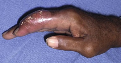
Gout, the most common of the crystalline deposition diseases, is also the most likely to be misdiagnosed as an infection in the upper limb (Fig. 16.1). Gout can be a primary metabolic disease or may present as a secondary manifestation of another primary disease process, such as myeloproliferative or renal disease.22 In acute cases of gout the hand may have swelling, erythema, pain with motion and, occasionally, fever, especially if secondarily infected. It is not typical, but the hand and upper extremity may be the initial presenting ground for the diagnosis of gout. The diagnosis is made by aspiration or procurement of the crystals from the tissue. The treatment is dependent on severity, and may include splinting, oral anti-inflammatory medications, colchicine, and in select cases, surgical extirpation of tophaceous material.
Pyogenic granuloma
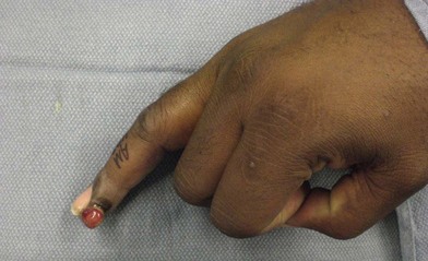
Pyogenic granuloma is a proliferative tissue in the hand that consists of inflammatory cells and exuberant granulation tissue. It is reddish, friable, and bleeds easily (Fig. 16.2). It is usually found following a penetrating injury that fails to heal. It may be treated with silver nitrate or surgical excision for larger lesions.
Pyoderma gangrenosum
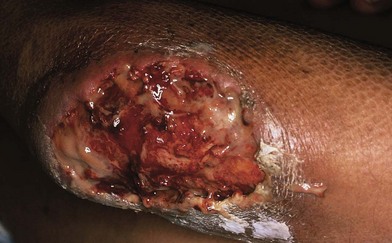
This is a rare skin lesion that starts as a small, painful ulcer. The central region becomes necrotic and spreads in a centrifugal pattern with increasing peripheral creep. The lesion is associated with an underlying systemic disease process, most commonly ulcerative colitis, but may also be seen with Crohn’s disease, carcinoid syndrome, polyarteritis nodosa, rheumatoid arthritis, and diabetes mellitus. The lesions are treated conservatively with local wound care. Surgical excision is contraindicated as it can lead to additional problems. The lesion has a very characteristic appearance (Fig. 16.3).
Brown recluse spider bite
Brown recluse spiders (genus Loxosceles), endemic to the Midwest and southeastern US, are identified by the violin-shaped pattern on the back of their cephalothorax. The initial bite is usually trivial-appearing; however, the area of injury may progress to a varying degree, from simple pruritus to full-thickness tissue necrosis. A severe systemic reaction, known as loxoscelism, is a rare life-threatening systemic symptom that can occur after a brown recluse spider envenomation and may include pyoderma gangrenosum, intravascular hemolysis, renal failure, pulmonary edema, and systemic toxicity. Because the initial injury is not usually witnessed, one should have a high index of suspicion with a history of working around warm, dark, dry environments. Most brown recluse spider bites can be treated with rest, ice, compression and elevation and will heal without further treatment. The true benefit of adjunctive therapy, such as dapsone, hyperbaric oxygen, and electric shock therapy, have also been called into question.23,24
Types of infection
Cellulitis
Cellulitis is characterized by a spreading diffuse hyperemia and edema of skin and subcutaneous tissue with infiltration of leukocytes. It is often accompanied by acute lymphangitis. Cases of cellulitis associated with abscesses, carbuncles, or furuncles are most often caused by Staphylococcus aureus. Diffuse cellulitis, or cellulitis without a defined entry point, is most commonly seen with streptococcal infections. Other questions to include in the history should center on recent physical activities or traumas, water and travel exposures, and bites by insects, animals, and humans.25 Treatment for uncomplicated cases of cellulitis include a first-generation cephalosporin, unless it is common in the community to have resistance to these agents. For penicillin-allergic patients, clindamycin or vancomycin is a good choice. If a lack of clinical response is noted, resistant strains, unusual organisms, or a deeper infection should be suspected. In those patients who are becoming increasingly ill, toxic shock syndrome, myonecrosis, or necrotizing fasciitis should be considered.20
Paronychia
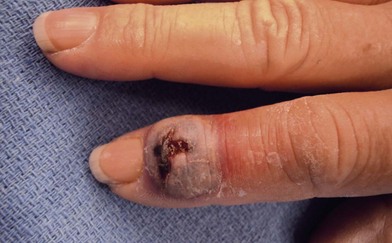
The significance of nail loss or deformity can be both aesthetic and functional. It is, therefore, important for those treating hand conditions to understand the anatomy and physiology of the fingertip and its relationship to the nail in order to provide optimum care26 (Fig. 16.4).


Stay updated, free articles. Join our Telegram channel

Full access? Get Clinical Tree


