Indications and Techniques for Buccal Fat Pad Excision
Alan Matarasso
Sammy Sinno
DEFINITION
The buccal fat pad is prominent in the infant face, where it contributes to the suckling function of the cheek.1
Typically, as the face matures, the buccal fat pad becomes less noticeable, but in some patients, it persists and serves as a primary contributor to a “round” face.
An attractive, angular, and youthful appearing face is characterized by malar convexity juxtaposed with submalar concavity.
Additionally, due to its intermuscular location, the buccal fat is typically spared from atrophy.2
ANATOMY
The buccal fat pad is a stellate-shaped structure with four extensions.
It is located deep to the masseter muscle and superficial to the buccinators in the buccopharyngeal membrane.
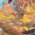
The parotid duct pierces the fat pad as it courses to the papilla across the second maxillary molar (FIG 1).
The fat pad extends to the anterior edge of the masseter muscle; it is at this location where the gland can protrude from overaggressive liposuction or facelift dissection among other causes, a phenomenon known as buccal fat pad pseudoherniation.3,4
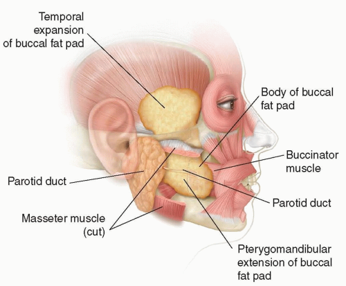
FIG 1 • Anatomy surrounding the buccal fat pad.
Posteriorly, the fat pad extends deep to the angle of the mandible.
Superiorly, the fat pad extends to the temporalis muscle and deep to its fascia.
Another superior extension continues along the zygomatic bone’s contribution to the lateral orbital wall.
PATIENT HISTORY AND PHYSICAL FINDINGS
The buccal fat pad can contribute to a “rounded” facial appearance, obliterating submalar concavity in certain patients. This feature is seen most prominently in the anterocaudal quadrant of the cheek, which is typically targeted by excision of the gland.
The buccal fat pad is above the mandible and posterior to the nasolabial fold, an area distinct from the jowl.
In contrast to a normally situated buccal fat pad, pseudoherniated fat can be reduced with gentle pressure.
IMAGING
When attempting to palpate the buccal fat pad, a firm, pulsatile, or subtly pigmented palpable mass warrants further workup with an MRI.
SURGICAL MANAGEMENT
Preoperative Planning
Patients undergoing buccal fat pad excision can have it as an isolated procedure or more commonly undergo other facial surgical procedures simultaneously.
Indications for excision include patients with a full, round face.
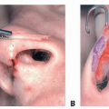
Excision is typically performed intraorally before other procedures (ie, facelift) under nonsterile conditions, to decrease the incidence of infection and risk of facial nerve injury.5Related posts:

Stay updated, free articles. Join our Telegram channel

Full access? Get Clinical Tree