div class=”ChapterContextInformation”>
11. Dermostyx (IB1): High Efficacy and Safe Topical Skin Protectant Against Percutaneous Toxic Agents
Keywords
Sulfur mustardVXEthyl parathionAcroleinPigsTopical skin protectantIntroduction
Development of a topical skin protectant is aimed to prevent the penetration of toxic agents through the skin and is essential for both military troops and civilian populations. The list of percutaneous toxic agents includes chemical warfare agents (CWAs) and toxic industrial chemicals (TICs) such as pesticides and irritants. The hazard of exposure to TICs has become a realistic threat since many TICs are routinely used in industry, agriculture, and research. Currently, the available therapy is limited and removal of the chemical substances from the body (skin and eyes) is the only way to reduce injury for some of the toxic agents.
A topically applied pretreatment that will act as a skin barrier may prevent the absorption of these toxic agents, as well as their respective toxicity [1].
The topical skin protectant strategy has been adopted by many countries including the USA, UK, and Israel [2]. The requirements from such preparation are to yield a significant protection, to be nontoxic and safe for human use, to remain effective on the skin for a reasonable time following application, to cause minimal discomfort to the user, and to be compatible with decontamination procedures.
We have developed a topical skin protectant that is applied onto the skin before exposure and prevents the penetration of warfare agents such as SM and VX through the skin and thus confers a highly efficient protection against the hazard they pose [2]. The formulation coded IB1 is a hydrophilic water-based solution comprised of magnesium sulfate and glycerin and it was shown to be safe in preclinical and Phase I studies [3]. IB1 was registered and approved by the Israel Ministry of Health as Dermostyx protective solution and is commercially available (Rekah Pharm, Israel).
The present study aimed to further test the efficacy of IB1 against CWAs, pesticides, and irritants and to elucidate the mechanism of protection/to investigate the role of the ingredients in the protection. The efficacy against the various toxic agents was evaluated in the well-established pig model [4], using clinical, histological, and biochemical monitoring. A representative example from each of these groups was chosen to demonstrate the protection afforded by this unique preparation. The list includes sulfur mustard (SM), VX [O-ethyl-S-(2-diisopropylaminoethyl)methylphosphothiolate], parathion, and acrolein as detailed below.
Sulfur mustard is a potent cutaneous vesicant that penetrates rapidly through the skin, causing severe prolonged injuries. Since its first use in World War I (WWI) and despite vast efforts, only limited success has been attained to find an effective treatment to prevent the injury [5–8]. VX is the most potent nerve agent that due to its low volatility and high persistence penetrates through the skin resulting in cholinergic toxicity and lethality [9]. The pesticide parathion is widely used in agriculture and is known to produce poisoning after dermal exposure in man and animals [10, 11].
Acrolein is a well-known strong irritant that was used by the French during WWI as a warfare agent “Papite” [12]. It is produced commercially worldwide since 1938 and is used mainly as an herbicide in water systems and as an intermediate product in many manufacturing processes [12–14]. According to the EPA reports, about 500 tons of acrolein are sold annually in the USA to use as an herbicide. Therefore, there is a risk of accidental exposure to this chemical. At present, therapy against the deleterious effects of SM and acrolein is only partially effective and for VX and organophosphate pesticides, the efficacy of treatments depends on the dose of the toxicants and on time elapsed from exposure. In addition to the efficacy studies, we hereby present data regarding the mode of protection, focusing on the role of glycerin and magnesium sulfate and their interaction with the stratum corneum.
Material and Methods
Animals
Young female pigs (commercial strain, Institute of Animal Research, Kibbutz Lahav, Israel) weighing 12–13 kg at the beginning of the experiment were used. Animals were housed in individual pens in a ventilated room with artificial lights from 06:00 to18:00 and the environmental temperature was adjusted to 21 ± 1 °C. They were fed with commercially available complete diet for pigs (Kibbutz Lahav) twice a day and had free access to tap water via automatic drinkers (drinking nipples).
All procedures involving animals were in accordance with the Guide for the Care and Use of Laboratory Animals, National Academy Press, Washington, DC, 1996, and were approved by the Institutional Animal Care and Use Committee.
Materials
Toxic agents:
Neat sulfur mustard (1250 μg μl−1, purity 95%: MW – 159.08, vapor pressure – 0.11 mmHg at 25 °C, solubility – 0.06% in water at 20 °C, logP – 1.37), neat VX, purity level of at least 95% (using gas chromatography; MW – 265.4, vapor pressure – 0.0007 mmHg at 25 °C, solubility 3% in water at 20 °C, logP – 0.675), Polidol 605E (commercial product, 467 mg/ml ethyl parathion in a diluting and wetting solution, Lidor Chemicals Ltd., Israel), and acrolein (90%, Sigma, Israel: MW −56.06, vapor pressure – 209.4 mmHg at 20 °C, solubility – 208 g/L water at 20 °C, log octanol-water partition coefficient – 0.9) were used for the exposure procedures. Decontamination was conducted using Fuller’s Earth powder (Shalon, Israel).
Topical skin protectant (TSP):
IB1 – a mixture of magnesium sulfate and glycerin (1:3).
Exposure Procedure
Pigs were restrained on specially made carts and the backs of the pigs were closely clipped. For the exposure of SM and acrolein: 8–10 fields were marked (4–5 fields of 35 cm2 each on each side) that served as exposure sites. All exposure procedures were performed within a hood at room temperature.
Liquid:
SM: Three droplets of 1 μl were applied in each exposure field, VX: a dose of 2LD50 in 1 μl droplets was applied along the back, ethyl parathion: droplets of 20 of 250 μl (for dosing of 40 and 250 mg/kg, respectively) were applied along the back, acrolein: a droplet of 50 μl on each exposure site. All droplets were applied to the pigs’ back skin using a digital microliter positive displacement pipette.
Vapor:
Toxic agents (5 μl of SM, 50 μl of acrolein) were placed onto filter paper disks (25 mm diameter) within the top inner surface of glass cups that were attached to the animals’ back and were sealed for the duration of the exposure (10 glass cups, 5 on each side of the back). At the end of the exposure, procedure pigs were decontaminated with fuller earth. A large amount of powder was applied to the skin to cover the exposed areas and then was removed with a spatula. This was repeated three times and then pigs were rinsed thoroughly with tap water.
Experimental Outline
IB1 (~1 ml/100 cm2) or water was applied on the skin as a single dose 10 min–12 h before exposure.
SM:
Pigs were exposed for the duration of 10, 30, and 60 min (liquid) or 15–60 min (vapor).
VX:
Pigs were exposed to 2LD50 (1.3 mg/kg) for the duration of 1 and 4 h (The LD50 value was determined over a period of 24 h).
Ethyl parathion:
Pigs were exposed to 40 mg/kg for 6 h and to 250 mg/kg for 2 h.
Acrolein:
Pigs were exposed for the duration of 30 and 60 min (liquid) or 15–60 min (vapor).
Each experimental group consisted of 2–4 pigs.
Quantitative Assessment of Lesions Area
The extent of the damage to the skin following exposure to SM and acrolein was measured using morphometric analysis of the damaged skin area. The exposed sites on the pigs’ backs were photographed using a digital camera (Coolpix 950, Nikon, Tokyo, Japan). Pictures were analyzed for the damaged area using an image analysis system (Image-Pro Express 4.0).
Erythema Measurements
The degree of erythema on the skin following exposure to SM or acrolein was measured using a reflectance color meter (Minolta Chroma-Meter Model C-300, Minolta Corporation, Ramsey, NJ), before the exposure (baseline) and at 2, 4, 6, 24, and 48 h following exposure to SM. The chromameter uses reflected light and reads color in a three-dimensional format L∗, a∗, and b∗. The a∗ coordinate is the balance between red and green and this chromaticity parameter was used as the indicator of the degree of erythema. Three replicate readings were taken on each exposed site and averaged.
Cholinesterase Inhibition in Whole Blood
Blood samples (0.5–0.7 ml) were collected from the jugular vein at various time points following exposure to VX and ethyl parathion. One hundred microliters of blood was immediately transferred into a heparinized tube containing 1.2 double-distilled water and mixed gently to cause hemolysis of red blood cells. Samples were stored at −4 °C until further analyzed.
The inhibition of the enzyme cholinesterase was determined using the radiometric method [15]. The blood samples were incubated with the substrate, H3-acetylcholine, at a temperature of 25 °C for 30 min. The reaction was ended with a reaction stopper that separated between the radioactive acetate and the choline. Scintillation solution was added and the radioactivity of the acetate in the organic phase was measured using a β counter.
Histology
Skin biopsies were taken at 1 week following exposure for fixation in 4% neutral buffered paraformaldehyde. Six micron paraffin sections were stained with hematoxylin and eosin (H&E) for light microscopy observations.
For studies of the interaction between IB1 and the stratum corneum, skin biopsies were quickly frozen in liquid nitrogen and 12 micron frozen sections were cut and stained for phospholipids by Sudan Black B.
Statistics
Results are expressed as mean ± standard error of the mean (s.e.m.). The quantitative data were subjected to statistical analysis using t-test or two-way ANOVA utilizing GraphPad Prism 4 software. Post hoc comparisons were performed using the Bonferroni test for multiple comparisons.
Results
Protection Efficacy Against SM Liquid and Vapor
Typical SM skin lesions developed on all exposed sites in a dose-dependent manner.
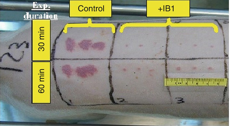
The efficacy of IB1 in pigs, 24 h following exposure to SM (1 μl droplets) for exposure durations of 30 and 60 min. Note the minor damage in the treated skin, compared to the severe lesions in the untreated control areas. IB1 was applied 1 h before exposure
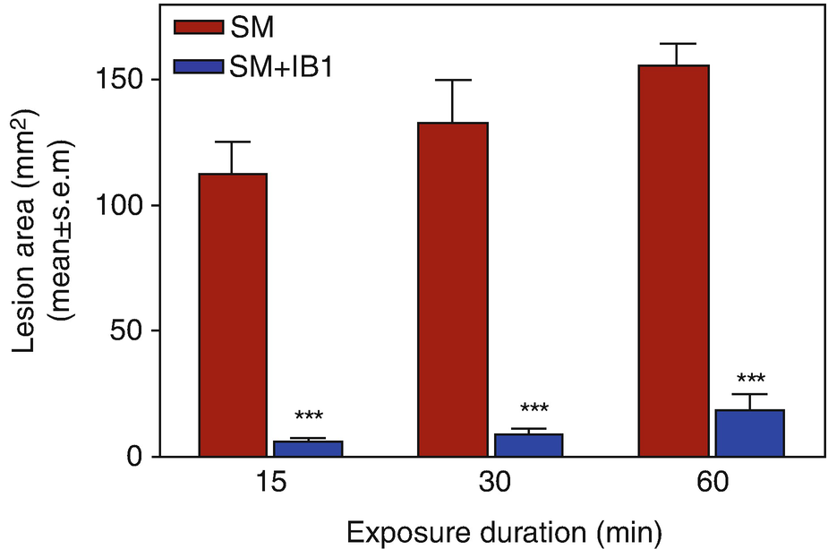
Quantitative analysis of the area of the skin lesions 24 h following exposure to SM (1 μl droplets) for 15–60 min. IB1 afforded about 90% protection (0% protection assigned to SM controls, ∗∗∗p < 0.001 vs SM) when applied 1 h before exposure
Related posts:
 of Chemical, Biological, Radiological, and Nuclear (CBRN) Skin Decontaminants: Toward Tests Standardization
Knowledge: Human/Animal Skin Decontamination
of Chemical, Biological, Radiological, and Nuclear (CBRN) Skin Decontaminants: Toward Tests Standardization
Knowledge: Human/Animal Skin Decontamination
 Human and Animal Stratum Corneum As a Partial Model for the 15 Steps of Percutaneous Absorption: Emphasizing Decontamination, Part I
 Affinity and Decontamination of Dermal Decontamination Gel (DDGel) to Model Chemical Warfare Agent (CWA) Simulants
 Human and Animal Stratum Corneum as a Partial Model for the 15 Steps of Percutaneous Absorption: Emphasizing Decontamination Part II
 Earth: Old and Faithful Skin Decontaminant Against Toxic Agents
Stay updated, free articles. Join our Telegram channel

Full access? Get Clinical Tree


