div class=”ChapterContextInformation”>
10. Binding Affinity and Decontamination of Dermal Decontamination Gel (DDGel) to Model Chemical Warfare Agent (CWA) Simulants
Keywords
Binding AffinitySkin DecontaminationPartition coefficientCWA simulantsDermal decontamination gel (DDGel)Introduction
Chemical absorption into and through skin is important in dermato-pharmacology and dermato-toxicology. Stratum corneum (SC), the outermost skin layer, constitutes a main rate-limiting barrier to transport of most chemicals across the skin [11, 29, 30, 38]. The chemicals first partition into the SC before penetrating the remaining epidermal layers, dermis, and vascular system to gain access to system circulation. Chemical partitioning proceeds much faster than complete diffusion through whole SC, and the partitioning process quickly reaches equilibrium [31]. In partitioning, chemicals may bind to components of the SC and be retained by SC, which serves as a reservoir [44]. Therefore, understanding the processes behind chemical partitioning into the SC is essential to gaining insight into the barrier properties of the SC and chemical transport across skin.
Human SC has been used as an in vitro model to explore percutaneous absorption and local or systemic risks associated with dermal exposure to toxic chemicals [24, 32]. Driven by applications in transdermal drug delivery and safety assessment, there has been a continuous interest in revealing a chemical’s partition and binding to the SC. Studies on chemical partitioning into SC lipids were broadly published [2, 5, 14, 18, 21, 24]. A good correlation exists between overall partitioning of chemicals into SC and chemical partitioning into SC lipids. But evidence shows that chemicals diffuse into SC corneocytes and also bind to SC proteins [1, 2, 19, 25]. A two-phase partitioning model has been proposed for analysis of the heterogeneous structure of the SC [2, 22, 25].
Penetrant’s binding to SC components can lead to differences in the effective and true diffusion and partition coefficients, a retardation of the absorption process, and a sustained presence of the bound compound. Therefore, accurate estimates of binding coefficients become essential to improve understanding of binding mechanisms to the major stratum corneum components. Stratum corneum study shows the relative role of lipids and proteins and thus provides a mechanistic insight into the relationship between the SC lipids and proteins as well as dermal penetration, absorption prevention, and skin decontamination.
An effective skin decontaminant should: (i) decrease the quantity of contaminant absorbed into skin (from stratum corneum to receptor fluid), (ii) significantly delay absorption time (if overall absorption could not be significantly decreased (holding), and/or (iii) have an ability to degrade the contaminant present on and/or in the skin. The nonabsorbed fraction (total quantity of chemical present on the skin surface) is the fraction considered as the removable amount from the skin surface. On the other hand, the absorbed fraction (i.e., sum of the quantities from the stratum corneum to receptor fluid) is the quantity of chemical which can potentially move into the blood. This is why, for each decontamination experiment, quantities of chemical in the nonabsorbed fraction and in the absorbed fraction have been compared.
Chemical weapons pose significant threats to society. In response to these threats, scientists have long searched for environmentally benign approaches to their decontamination. Knowledge to decontaminate chemical warfare agents (CWAs) such as distilled mustard, lewisite, tabun, sarin, soman, and VX is thus important. However, it is often necessary to conduct studies examining chemical agent behavior using simulants due to the toxicity of the agents and usage restrictions [4]. An ideal chemical agent simulant would mimic all relevant chemical and physical properties of the agent without its associated toxicological properties. Although many chemicals have been used as simulants, no individual compound is perfect because a single simulant cannot satisfactorily represent all properties of a given CWA. Thus, many chemicals have been used as simulants, depending on the physical–chemical properties of interest. Mustard gas (HD) simulates include DEA, DMA, CEMS, CEPS; yet TMP and DES are used as G-agents and VX simulants, respectively. The volatility of CWAs and their simulants are different. HD and G agents have high volatility and VX has a relatively low volatility. There are no experimental volatility data for these simulants; most are volatile; TMP is the most volatile [9, 16, 33, 36].
A successful skin decontamination strategy, for example, should not only remove CWAs from the skin surface, but also back-extract chemicals from the SC reservoir and reduce further penetration and systemic absorption. A dermal decontamination gel (DDGel) has shown excellent decontamination efficiency in removing, and more importantly, back-extracting topically applied chemicals such as chemical warfare simulants and other model chemicals.
Here, we use human stratum corneum as an in vitro model to assess relationship between physicochemical properties of simulants and their perspective partition mechanisms that govern partitioning between (a) the SC and water and (b) DDGel and water. Then we determined whether or not the partition coefficient of PCDDGel/w correlated to PCsc/w and/or to PCo/w. These values were further correlated with SC protein and SC lipid binding rates, desorption rates (off-rates), and skin decontamination efficiencies of those simulants. Also, skin decontamination experiments were performed to test decontamination efficacy of DDGel with these chemicals using in vitro human skin models. Those studies serve to verify the theoretical estimation of the relationship between chemical penetration and SC domains of lipid, protein, and water [41].
Materials and Methods
Test Chemicals and Reagents
Radiochemical purities of all carbon-14 labeled chemicals (hot) were greater than 95%. [C14]-trimethyl phosphate (TMP) (50 mCi/mmol), [C14]-dimethyl adipate (DMA) (50 mCi/mmol), [C14]-2-chloroethyl methyl sulfide (CEMS) (55 mCi/mmol), [C14]-diethyl adipate (DEA) (50 mCi/mmol), [C14]-chloroethyl phenyl sulfide (CEPS) (50 mCi/mmol), and [C14]-diethyl sebacate (DES) (100 mCi/mmol) were custom synthesized by American Radiolabeled Chemicals, Inc. (St. Louis, MO, USA). Reagent grade ethanol was purchased from Fisher Scientific (Pittsburg, PA, USA). Soluene-350® tissue solubilizer and ULTIMA GOLD™ scintillation cocktails were manufactured by PerkinElmer Life and Analytical Sciences (Boston, MA, USA). Nonradiolabeled (cold) trimethyl phosphate, dimethyl adipate, 2-chloroethyl methyl sulfide, diethyl adipate, chloroethyl phenyl sulfide, and diethyl sebacate were obtained from American Radiolabeled Chemicals, Inc. (St. Louis, MO, USA). Dosing solutions were prepared in ethanol with radiolabeled compounds and 20 mg/ml of nonradiolabeled chemicals to achieve 0.05 m/Ci/ml specific radioactivity. Receptor fluid was 0.01mole/liter phosphate-buffered saline (PBS) aqueous solution containing 6% (v/v) polyethylene glycol prepared using PBS tablets obtained from Diagnostic BioSystems (Pleasanton, CA, USA) and polyethylene glycol from Sigma-Aldrich (St. Louis, MO, USA).
Preparation of DDGel
DDGel is a gel-quickly-dried-film formulation prepared with polymers (Kollidon SR, lutrol, and carboxymethyl cellulose), absorbent clay (Fuller’s earth and bentonite), and solvents (ethanol and water). Weight Kollidon SR (3 g), lutrol (1 g), carboxymethyl cellulose (0.3 g), Fuller’s earth (2.5 g), and bentonite (0.5 g) were dissolved with 2 ml of water and 10 ml of ethanol and mixed.
Preparation of Intact Stratum Corneum Membranes and DDGel Sheets for Chemical Binding Experiments
Skin samples were obtained from the thigh of adult human cadavers at the Pathology Department of the University of California, San Francisco School of Medicine. Skin samples at a target thickness of 400 μm were prepared using a Padgett Electro-dermatome (Padgett Instruments, Inc., Kansas City, MO, USA), and skin samples were stored at 4 °C until use. Skin sample physical conditions were examined visually to exclude samples with surface damage or abnormal appearance. Skin samples were processed to obtain SC membranes by employing a modified method [43]. After submerging the skin sample in 60 °C water for 1 min, the epidermis was carefully peeled from dermis and placed on a cotton pad soaked with 0.5% (wt/wt) trypsin solution with the dermal side down at 37 °C for 15 to 24 hrs during skin digestion. The pH of the trypsin solution was adjusted to 8.0–8.6 with 5% (wt/wt) sodium bicarbonate. When digestion was completed, SC was thoroughly washed thrice with water and then dried under vacuum overnight at room temperature. Prepared SC membranes were stored at room temperature until use (less than 6 months). Stratum corneum thickness was measured by digital caliper (Marathon Watch Company Ltd., Ontario, CA) before experiments.
DDGels were prepared as a film on glass, then punched into discs of the same size (diameter = 10 mm). Weights of DDGel disc sheets were measured by analytical balances (Sartorius AG, Goettingen, Germany).
Stratum Corneum and DDGel Chemical Binding Experimental Procedures
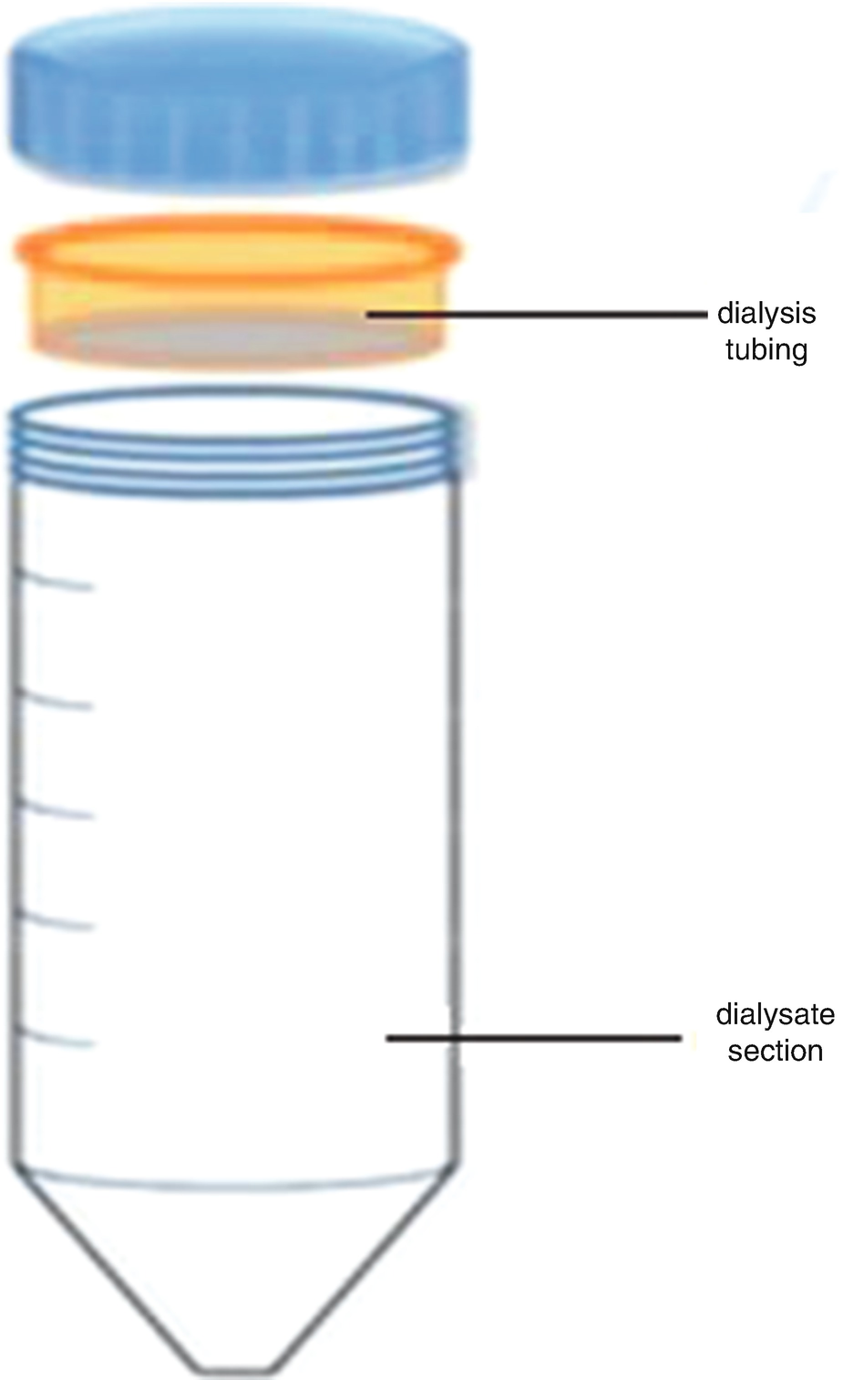
Dialysis system for binding test. This shows the two-part dialysis system
Stratum corneum water uptake capacity was calculated by the increase of the post-equilibrium sample weight.
In Vitro Skin Decontamination Efficacy
Adult human cadaver skin samples were stored at −20 °C. On the day of experiment, skin samples were thawed at room temperature and the skin was cut into 6.25 cm2 circular pieces. Full-thickness unclipped skin was used. Thickness was measured with a digital caliper (Marathon watch company Ltd., Richmond Hill, Ontario, CA). Human skin membrane thicknesses were 300–500 μm. Skin discs were mounted onto Flow-Through diffusion cells and maintained at 37 °C in a water bath in order to obtain a skin surface temperature of 33–34 °C as observed in vivo in humans. Skin samples were divided into groups of six biological replicates from three donors (69, 71, 72 years, White, male). Skin area available for chemical diffusion was 1 cm2. After 30 min equilibration with receptor fluid, skin integrity was assessed by measuring the Trans-Epidermal Water Loss (TEWL) using a Vapometer (Delfin Technologies, Ltd. Kuopio, Finland) before any drug or chemical dosing. Following the OECD guidelines [23], only skin with TEWL values ranging between 3 and 10 g/h/m were used in the penetration studies. This permeability is comparable to a fresh human skin specimen [10]. The donor compartment of the diffusion cell apparatus remained open during the experiment. An aliquot of 10 μL of a simulant was evenly applied to the center of the unclipped human skin surface, resulting in an applied surface dose of 1 mg/cm2 [17]. Decontamination procedures were performed at 0.5 hrs post chemical dosing following the procedure as described: An aliquot of 150 mg of DDGel was massaged onto the surface in a circular motion of each skin disc for 5 seconds with a glass rod. DDGel was then peeled off from skin surface after 30 mins from DDGel deposition time. The skin surface was softly wiped one time with a dry cotton ball to remove the residual DDGel. Simulants permeation assay was continued for up to 24 hrs post chemical dosing to ascertain the impact of the skin decontamination process on dermal penetration and percutaneous absorption. Skin samples without any decontamination were used as controls. Receptor fluid (RF) was collected at 30 min, 1 hr, and then every 2 hrs thereafter. RF samples were handled according to established procedures (mixed with liquid scintillation cocktail at volume ratio of 1:100). At the end of the experiment, skin discs were removed from the diffusion cells. The surface of control skin samples was gently wiped two times with a dry cotton ball to recover the fraction of chemical remaining on the skin surface. Stratum corneum was removed by tape stripping for ten times using D-Squame standard sampling disc (Cuderm). The viable epidermis was separated from dermis by heat treatment (in 60 °C water for 45 sec) [28]. Dermis and viable epidermis were cut into small pieces and placed individually into vials with 2 mL of Soluene 350 Tissue Solubilizer (PekinElmer Life and Analytical Sciences, Boston, MA, USA) for total tissue digestion. The test chemical was extracted overnight at room temperature without shaking.
One-hour in vitro skin penetration experiment of CEPS was performed. Skin samples were divided into groups of three biological replicates from one donor (80 years, White, male). The experiment method was the same as for the 24 hrs decontamination experiment. After 30 min exposure, DDGel was applied and then wiped before gel sheet formation (in 5 mins) to avoid DDGel’s SC peeling effect so that the back-extracting effect can be demonstrated. RF was collected every 5 mins.
Radioactivity Measurements
Collected test samples were measured for radioactivity with a PerkinElmer Tri-Carb 2900TR liquid scintillation spectrometer (PerkinElmer Life and Analytical Sciences, Inc., Waltham, MA, USA), which was calibrated with background control scintillation cocktail standards.
SC and DDGel samples were mixed with scintillation cocktails for radioactivity quantitation. After binding, the vehicle fluid and surface washes were mixed with scintillation cocktails (Ecolite (+)) for assay. Control and test sample scintillation counts were acquired into, and analyzed by, a computer program which comes with the liquid scintillation spectrometer to generate reportable data.
All in vitro experiments samples were collected and mixed with scintillation cocktail for radioactivity quantitation. Radioactivities in cotton ball and scintillation cocktails were measured.
Accuracy and reliability of liquid scintillation spectrometry were ensured weekly by calibration by running its manufacturer-provided internal quench and calibration standards. Liquid scintillation spectrometer is an automatically calibrated system. Before samples are counted, the system will be normalized and calibrated. Normalization, calibration, and Instrument Performance Assessment (IPA) occurs automatically by leaving the Self Normalization and Calibration (SNC) and IPA cassette (containing the 14C calibration standard, unquenched Tritium standard , and background standard) on the instrument counting deck at all times.
Data Analysis and Regression
- 1.
Partition coefficients of each test chemical between SC and water or between DDGel and water were measured following the method of Hui et al. [13], after a 24-hour incubation period at 37 °C to reach equilibrium. Partition coefficient (PC) of a test chemical between SC and water was calculated as:


- 2.
Efficacy of a skin decontaminant
Mass balance calculations were performed to ensure data accuracy. Total chemical amount in a given tissue was calculated based on the total sample volume or weight and chemical concentration in that sample. To enhance and ensure best data comparability across treatments (control vs DDGel) and across simulants, the assayed decontamination and absorption data were normalized by total recovery for each diffusion cell and then statistically analyzed and compared.
Amounts of chemical were expressed as percent of dose recovered from receptor fluid, skin surface, stratum corneum, viable epidermis, and dermis. Means of all data are given along with corresponding standard deviations (S.D., n = 6 replicates). A normality test (Kolmogorov–Smirnov) was conducted on all data acquired from the in vitro studies: the data were found not normally distributed (non-Gaussian). Because the quantities of chemical in each fraction in a given sample type (receptor fluid, skin surface, stratum corneum, viable epidermis, dermis, total, absorbed fraction, or nonabsorbed fraction) do not follow a normal distribution and variances were not equal, a Student’s t-test (P < 0.05) was applied to check if decontaminant of DDGel system significantly reduced the chemical amounts in various compartments of the skin and the perfused (mimics the blood in systemic circulation) when compared to the control.
Results
Binding Affinity of SC and DDGel
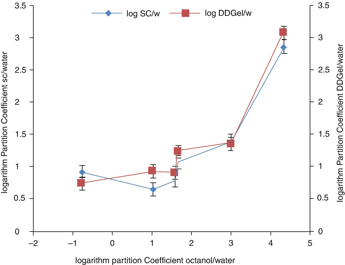
Correlation of log Po/w, log Psc/w, and log PDDGel/w of six CWA simulants. Data of partition coefficients (PCSC/w and PCDDGel/w) of each chemical were the results at equilibrium after a 24-hr incubation. Each logarithm partition coefficient data point in Fig. 10.2 (Log PSC/w or Log PDDGel/w) represents the mean ± S.D. (n = 5) of a CWA simulant tested. The corresponding data points of log Psc/w and log PDDGel/w are closely colocated at the corresponding log Po/w (octanol/water) value of a CWA simulant
Molecular weight (MW), hydrogen-bond (donor and acceptor) counts, log Po/w, log Psc/w, and log PDDGel/w of six CWA simulants tested
CWA simulants | MW | Hydrogen bond | Logarithm partition coefficient | |||||
|---|---|---|---|---|---|---|---|---|
Donor | Acceptor | Oct/w | SC/w | DDGel/w | ||||
Mean | S.D. | Mean | S.D. | |||||
Trimethyl phosphate ( TMP ) | 140.07 | 0 | 4 | −0.78 | 0.92 | 0.12 | 0.74 | 0.01 |
Dimethyl adipate (DMA) | 174.19 | 0 | 4 | 1.03 | 0.65 | 0.16 | 0.93 | 0.05 |
2-Chloroethyl methyl sulfide (CEMS) | 110.61 | 0 | 1 | 1.59 | 0.78 | 0.08 | 0.9 | 0.07 |
Diethyl adipate (DEA) | 202.25 | 0 | 4 | 1.67 | 1.07 | 0.05 | 1.23 | 0.12 |
Chlorethyl phenyl sulfide (CEPS) | 172.67 | 0 | 1 | 3.01 | 1.4 | 0.08 | 1.35 | 0.1 |
Diethyl sebacate (DES) | 258.35 | 0 | 4 | 4.33 | 2.86 | 0.13 | 3.08 | 0.06 |
Related posts:
 of Chemical, Biological, Radiological, and Nuclear (CBRN) Skin Decontaminants: Toward Tests Standardization
Knowledge: Human/Animal Skin Decontamination
of Chemical, Biological, Radiological, and Nuclear (CBRN) Skin Decontaminants: Toward Tests Standardization
Knowledge: Human/Animal Skin Decontamination
 Human and Animal Stratum Corneum As a Partial Model for the 15 Steps of Percutaneous Absorption: Emphasizing Decontamination, Part I
 Human and Animal Stratum Corneum as a Partial Model for the 15 Steps of Percutaneous Absorption: Emphasizing Decontamination Part II
 Earth: Old and Faithful Skin Decontaminant Against Toxic Agents
 Mass Decontamination Paradigm: Response Relating to Gas Phase Exposures and Skin Decontamination
Stay updated, free articles. Join our Telegram channel

Full access? Get Clinical Tree


