div class=”ChapterContextInformation”>
7. Fuller’s Earth: Old and Faithful Skin Decontaminant Against Toxic Agents
Keywords
Fuller’s earthRSDLM291Sulfur mustardVXDecontaminationIntroduction
Skin exposure to warfare agents is a major problem during nonconventional war [1] or terrorist attack. The skin, being the largest organ of the body and having the largest surface area in contact with the outside environment, is one of the principal targets for warfare agents that might either damage the skin or penetrate through it. Therefore, removal of the warfare agents by decontamination is a most important component in the individual defense doctrine against exposure to such compounds.
Liquids and solids are the only substances that can be effectively removed from the skin while it is generally not possible and not necessary to decontaminate vapor. Removal from the atmosphere containing the vapor is all that is required [2].
The purpose of the decontamination procedure performed after exposure is the removal of maximal amount of warfare agent from the skin, prevention of agent’s penetration through the skin, and avoiding secondary contamination of the medical team that comes in contact with the injured individuals [3–5].
Although decontamination issues have been explored since the beginning of modern chemical warfare, currently there is no an ideal decontaminant. Many substances have been evaluated for their efficacy for skin decontamination, but only few became practical.
At present, decontamination of CWA from the skin is still considered as a difficult problem due to the necessity for both rapid action and nonaggressive performance. As yet, there is no consensus as to the optimal procedure for removing the toxic agents from the skin.
There are three basic methods of decontamination : physical removal, chemical deactivation, and biological deactivation of the agent. Up till now, biological decontamination has not been developed to the point of being practical.
An ideal decontaminant is required to have several essential characteristics : neutralize the agent, safety (nontoxic and noncorrosive), easy to apply, readily available, rapid action, will not produce toxic end-products, stable in long-term storage, will not enhance percutaneous agent absorption, and easy to dispose [6, 7] . Nevertheless, the most common problems with potential decontaminants are irritation of the skin, toxicity, ineffectiveness, or high cost.
The two most common chemical warfare agents (CWAs) that constitute a threat for skin exposure are sulfur mustard (HD) and VX (O-ethyl-S-(2-diisopropylaminoethyl-methylphosphothiolate).
Sulfur mustard is a potent cutaneous vesicant that penetrates rapidly through the skin, causing severe injuries. Being a blistering agent, it causes inflammation, blisters, and severe damage to the skin that might result in incapacitation of the victims. Healing time from such injuries is very long and often the healing is pathologic [8–11]. Since its first use in World War I and despite vast efforts, only limited success has been attained to find an effective treatment to prevent or reduce these lesions.
VX, on the other hand , is a very toxic nerve agent, an organophosphate and an extremely potent cholinesterase inhibitor. It is relatively stable, has a very low volatility, efficiently penetrates through the skin, and might cause death at very low doses (dermal human LD50 ≈ 0.04–0.14 mg/kg) [12–14].
Removal of the CWA from the skin soon after exposure may reduce significantly the extent of damage following exposure to HD and the mortality rate and systemic clinical symptoms after exposure to VX.
The present article aimed to explore the efficacy of Fuller’s earth powder as a universal decontaminant against CWA and to compare its efficacy against the two representative agents to other common decontaminants.
Fuller’s earth powder is a natural substance containing silica of aluminum and magnesium which has very high adsorption properties. In addition, it contains small amounts of calcite, dolomite, and quartz.
Fuller’s earth has been demonstrated to be a nontoxic and effective absorbent of classical liquid CWAs [15]. The powder is chemically inactive, is relatively cheap, and has long shelf life. Fuller’s earth has been fielded by the Israeli Defense Forces and in by several North Atlantic Treaty Organization countries [6]. The decontamination doctrine for percutaneous toxic agents in Israel is based on a combination of application of Fuller’s earth (×3) followed by wet decontamination (copious amount of water), which will be usually performed a few hours after Fuller’s earth decontamination by the medical personnel.
A comparison between Fuller’s earth and other common decontaminants is presented following exposure of pigs to HD and VX.
Experimental Models and Experimental Design
Cutaneous exposure was performed in the well-established pig model which is the preferred model for skin injury research due to its anatomical, physiological, and biochemical similarity to human skin [16–18].
Young female white pigs (supplied by the Institute of Animal Research, Kibbutz Lahav, Israel, and Topig20, the Netherlands) weighting 10–11 kg were housed in individual pans located in a temperature-controlled (21 ± 1 °C) room, with lights on from 06:00 to 18:00. Pigs were fed twice a day and had free access to tap water.
All procedures involving animals were in accordance with the Guide for the Care and Use of Laboratory Animals, National Academy Press, Washington, DC, 1996, and were approved by the Institutional Animal Care and Use Committee.
Exposure Procedure
Exposure to HD – Droplets
Ten fields were marked on the female pigs’ back skin (5 on each side), and three droplets of neat HD (1 μl each) were placed on the skin in each of the fields. Exposure durations varied between experiments and were 1, 2, 3, 5, 15, 30, and 60 minutes.
Exposure to VX
Female pigs were exposed to 1-μl droplets of VX (0.4 mg/kg or 1.3 mg/kg) which was equivalent to 2LD50 for 24 hours (unpublished data). The shift in the LD50 dose is due to the changes in the exposure setup and newly imposed safety regulations.
VX was placed on the pigs’ back skin along the midline. Exposure durations were 20 minutes and 1, 2, and 5 hours. Control pigs were exposed to VX for the duration of 12–24 hours.
The exposure procedures of both HD and VX were performed in a hood at room temperature and were terminated, after a predetermined period by decontamination with one of the tested materials.
All exposed pigs, independent of their decontamination methods, were washed with the decontamination solution IPP2 at the end of the day, before returning to their home cages (about 10–12 hours following exposure). This was a safety precaution required by our institution safety procedures. IPP2 decontamination solution was prepared by dissolving chloramine T (38 g /100 ml, (Axcentive, the Netherlands) and zinc chloride (5.5%) in water (56%) and ethanol (44%) (Sigma, Israel).
Decontamination efficacy was determined by morphometric analysis of the lesion areas (exposure to HD) or by the extent of ChE inhibition in whole-blood samples, clinical signs, and the occurrence of death (exposure to VX).
Quantitative Assessment of the Lesion Area
The extent of the injury following exposure to HD droplets was measured using morphometric analysis of the damaged skin area. The exposed sites on the pigs’ backs were photographed using a digital camera (Coolpix 950, Nikon, Tokyo, Japan). Pictures were analyzed for the damaged area using an image analysis system (Image-Pro Express 4.0).
Cholinesterase Inhibition in Whole Blood
Blood samples (0.5–0.7 ml) were collected from the jugular vein at various time points following exposure to VX. One hundred microliters of blood was immediately transferred into a heparinized tube containing 1.2 DDW and mixed gently to cause hemolysis of red blood cells. Samples were stored at −4 °C until further analyzed.
The inhibition of the enzyme cholinesterase was determined using the radiometric method [19]. The blood samples were incubated with the substrate, H3-acetylcholine, at a temperature of 25 °C for 30 minutes. The reaction was ended with a reaction stopper that separated between the radioactive acetate and the choline. Scintillation solution was added, and the radioactivity of the acetate in the organic phase was measured using a β counter.
Results
The Efficacy of Fuller’s Earth
Fuller’s earth is a clay material containing nonplastic form of kaolin with aluminum-magnesium silicate [20] that has the capability to absorb oily liquids and other liquids. Fuller’s earth is used in the industry as absorbents for oil, grease, and animal waste and is widely used in cosmetics. It is also recommended for personal decontamination of warfare agents.
Decontamination of experimental animals was performed by applying a large amount of powder to the skin to cover the exposed areas and then was removed with a spatula. This was repeated three times and then was followed by thorough rinsing with copious amount of tap water before returning the animals to the pens.
Exposure to Liquid HD
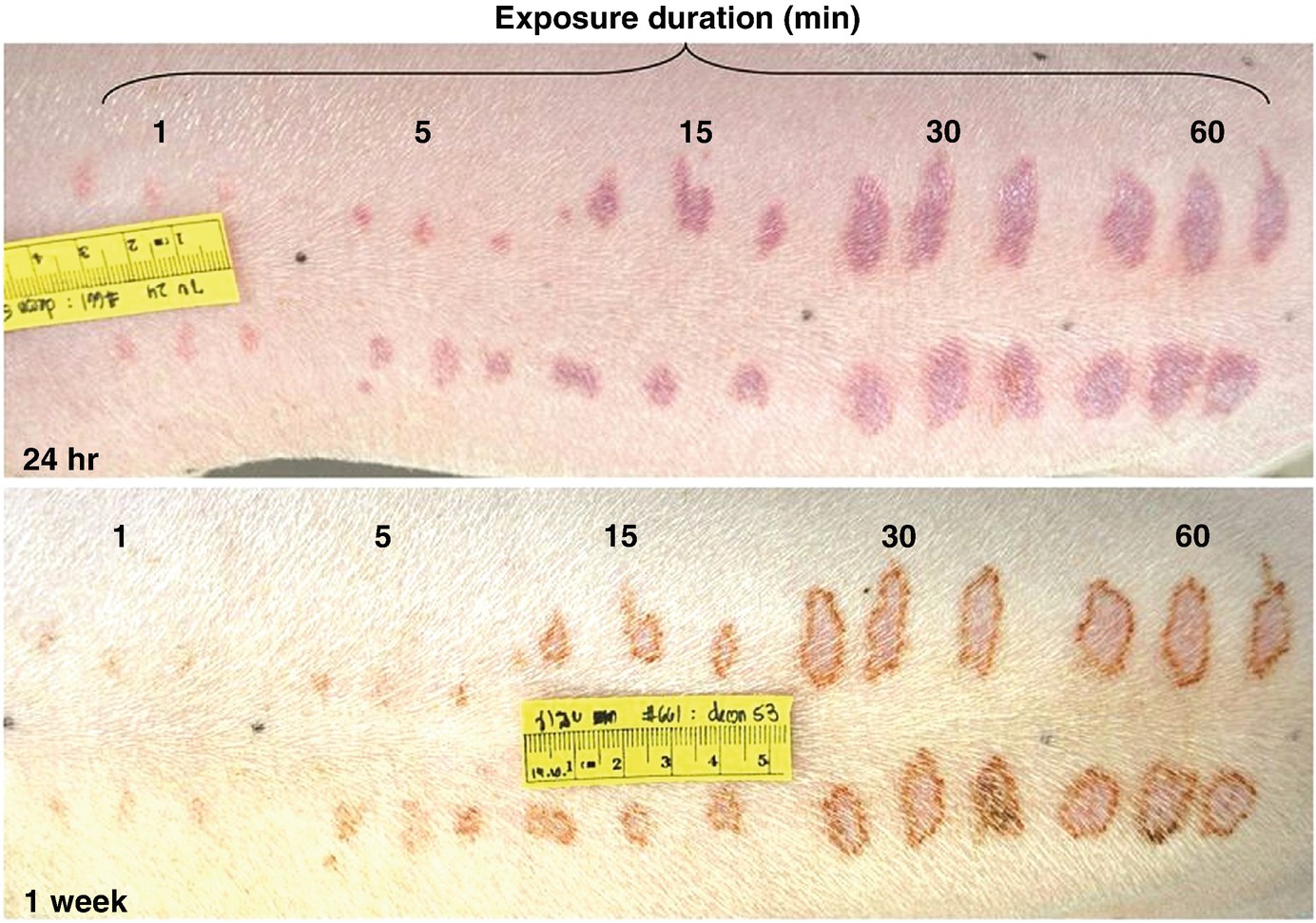
The effect of decontamination with Fuller’s earth 24 hours and 1 week following dermal exposure to HD droplets (1 μl) on the extent of the damage. Note the time-dependent effect on the skin lesions (exposure durations of: 1, 5, 15, 30, and 60 minutes)
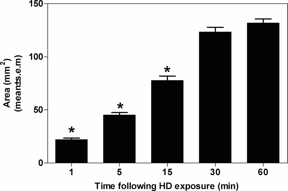
A morphometric analysis of skin lesion area 24 hours following exposure to HD droplets (1 μl) and decontamination with Fuller’s earth at various time points after exposure. Note the time-dependent response and the significant reduction in lesion area even when decontamination was performed 15 minutes post exposure (∗p < 0.001 vs all other time points, n = 21 pigs)
Exposure to VX
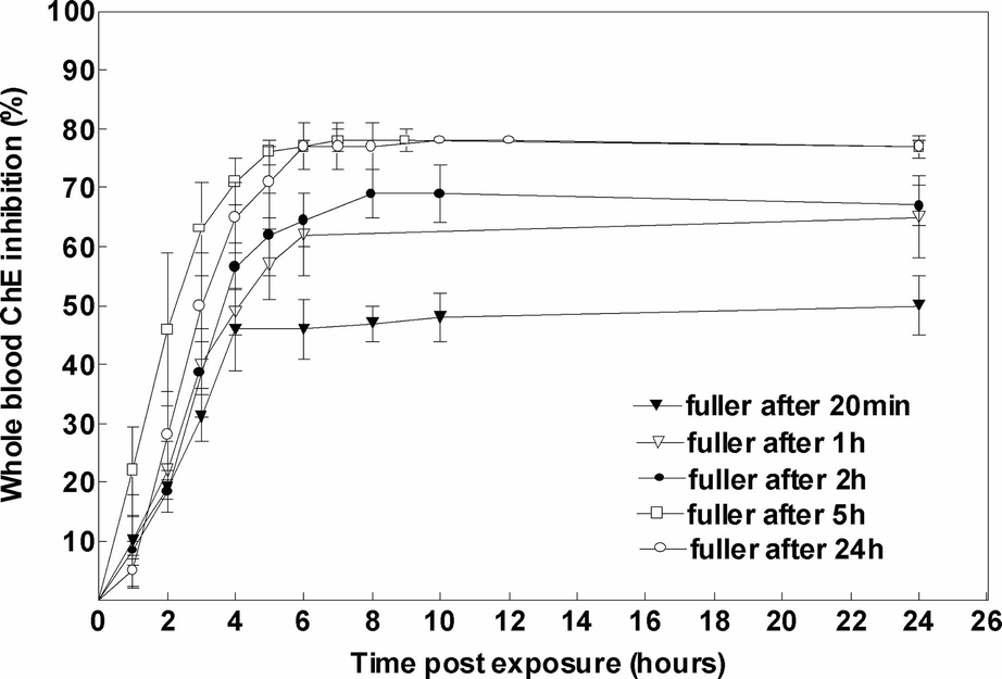
Profile of ChE inhibition following dermal exposure to 2LD50 of VX and decontamination with Fuller’s earth at various time points post exposure (20 minutes – n = 3, 1 hours – n = 6, 2 hours – n = 2, 5 hours – n = 4, 24 hours – n = 4)
Interestingly, the time of decontamination affected the rate of ChE inhibition development, which by itself influenced the severity of clinical symptoms. When decontamination was performed up to 2 hours post exposure, the slope was more moderate than the slope following longer periods of exposure, leading to a less severe clinical outcome (Fig. 7.3).
Another characteristic of percutaneous exposure to VX is the continuous decrease in ChE activity in blood after decontamination was performed (at least up to 2-hour exposure duration), suggesting a possible release of VX from a skin depot. When decontamination was performed after 5 hours of exposure, the inhibition was maximal most likely because all the amount of VX already reached the circulation.
To summarize, Fuller’s earth decontamination was found beneficial, in reducing skin lesions up to 30-minute exposure durations to liquid HD and in preventing most of the VX clinical signs when performed up to 2 hours following exposure.
The Effect of Fuller’s Earth Compared to Water
Rinsing the skin with water can remove or dilute substantial amounts of the agent and produce significant results. Moreover, water has the capacity to remove chemical agents not only through mechanical force but also via slow hydrolysis; however, the low solubility and slow rate of diffusion of HD and VX in water significantly limit these agents’ hydrolysis rate.
Water or a solution of soap and water were suggested for decontamination if other decontaminants were not available. The solutions are well suited for mass casualty situations in which adequate water supplies are available, and they provide some protection against CWA mostly by physical removal of the agents. The following section compares the efficacy of water as a single decontaminant to the efficacy of dry decontamination with Fuller’s earth.
Exposure to Liquid HD
A comparison between the efficacy of water and Fuller’s earth was performed following exposure to HD. The decontamination regimens that were tested were tap water (1 liter/400 cm2), soap + water, Fuller’s earth × 3, Fuller’s earth × 1 + water. Analyzing all parameters that were tested it was found that Fuller’s earth × 3 was the most effective decontamination method over time. The decontamination with water alone or soap + water exacerbated the effect of HD and resulted in an increase in the damaged area probably because in this experiment pigs were not rinsed with a copious amount of water but only washed with a rather small amount. Wetting the area of exposure is not efficient in removing HD from the skin.
Related posts:
 of Chemical, Biological, Radiological, and Nuclear (CBRN) Skin Decontaminants: Toward Tests Standardization
Knowledge: Human/Animal Skin Decontamination
of Chemical, Biological, Radiological, and Nuclear (CBRN) Skin Decontaminants: Toward Tests Standardization
Knowledge: Human/Animal Skin Decontamination
 Human and Animal Stratum Corneum As a Partial Model for the 15 Steps of Percutaneous Absorption: Emphasizing Decontamination, Part I
 Affinity and Decontamination of Dermal Decontamination Gel (DDGel) to Model Chemical Warfare Agent (CWA) Simulants
 Human and Animal Stratum Corneum as a Partial Model for the 15 Steps of Percutaneous Absorption: Emphasizing Decontamination Part II
 Mass Decontamination Paradigm: Response Relating to Gas Phase Exposures and Skin Decontamination
Stay updated, free articles. Join our Telegram channel

Full access? Get Clinical Tree


