History
Patient presents with the absence of a distal portion of the limb with an otherwise normal proximal portion.
Exam
This disorder is usually 98 % unilateral, mostly at upper forearm level. If it is bilateral, it may be an AR trait. It is nonsyndromic and is reported to appear with hydrocephalus, spina bifida, meningomyelocele, and clubfoot. It is associated with hypoplasia of proximal muscles which differentiates it from constriction band syndrome.
Treatment
Treatment is mostly nonsurgical. Prosthesis adapted to normal hand function according to the age of the patient.
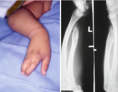
2.1.2 Radial Clubhand
History
A form of preaxial deficiency that is typically sporadic and unilateral. Radial dysplasias are more frequent in males, on the right side, and are frequently associated with syndromes including Fanconi anemia, TAR syndrome, Holt-Oram syndrome, and VATER syndrome.
Exam
A foreshortened forearm with radial deviation at the wrist. The deformity affects all structures including the bone, tendons, joints, and soft tissue with the absence of the radius as the most severe and most common type.
Treatment
Mild forms of dysplasia are treated with splinting. Evaluation and treatment of associated syndromes must be considered prior to surgical reconstruction. Distraction lengthening is recommended for a short or hypoplastic radius. Centralization is performed in cases of severe hypoplasia or the absence of a radius.
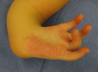
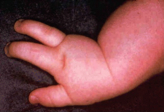
2.1.3 Ulnar Deficiency
History
Patient presents with shortened, bowed forearm.
Ulnar longitudinal deficiency (ULD) (also known as ulnar clubhand) is a lack of formation of the pinky side of the upper extremity. It usually affects the forearm but can affect the hand, forearm, and upper arm. It can affect the bone, muscle, tendon, nerves, and blood vessels. The severity is different in each affected child.
Exam
It usually affects the forearm, but the hand and upper arm can be involved as well. The first webspace is often narrowed. There can be syndactyly between other digits, and in more severe cases, a variable number of digits may be absent. Abnormal bony connections between the carpal bones and metacarpals may exist. The ulna is shortened and bowed or absent. The radius usually is usually normal or fused to the humerus (radioulnar synostosis).
Treatment
Early stages of treatment include splinting. Surgical treatment entails syndactyly release, digital rotation osteotomies, resection of radial head, and osteotomy of the synostosis.
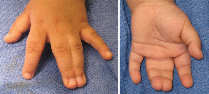
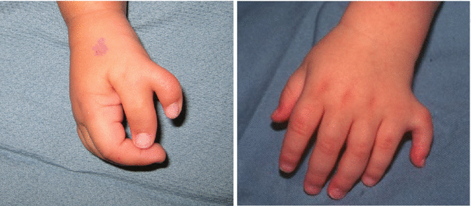
2.1.4 Cleft Hand
History
It is associated with other anomalies including cleft lip and palate congenital heart disease and significant deformities of the upper and lower extremities. It has been named various medical and layperson names such as ectrodactyly, split hand, or lobster claw hand. It is a type I failure of formation—longitudinal arrest. Classified as typical (true cleft hand) or atypical based on physical findings and genetic predilection.
Exam
Patients with typical central deficiencies have an absence of the third ray, often with bilateral findings.
Treatment
Improving the function and cosmetic appearance of the hand is the goal when the thumb or first webspace is absent. Different procedures have been described: Snow-Littler procedure is designed to create a wide first webspace and to minimize the limitations of a cleft in the hand. Ueba, Miura, and Komada described additional procedures with modifications or simplifications of the Snow-Littler procedure.
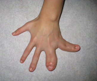
2.1.5 Diagnosis: Syndactyly
History
Syndactyly is the second most common congenital hand anomaly in which 1:2,000 of live births are affected. Syndactyly represents a form of failure of differentiation in the recession process at the webspaces. It commonly affects Caucasian males with the third webspace being the most common site of fusion.
Exam
In complete syndactyly, the fusion encompasses the entire webspace. Complex syndactyly implies bony fusion in addition to skin involvement. In complicated syndactyly, other findings are found on the exam, such as polydactyly, symphalangism, or brachydactyly.
Treatment
Border digits and complex syndactyly fusions should be addressed early. Separation should not be performed simultaneously on adjacent webs. Separation is carried out with the creation of interdigitating flaps, while the residual raw areas are reconstructed with a skin graft.
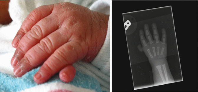
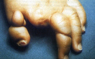
2.1.6 Duplicated Thumb (Polydactyly)
History
A radial-sided (preaxial) polydactyly is commonly found in Caucasians, as opposed to the postaxial polydactyly which is more common in African Americans.
Exam
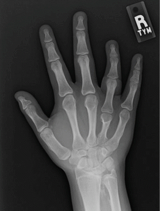
The supernumerary digit can be well developed or pedunculated and rudimentary. The Wassel classification (seven types) is used to determine the level of differentiation of the duplicated thumb, and it ranges from a bifid distal phalanx to complete duplication at the CMC joint (types I–VII). Type VII is a triphalangeal thumb.
Treatment
Types I–II benefit from a Bilhaut procedure or resection of the radial thumb. In types III–IV, the best phalangeal portions of both thumbs are incorporated to create a thumb. In types V–VI, the radial thumb is amputated with RCL reconstruction.
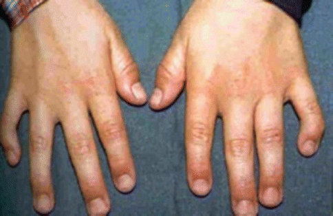
2.1.7 Macrodactyly
History
It is an overgrowth congenital anomaly. Etiology is mostly unknown, but some believe it is related to nerve-stimulated pathology with abnormal neural control in sensory distribution of the median nerve.
Exam
Four subtypes have been described. Patients can be born with an enlarged digit. The enlargement of the digit may be associated with signs of neurofibromatosis.
Treatment
Skin and subcutaneous resection, extensive neurolysis and resection, epiphysiodesis, arthrodesis, and amputation. Amputation is reserved for the digit that is of no use, significantly enlarged, and a source of embarrassment.
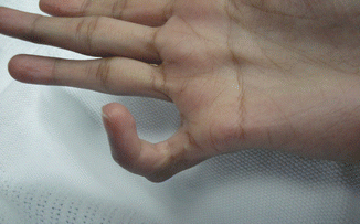
2.1.8 Hypoplastic Thumb
Diagnosis
Thumb hypoplasia.
History
It is a variant form of undergrowth (class V) involving the radial half of the upper limb and often associated with radial clubhand. Other anomalies are to be ruled out, such as the Holt-Oram syndrome, VACTERL, TAR, and Fanconi anemia.
Exam
The thumb presents with small to absent tendons, muscles, bones, and joints. Based on the modified Blauth classification, five types have been described. Type 1 implied a grossly diminished size. In type 2, the first webspace is affected. In type 3, it is necessary to determine if an unstable thumb CMC joint is present as in type 3B in order to guide treatment. Type 4 is a floating thumb (pouce flottant) indicating rudimentary phalanges. In type 5, the thumb is completely absent.
Treatment
Types 1–3A require an opponensplasty procedure combined with a first webspace deepening procedure such as a four-limb Z-plasty. In types 3B–5, complete reconstruction with pollicization of the index finger is required.
2.1.9 Brachydactyly
History
Brachydactyly can occur as an inherited condition with an autosomal dominant pattern, as a sporadic anomaly, or as part of a syndrome. Brachydactyly can also result as a condition following trauma, infection, or frostbite.
Exam
Short digits in which parts of the skeleton are present but reduced in size. The middle phalanx is most affected, while the index and small finger are the most commonly affected digits. Associated findings on exam are syndactyly, clinodactyly, and symphalangism. In Poland syndrome, additional upper extremity abnormalities and chest wall findings are expected.
Treatment
Management is based on the degree of shortening and the existing functional deficit. Hand function is usually intact in brachydactyly patients, and the surgical outcome is often disappointing. It is therefore preferred to avoid surgery in patients with short phalanges. Lengthening techniques have been used in selected cases with distraction osteogenesis or immediate lengthening with bone grafts.
2.1.10 Clinodactyly
Clinodactyly is a congenital anomaly relating to the failure of differentiation. It is defined as curvature of a digit in a radioulnar plane. The etiology is unknown. It can be inherited in an autosomal dominant fashion with variable expression.
History
Deformity may be associated with syndromes such as Apert.
Exam
An ulnar angulation of the small finger is seen. A delta phalanx is identified on an X-ray, which is the triangular bracketed-shaped bony wedge in angulated digit. Limited motion is noted when a bracketed phalanx is present.
Treatment
Bracket resection with preservation of the growth plate and fat graft or wedge osteotomy. Skin deficit after correction may require Z-plasties. A K-wire is used to hold correction in place.
2.1.11 Diagnosis: Camptodactyly
History
Camptodactyly is a congenital constriction deformity of the PIP joint and represents a form of soft tissue failure of differentiation. It is present in approximately 1 % of the population with unclear etiologic factors. The function of the hand is rarely impaired. Most patients seek medical attention due to appearance.
Exam
Abnormal flexion posture of the PIP joint, most commonly of the little finger.
Treatment
Complete correction is difficult to achieve, and surgical management should be discouraged. Nonoperative treatment consists of splinting and stretch exercises. If the contracture is more than 30°, consider release of the involved lumbricals, FDS, and accessory collateral ligaments.
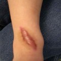
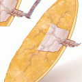
2.1.12 Constriction Band Syndrome (CBS)
History
A congenital upper extremity deformity manifested as an intrauterine amputation of the digits, hand, or limb. Amniotic constriction band syndromes have no known genetic transmission. It is frequently associated with anomalies such as clubfoot, cleft lip and palate, craniofacial defects, hemangioma, and meningocele.
Exam
The presentation of CBS varies greatly. Patients may have a spectrum of findings that vary from superficial grooving to complete amputation of a limb.
Treatment
Simple constrictions may not require any surgical treatment. More severe cases can be managed with Z-plasties in a staged fashion for release of the soft tissue bands and separation of fused digits to allow for unimpeded grow. Distractions lengthening with bone grafting and toe-to-hand transfers have also been described.
2.2 Hand, Trauma
2.2.1 Soft Tissue
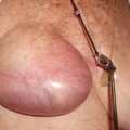
2.2.1.1 Subungual Hematoma
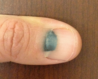
History
Trauma to the nail bed.
Exam
Throbbing pain is present on the exam because of pressure in the closed space between the nail plate and nail bed.
Treatment
Hematoma drainage (trephination) can be done with microcautery device or heated sterile paper clip. The hole should be large enough to allow for prolonged drainage. Further injury to the nail bed should be avoided. Repair of the nail bed may be required, and this can be done by removing the torn nail plate to provide exposure for the repair.
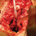
2.2.1.2 Radial Collateral Ligament (RCL) Injury
A partial or complete tear of the RCL of the thumb at the MP joint.
History
Occurs with forced adduction on the thumb when the MP joint is flexed. The RCL can be torn at its proximal or distal end, but no aponeurosis displaces the lesion and prevents interposition.
Exam
Partial tears are characterized by swelling, tenderness, and possibly ecchymosis on the radial side of the metacarpal head and MP joint. When compared with the contralateral side, a laxity of 30° in flexion or extension or 15° of increased laxity is suggestive of a complete tear.
Treatment
Acute RCL ruptures are managed conservatively if a partial tear is suspected. Surgical intervention is reserved for complete tears that are associated with volar subluxation of the proximal phalanx. If an RCL reconstruction is indicated, a free tendon graft is used. The thumb is immobilized in a thumb spica for 6 weeks postoperatively.
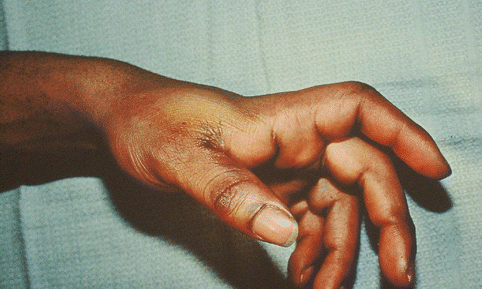
2.2.1.3 Ulnar Collateral Ligament (UCL) Injury
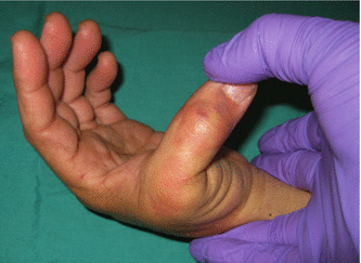
UCL injuries of the thumb MP joint occur ten times more frequently than RCL injuries.
History
The injury is from a forced radial deviation of the joint. This occurs during a fall on an outstretched hand while the thumb is abducted. Patients complain of pain and swelling on the ulnar aspect of the MP joint.
Exam
Tenderness, swelling, and ecchymosis over the ulnar aspect of the thumb at MP joint with unopposed deviation to the radial side with manipulation. The classic findings of a Stener’s lesion, the UCL avulses and retracts proximally, and the interposed adductor aponeurosis interferes with primary healing. When avulsion occurs with a fracture, a mass can be palpated. The thumb demonstrates laxity of the ulnar capsule.
Treatment
Incomplete ligament tears not associated with instability are treated conservatively. Partial tears are managed surgically if the UCL instability is greater than 30°. Stener’s lesions are treated with surgical debridement of the fracture fragment, division of the adductor aponeurosis, and stabilization of the residual UCL to the proximal phalanx.
2.2.1.4 Flexor Tendon Laceration
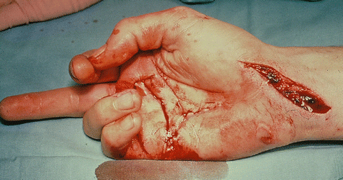
History
Flexor tendon lacerations following sharp injuries require evaluation of the patient’s age, occupation, mechanism of injury, and hand position during the time of injury. The five clinical tendon zones in the hand are based on anatomic landmarks and influence the prognosis of repair.
Exam
The normal hand cascade is inspected. Active flexion of the DIP joint while stabilizing the PIP joint tests the function of the profundus tendon. Blocking the movement of the other fingers and instructing the patient to flex at the PIP joint will test the function of the sublimis tendon.
Treatment
Different tendon retrieval techniques exist for each injury zone. Delaying the definitive repair up to 14 days has not been shown to have an adverse outcome. Tendon lacerations of 50 % or less of the cross-sectional area need not be repaired unless they cause triggering in a pulley. Early mobilization is encouraged.
2.2.1.5 Extensor Tendon Injury
History
A patient presents with deformity of the finger after laceration/trauma.
Exam
Patient is unable to extend the affected finger.
Treatment
Nonoperative treatment consists of immobilization of the affected joint with extension splinting. Operative treatment includes tendon repair, fixation of bony avulsion, tendon reconstruction, central slip reconstruction, or tendon transfer.
2.2.1.6 Jersey Finger
History
The injury pattern of a sudden failure of the fingertip to grasp. Common mechanism of injury is of a football player attempting to tackle an opponent by the jersey. Subsequently, the patient is unable to flex the tip of his finger. Injuries are classified based on whether this is a pure soft tissue allusion of the FDP tendon or a certain portion of the volar distal phalanx bone is attached to the ruptured tendon.
Exam
No active flexing of the DIP joint, however, the DIP joint can be passively flexed from zero degrees to full flexion position.
Treatment
For soft tissue injuries alone, immediate retrieval and repair is recommended. When the bony fragment is held at the A3 pulley, it is possible to delay treatment for up to 3 weeks and achieve osteosynthesis with the use of stable bony fixation. Bony fragments held at the A4 pulley prevent tendon retraction and maintain the fragment close the fracture site. Hence, repair can be delayed almost indefinitely. Repair is performed with a Bunnell suture and tied dorsally over a button.
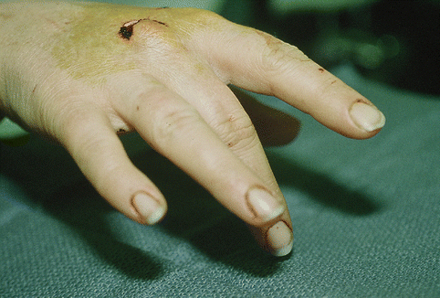
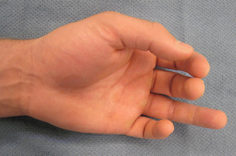
2.2.1.7 Mallet Finger
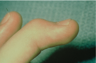
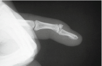
Definition
Disruption of the terminal extensor tendon.
History
There are a variety of mechanisms of injury that can lead to a mallet finger. Typically a mallet finger is the result of a closed injury that occurs when sudden flexion is applied to an extending DIP joint. Forceful hyperextension of the DIP joint can lead to a mallet finger and typically a fracture associated with this type of injury. Open injuries to the distal phalanx can also lead to a mallet deformity. Mallet fingers most commonly involve the ulnar digits. If untreated the patient will go on to develop an extensor lag and potentially compensatory hyperextension of the PIP resulting in a swan neck deformity. Injuries presenting within 4 weeks are considered acute, while a chronic mallet finger is greater than 4 weeks out from initial injury.
Exam
Extensor lag of the involved DIP joint. There may be associated tenderness over the distal phalanx depending on timing of presentation. Plain films may demonstrate associated fracture.
Treatment
When a mallet finger is diagnosed acutely, then the DIP joint of the affected digit is splinted in extension. The patient is to wear the splint continuously for 8 weeks. They are then transitioned to night splinting for an additional 4 weeks. Surgery is indicated in the acute setting if there is associated fracture with a fragment greater than 1/3 of the articular surface. Open injuries usually require operative intervention. Suture repair that incorporates both the skin and tendon eliminates need for extensive dissection that could compromise blood supply and healing. Chronic mallet injuries may require a Fowler tenotomy or spiral oblique retinacular ligament reconstruction depending on the amount of extensor lag that is present.
2.2.2 Skeletal
2.2.2.1 Phalanx Fractures
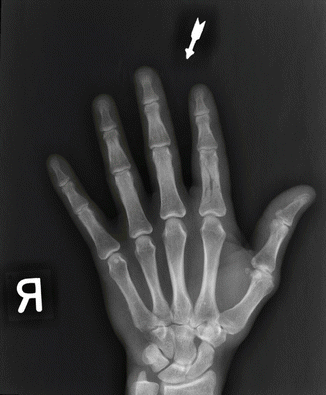
History
Trauma to the hand; fracture of the phalanx is the most common skeletal injuries, accounting for 10 % of all fractures. Distal phalanx is the most common fractured bone in the hand.
Exam
Physical findings include local tenderness and deformity, with or without an open wound.
Treatment
Proximal and middle phalanx fracture can be treated with buddy taping, or close reduction with percutaneous pinning, or open reduction internal fixation. Distal phalanx fracture can be treated with close reduction and splinting as well. However, if there is concurrent nail bed injury, nail bed repair is warranted as well.
2.2.2.2 Pediatric Hand Fracture
History
The mechanism by which pediatric fractures are sustained is not different from adult fractures. These include a fall, crush injuries, or other accidental means. The difference is in the prognosis after the injury, given the susceptibility of the injured epiphyseal plate to poor healing. The Salter-Harris (SH) classification system describes fractures involving the epiphyseal plate in children. The degree of growth disturbance correlates directly with the level of fracture classification.
Exam
A pediatric hand fracture can present as an obvious deformity with irregularity in the bony surface, tenderness, swelling, ecchymosis, and reduced range of motion secondary to pain. According to the SH classification, the epiphysis is separated from the metaphysis in type I fractures. Type II fractures involve the metaphysis and growth plate as opposed to the type III fractures which involve the epiphysis and growth plate. In SH IV, the fracture is through the epiphysis, growth plate, and metaphysis. In type V fractures, the epiphyseal plate is crushed with no involvement of the metaphysis.
Treatment
In children with types I and II fractures, conservative treatment with closed reduction, casting, or pin fixation is recommended. Types III, IV, and V fractures are managed with similarly but also include reconstruction of the articular surface.
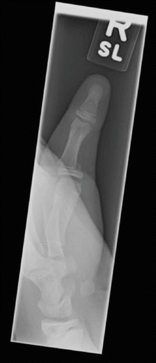
2.2.2.3 PIP Dislocation
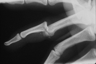
History
A patient presents with deformity at the PIP joint after trauma.
Exam
PIP joint injuries can be categorized as dorsal dislocations, dorsal fracture dislocations, volar dislocation, volar fracture dislocation, and rotatory dislocations. Finger and hand X-rays must be obtained to evaluate the extent of injury.
Treatment
Nonoperative treatments include close reduction with buddy taping and dorsal extension block splinting. Surgical options include open reduction internal fixation, dynamic distraction external fixation, and volar plate arthroplasty or arthrodesis.
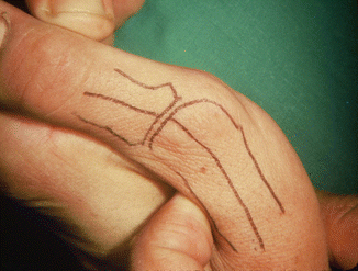
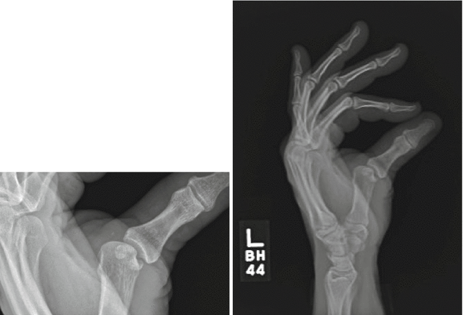
2.2.2.4 MCP Dislocation
History
A patient presents with deformity at the MCP joint after trauma.
Exam
Skin dimpling may be seen in complex dislocations. X-rays show that joint space widening may indicate interposition of volar plate.
Related posts:
Stay updated, free articles. Join our Telegram channel

Full access? Get Clinical Tree


