Hair Transplantation


Hair Transplantation


Carlos K. Wesley

Robin H. Unger

Walter P. Unger


Since initial reports on grafting human scalp hair were first made in Japanese literature in 1939, advancements in hair restoration surgery (HRS) have benefitted all patients, including both men in the early stages of hair loss and female patients. Incorporated in 1993, the International Society for Hair Restoration Surgery (ISHRS) now consists of over 1,000 members who performed approximately 279,381 HRS procedures in 2010, an increase of 11% over the previous 2 years.1

Modern day hair transplantation evolved over three distinct eras: the “plug” era, the transition period of progressively smaller unit minigrafting and micrografting, and the current period of follicular unit transplantation (FUT).2 This current method capitalizes on findings that human scalp hairs grow naturally in individual bundles, called follicular units (FUs), comprised of clusters of one to four follicles surrounded by concentric layers of collagen fibers.3 When performed properly, FUT consistently results in a cosmetic appearance indistinguishable from natural scalp hair growth (Figure 52.1). In addition to the more natural appearance of the transplanted follicles with FUT, the ratio of the donor-to-recipient area has effectively expanded. In the plug era this relationship was at best 1:1, but FUT now favors a ratio of 1:2 or 1:4, meaning 1 cm2 of donor scalp harvested may yield a sufficient number of FU (also referred to as grafts) to cover a 2 to 4 cm2 recipient area with adequate cosmetic density. Smaller, more superficial recipient sites can be spaced much more closely, while causing minimal damage to the preexisting hair in the recipient area. Despite these refinements, the unavoidable progression of hair loss over time and the limitation of donor hair for transplantation remain of paramount importance when determining the surface area and hair density that can reasonably be treated with hair transplant surgery.


INITIAL CONSULTATION AND EVALUATION

Patient candidacy is determined by a careful evaluation of the donor area’s capacity to effectively address the current and future areas affected by hair loss. In addition to providing information to the prospective patient and establishing a rapport with the patient, the physician should set realistic and prudent short- and long-term goals. Drawing from an examination of the patient’s hair phenotype, a thorough review of the patient’s family history of androgenetic alopecia (AGA) (including both the paternal and maternal lineages), and a familiarity with the progressive nature of male pattern baldness (MPB) and female pattern hair loss (FPHL), the physician can propose a surgical recipient pattern that will be appropriate even when having to “stand alone” at any future age. It is advisable to estimate the number of procedures or FUs the patient will likely have available over his or her lifetime based on the projected permanent donor fringe. Especially for young male patients, individual projections should be based on the worst possible scenario of MPB that can be realistically foreseen. This can help even young patients wanting to replicate their teenage level hairlines to modify their unrealistic expectations.

A thorough evaluation of the donor area is the critical initial step. The following elements should be considered: density of FUs per surface unit, number of hairs per FU, anagen-telogen ratio, diversity of hair caliber, color contrast between the hair and the scalp, hair texture (e.g., wave, curl, and frizz), and scalp laxity. While experienced hair restoration surgeons may be able to evaluate these characteristics with the naked eye, many practitioners prefer diagnostic tools to quantitatively assess the donor area (Table 52.1).

Screening technology that can help includes quantitative microscopic donor area measurements (e.g., Folliscope) as well as qualitative analysis of non-androgenetic forms of alopecia (e.g., PhotoFinder) that may benefit more from medical therapy (Figure 52.2). The Folliscope, capable of discerning between terminal and vellus hairs, is used by some practitioners to “rule out” patients who possess more than 20% miniaturized hairs within their donor area.4 Two separate measurements several weeks apart are required to differentiate between a hair that is miniaturized and one that is simply in early anagen (the growth phase in the hair cycle) and has only its thin, tapered edge protruding from the skin.

Once a patient’s surgical candidacy is determined, he or she is informed of the risks of the procedure. The authors emphasize three caveats: 1) postoperative edema that is minimal in most, but in approximately 2% of patients, may be severe enough to cause ecchymosis around the eyes; 2) scalp hypoesthesia resulting from severed sensory nerves during the processes of both donor harvest and recipient site creation, which may take 3 to 18 months to return to normal, and 3) telogen effluvium, or temporary hair thinning, that may be experienced by approximately 10% to 20% of male patients and 40% to 50% of female patients.5 If a patient is not emotionally prepared for this likelihood, they should not undergo HRS.


THE PREOPERATIVE PERIOD

Preoperative instructions include the discontinuation of herbal medications that may increase bleeding tendency, such as vitamin E and fish oil, 3 weeks prior to the date of surgery. Ten days prior to surgery, acetylsalicylic acid (ASA), or any drugs containing ASA that influence platelet activity, should be discontinued and the patient should abstain from alcohol consumption. The authors also recommend a twice-daily application of topical minoxidil 2% to 5% to the recipient area beginning 1 week prior to surgery due to the theoretical decreased likelihood of temporary hair loss.6 Patients with a relatively tight scalp are instructed how to massage their scalp during the final 4 weeks prior to surgery in order to increase scalp laxity within the donor area, thus enabling a wider strip harvest and a greater FU yield.7

The authors prescribe oral Cefdinir (300 mg) to be taken 1 hour prior to surgery. However, many surgeons avoid prophylactic antibiotics because they believe that the risk of adverse drug reaction outweighs the risk of infection. Written consent for the procedure, anesthesia, and photography must be obtained from the patient on the morning of the surgery.


SURGICAL PREPARATION AND ANESTHESIA

A comfortable operative experience greatly enhances a patient’s overall impression of the practice. Levels I and II sedation are commonly utilized. Preoperative sedation is often in the form of an oral benzodiazepine (diazepam 10 to 20 mg or lorazepam 2 mg) along with an analgesic (hydrocodone/acetaminophen 5/325 mg).

Photographic results can be optimized and reproduced when standardized. Generally, a ceiling light immediately
behind the photographer with a uniformly bright background color enhances the viewing of the scalp by creating contrast, thus outlining the peripheral boundaries of the hair. Highdefinition video is often a more convincing media for viewers leery of misleading photographic tricks.


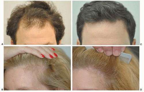
FIGURE 52.1. A 32-year-old man shown before (A) and 16 months after (B) treatment with 2,450 FU throughout the frontal third of his scalp. A 52-year-old female as seen before (C) and 14 months after treatment with 1,573 FU (D).


TABLE 52.1 METHODS OF MEASURING HAIR CHARACTERISTICS. INVASIVE, SEMI-INVASIVE, AND NON-INVASIVE TECHNIQUES CAN HELP EVALUATE SUBTLE DIFFERENCES IN HAIR DENSITY, HAIR CALIBER, AND EVEN HAIR GROWTH RATES


TECHNIQUE


STRENGTHS


WEAKNESSES


INVASIVE


Scalp biopsy


Interfollicular inflammatory changes


Subtle structural skin changes


Does not evaluate dynamic process of individual hair follicles


SEMI-INVASIVE


Trichogram, unit area trichogram (UAT)


Hair root analysis


Enables measurement of hair density, anagen percentage, hair length and diameter


Poor indicator of disease activity and/or severity


Not easily reproducible


UAT involves several (often painful) epilations


NON-INVASIVE


Global methods:


Global photographs, hair pull test, hair weight/count methods, calibrated scalp coverage scoring, categorical classification systems


Ease of use by investigators


Measures and defines the continuum of scalp coverage


Often difficult to standardize


Inability to resolve subtle details of hair loss for critical analysis


Analytical methods:


Trichoscan, Contrast-enhanced phototrichogram, Folliscope: hand-held phototrichogram


Accurate analysis of all important parameters of hair growth (density, diameter, growth rate, vellus/terminal hair density)


Quantifies subtle hair changes


Reproducible results


May require a hair dye for contrast enhancement


Measurement area must be clipped before analysis


Requires follow-up imaging for comparison


UAT, unit area trichogram.


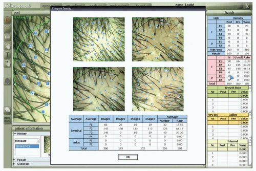
FIGURE 52.2. Folliscope evaluation for density comparison as well as total vellus and terminal hair number. This figure displays four digital images captured at 50-fold magnification and includes the area of 0.34 cm2 (ovals in box) in which quantitative analyses are performed. Calculations of hair density, growth rate, caliber, and spacing are illustrated in numerical format (right-hand panel).

The surgical design is drawn and the hairs in the donor area are trimmed to 2 to 3 mm in length. This hair length is chosen to facilitate correct angling of donor area incisions and to assist in determining directionality within the graft during implantation. The donor region is prepped with Betadine (povidone-iodine) prior to intravenous administration of an anti-anxiolytic (midazolam 2 mg, diazepam 5 mg) coupled with an analgesic, such as fentanyl (50 µg).

Local anesthesia of the donor site is achieved by creating a field block inferior to the donor region using 1% lidocaine HCl with epinephrine (1:100,000) followed by longer acting 0.25% to 0.5% bupivacaine HCl with epinephrine (1:100,000). This local ring block effectively anesthetizes the greater and lesser occipital nerves as well as the postauricular nerve. Further infiltration of 1:50,000 epinephrine is used not only to minimize diffusion of the anesthetic but also to enhance vasoconstriction, making surgical excision easier due to improved hemostasis. Additionally, sterile saline tumescence may be used to increase separation between the layer of hair follicle bulbs and the underlying nerve and vascular plexus within the deeper subcutaneous plane.

A field block anterior to the anticipated recipient area is then performed and reinforced in the same manner. Regional nerve blocks of the supraorbital and supratrochlear nerves are an alternative method of anesthesia for the recipient site.


THE DONOR SITE

Ultimately, the objective of donor area evaluation is to determine the area from which hair is most likely to be permanent and thus will ostensibly persist in the recipient area long after transplantation. To this end, the senior author conducted a study of 328 men aged 65 years or older in which areas containing at least 8 hairs per 4 mm diameter circle were measured.8 The dimensions established from this study represent the region of harvest that would be “safe” (i.e., the most hairs that would be most likely to persist) in approximately 80% of patients under the age of 80 years. This persisting region of the donor scalp was termed the “safe” donor area (SDA). These boundaries have since been modified for an alternative follicle-harvesting technique called follicular unit extraction (FUE), which will be discussed later in this chapter (Figure 52.3). Due to the less visible punctate scarring from this alternative technique, Cole’s FUE SDA is slightly expanded (203 cm2) and includes 14 subdivisions based on hair density.9

Although these accepted dimensions address the surface area of the donor area, they fall short of helping practitioners determine the quantity of “permanent” FU that may be transplanted over a patient’s lifetime. In order to provide guidelines regarding those limits, a survey of 39 of the world’s most experienced practitioners of HRS with a collective professional experience of nearly 1,000 years concluded that a 30-year-old male destined to develop Hamilton/Norwood type V or type VI MPB would most likely yield the numbers of FU as seen in Figure 52.4 based on the various degrees of donor hair density upon presentation.10 Respondents suggested that the aforementioned patient presenting with an average density donor area could yield a lifetime harvest of 6,404 or 5,393 FU when destined to develop MPB types V or VI, respectively.


Harvesting Techniques


Strip Excision.

Strip excision is unquestionably the most common method for donor harvesting, used in roughly 88.5% of HRS cases.11,12 The most important tenets for strip harvesting include minimizing the amount of hair follicle transection as incisions are made; extracting donor strip widths with caution in order to minimize closing tension; and producing only a single scar regardless of the number of sessions performed on a single patient.

In addition to using magnification to help follow the angle and direction of hair shaft exit from the skin, minimal follicle transection can be achieved by using a tumescent solution at the dermal level prior to incising in order to increase the inter-FU distance and align the follicle shafts more perpendicularly to the skin surface. Follicular transection may be reduced below the current 10% to 15% average by using a skin hook technique (Figure 52.5) to facilitate separating the edges of incision.13 Alternatively, a tissue spreader comprised of a modified iconoclast instrument introduced into the superficial wound edge incision may serve the same purpose and may prove especially useful in gray or curly hair, which pose an added challenge to minimizing follicular trauma during the donor harvest.


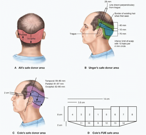
FIGURE 52.3. A. The safe donor area (SDA) according to Alt. B. Unger’s safe donor area for 80% of patients under the age of 80 years, as determined from a study of 328 men aged 65 years or older. C and D. Cole’s FUE safe area. (Images courtesy of J. Devroye, M.D. and J. Cole, M.D. Redrawn from Unger WP, Shapiro R, Unger R, Unger M, eds. Hair Transplantation 5E. London and New York: Informa Healthcare; 2011.)

The most important factor in achieving optimal donor closure tension is a careful preoperative evaluation of scalp laxity. In the majority of patients, the authors excise a donor strip of 0.8 to 1.2 cm width from the densest portion of the SDA. An excessively wide strip can lead to excess tension, which may result in unsightly scars, temporary hair effluvium (short-term loss), or tissue ischemia. Many HRS practitioners assess scalp laxity by moving the donor tissue superiorly and inferiorly, or pinching it between their fingers, using clinical experience to gauge the laxity. Others rely on mechanical devices such as the laxometer prior to incising or the intraoperative tensionometer to estimate scalp laxity and closing tension, respectively.

If donor wound closure requires an unanticipated degree of tension, undermining skin edges may sometimes be coupled with a two-layered closure using interrupted absorbable sutures (3-0 or 4-0 vicryl or monocryl). Over the mastoid region, where excessive tension is most frequent, wedgeshaped sutures may also help relieve tension (Figure 52.6).14 This technique eases the work (W) required for closure more effectively than sutures placed perpendicular to the wound edge by increasing the distribution of force (F) along the wound edge. With F and wound edge displacement (d) being constant in the formula for work (W = Fd cos θ), the magnitude of work is reduced as the angle (θ) increases.

Excising a prior scar as a part of any new donor strip has numerous aesthetic advantages over extracting a new strip inferior or superior to it. Most importantly, the harvest is then always removed from the densest portion of the donor area to maximize both longevity of transplanted hair and the number of grafts per given strip width. Second, the new wound will not
have its superior and inferior blood supply somewhat reduced because it is surrounded by virgin scalp tissue rather than bordered by previous scarring. Old scars also bind down the adjacent edge of the neighboring donor strip harvest, which may result in increased closing tension or a reduced available donor strip width. If a new strip is harvested superior (rather than inferior) to a linear scar from a previous surgery, lymphatic drainage will be compromised, leading to more severe and prolonged edema around the new wound. Compounding the probable less-optimal scar that results is the increased likelihood of telogen effluvium in the area between the old and new scar.


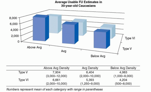
FIGURE 52.4. A total of 34 experts with over 900 years combined experience in hair restoration surgery responded to the question: “Keeping in mind that over the years, the hairs closest to the superior, inferior, and anterior borders of the fringe will be lost, how many FU containing very likely permanent hairs can be harvested from (a) a 30-year-old patient who you believe is destined to develop Norwood type V MPB and has: (1) higher than average hair density; (2) average hair density; (3) less than average hair density; (b) the same question but for a patient you believe is destined to evolve to type VI MPB.” While these data should not be viewed as dogmatic limitations, they may provide helpful guidelines when evaluating the lifetime donor yield of young Caucasian male patients.10


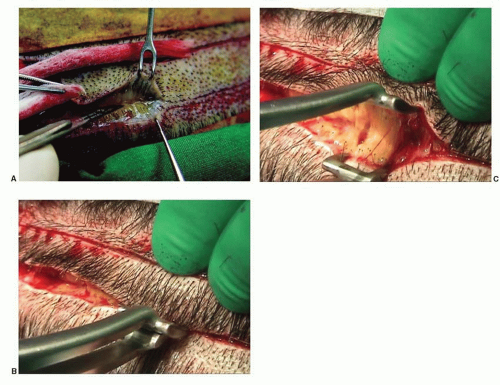
FIGURE 52.5. A. Two skin hooks for one assistant. B. Spreader inserted into scoring incision and (C) the spreader open, showing intact hair follicles at the edge of the wound. (Courtesy of D. Pathomvanich, M.D. and R. Haber, M.D. From Unger WP, Shapiro R, Unger R, Unger M, eds. Hair Transplantation 5E. London and New York; Informa Healthcare; 2011.)


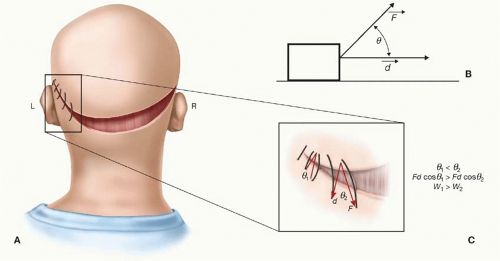
FIGURE 52.6. A. The mastoid region, where scalp tension is often greatest, is the zone in which a wedge suture may be advantageous. A magnified panel illustrating how the placement of wedge sutures is slightly wider along the interior edge of the donor area. B. A block is pulled by a force (F) at an angle (y) to a horizontal vector (d). The work (W) required to pull the block along the horizontal plane (W = Fd cos y) is reduced as y increases. C. The work of a suture to bring two wound edges together is minimized as the placement of sutures along the inferior donor edge widens, increasing the angle (y) along the superior edge.

The final component of achieving nearly imperceptible donor area scarring is the closure technique. Most physicians prefer nylon sutures over staples due to patient comfort throughout the postoperative recovery process using either a single-layer or two-layer closure. A “trichophytic” closure may be used to provide further camouflage of the donor scar.15 By excising the epidermis from one edge of the donor wound prior to closing (approximately 1 mm wide and 1 mm deep so that the bulge area of the follicle is not affected), one row of hairs effectively lie beneath the healing wound and eventually grow through the resultant scar. This technique can be used in any surgery performed in hair-bearing tissue to help prevent the appearance of an alopecic linear scar. As in other fields of plastic surgery, bioactive acellular matrix products have been investigated in HRS donor wound closures. The results of using such substances are still unclear: one recent case report suggests that the resultant donor scar might be slightly wider and more erythematous, outweighing the benefit of the improved texture of the scar tissue.16


Follicular Unit Extraction (FUE).

FUE is an alternative method of hair follicle harvesting that involves removal of individual FUs directly from the donor area one at a time, rather than from an excised strip that is subsequently dissected into FU (see below). Performed in approximately 11.5% of HRS procedures, this method holds the benefits of no linear scarring in the donor area as well as a more rapid and comfortable postoperative recovery for patients. Despite the overall increase in surface area of donor scarring with FUE compared with traditional strip harvests of large (1,000+ FU) sessions, when performed properly, the decreased visibility of scarring within the donor area may more often allow patients to wear their hair relatively short after undergoing FUE. FUE may also be utilized for beard and body hair follicle extraction as well as for removal of improperly placed grafts during corrective procedures of previous hair transplants.

Originally involving the use of a sharp 1-mm “cookie cutter”-like punch, hair follicles trimmed to 2 mm in length were extracted manually in a random distribution so as to avoid overharvesting any particular area, which may result in a “moth-eaten” appearance (Figure 52.7).17 Powered instruments for FUE have replaced manual punches by demonstrating increased extraction speed and efficiency.18 Increased speed, however, requires heightened attention to avoiding follicle transection or “decapitation” that may occur when a sharp punch is introduced at the improper angle.19 Variable hair characteristics such as follicle curvature, angle of exit from the skin, or splaying arrangement beneath the skin surface may further increase the challenge of avoiding graft transection in order to achieve intact hair follicle dissection. To minimize follicle transection, non-sharp motorized punches have been developed to perform “blunt” dissection of FU from the skin. Rather than cutting the deep segment of a follicle with an unforeseen curvature, a dull punch may push the follicle within the cylinder to reduce transection rates. Alternatively, a two-step manual process involving an initial sharp “scoring incision” at a 0.3- to 0.5-mm depth around the follicle followed by insertion of a blunt dissecting cylinder that reaches the full depth of the follicle (approximately 4 to 5 mm) enables full separation of the intact follicle from its native tissue prior to manual extraction using forceps. No matter what
the technique employed is, a surgeon’s ability to properly position the punch according to the follicle’s angle of exit from the skin surface typically requires 5× to 6.5× magnification. In addition, as mentioned earlier, use of sterile saline tumescence increases predictability of hair follicle angle deep to the skin surface by making the follicles more erect and separates the follicle bulb from the underlying vascular and nerve plexus.

Only gold members can continue reading. Log In or Register to continue

Stay updated, free articles. Join our Telegram channel

Jun 26, 2016 | Posted by in General Surgery | Comments Off on Hair Transplantation

Full access? Get Clinical Tree

Get Clinical Tree app for offline access