Genetic Disorders of Pigmentation


Genetic Disorders of Pigmentation


Jennifer A. Day

Travis Vandergriff

Adnan Mir


VITILIGO


Definition and Epidemiology

Vitiligo is an acquired pigmentary dermatosis characterized by a loss of cutaneous melanocytes resulting in depigmented patches on the skin. It is relatively common, with a frequency of 1% to 2% in the United States. There is variation in the occurrence worldwide, with a frequency of nearly 9% reported in one part of India.1 A family history is common, as is the coexistence of other autoimmune conditions. Concordance between monozygotic twins is 23%. Males and females appear to be equally affected. About 50% of patients experience onset prior to age 20, and those who experience childhood onset are more likely to have affected family members.2,3,4


Etiology

Although vitiligo occurs at a high frequency in patients with a family history of the disease, it is likely a polygenic trait that results in autoimmune destruction of melanocytes. Early linkage analyses and more recent genome-wide association studies have identified a number of associations, including immune mediators, melanocyte components, and regulators of apoptosis.5,6 Complex interactions of these pathways, likely coupled with environmental factors, lead to the development of the clinical phenotype seen in vitiligo.

The autoimmune nature of vitiligo is supported by genetic studies as well as several key pieces of evidence: (1) There are circulating antibodies targeting melanocyte proteins.7 (2) The clinical development of vitiligo in patients with melanoma has long been known to be a positive prognostic factor, suggesting that the antimelanocyte immune reaction evident in the skin depigmentation of these patients also targets malignant melanocytes.8 (3) Vitiligo often coexists in patients or families with other autoimmune conditions such as Hashimoto thyroiditis and Graves’ disease, type 1 diabetes mellitus, alopecia areata, and pernicious anemia.9,10 (4) Finally, vitiligo is among the autoimmune phenomena that occur in patients with rheumatologic and gastrointestinal inflammatory diseases treated with tumor necrosis factor-alpha inhibitors.11 Other hypotheses that have been put forth for the pathogenesis of vitiligo include dysregulation of the nervous system, cytotoxic mechanisms, oxidative stress-induced depigmentation, reduced melanocyte adhesion and survival, and others. Although each of these may play a role in individual patients, evidence is mounting that the underlying factor allowing or promoting melanocyte targeting is autoimmune in nature.


Clinical Presentation

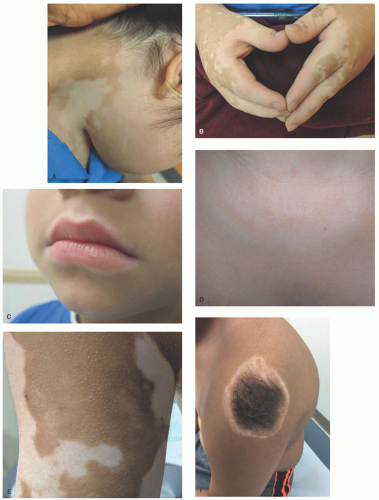
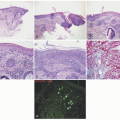
The clinical presentation of vitiligo is varied, but almost always includes depigmented, white patches and macules. Classically, it presents as symmetric, well-demarcated, depigmented patches that favor the scalp, face, neck, forearms, waist, genitalia, knees, and dorsal hands and feet (Figure 8-1A-C). It favors areas of repetitive stress or trauma

like joints or backs of the hands and can manifest the Koebner phenomena. Widespread cases may affect any surface, and in severe cases, near-total depigmentation may occur. In patients with light skin, depigmented patches may be subtle and difficult to detect without the aid of a Wood’s lamp.


FIGURE 8-1. Vitiligo. (A) Well-demarcated, depigmented, coalescing patches on the neck of a boy with Down syndrome. (B) Symmetric patches on the hands. Note linear depigmentation on the wrist, representing trauma-induced vitiligo, or Koebner phenomenon. Rough nails (trachyonychia) are also seen. (C) Perioral depigmentation. (D) Marginal inflammatory vitiligo with an erythematous, raised border. (E) Trichrome vitiligo, with three distinct shades of pigmentation. (F) A halo congenital nevus, with a depigmented patch around a typical, medium-sized congenital nevus. The patient had patches of vitiligo on his knees as well.

Segmental vitiligo, typically unilateral and confined to a single segment of the body with a sharp midline demarcation, shares many clinical features of nonsegmental vitiligo, but is likely predisposed by somatic mutation in a susceptibility gene, as described earlier. It is not typically associated with other autoimmune conditions, but may respond to standard therapies.

Inflammatory vitiligo may present with a raised, thread-like rim of linear erythema at the border (Figure 8-1D), which may indicate active spread, but often remains stable for many months. Trichrome vitiligo (Figure 8-1E), in which three distinct shades of pigmentation are visible within a body area, and vitiligo with confetti-like depigmented macules sometimes occur, and may portend a poor prognosis with rapid progression.12 Patches of vitiligo on hair-bearing areas may present with poliosis, or depigmented, white hair. Halo nevi (Figure 8-1F), which are melanocytic nevi with a surrounding depigmented patch, are often associated with vitiligo and are sometimes the first sign of disease.13


FIGURE 8-2. Vitiligo. There is a broad area of decreased pigmentation in the epidermis with occasional pigmented melanophages in the dermis. A Fontana-Masson stain highlights the marked reduced presence of melanin pigment in the epidermis. A Melan-A immunostain shows a marked reduction in the density of melanocytes throughout the lesion.

Spontaneous repigmentation sometimes occurs and can be hastened by a number of immunosuppressive treatment modalities. This repigmentation may be from peripheral extension (as occurs with topical steroid treatment) or may appear as perifollicular pigmented macules (as occurs with phototherapy) within the lesion from a follicular source of melanocytes.

The morbidity of vitiligo is primarily evident in its impact on the quality of life. Affected children and adults often suffer from significant psychosocial stress, depression, and anxiety.14,15


Histologic Findings

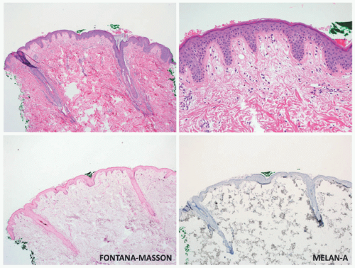
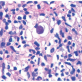
Although vitiligo is primarily a clinical diagnosis, biopsy may sometimes be necessary to differentiate it from other diseases of hypo-or depigmentation. At scanning magnification, biopsy specimens may appear completely unremarkable. Well-developed lesions are characterized by a complete absence of melanocytes and pigment (Figure 8-2). The absence of pigment may be confirmed by negative
Fontana-Masson staining, and a lack of melanocytes may be evident by immunohistochemical studies. Inflammation is sometimes present, both in specimens that appear clinically inflammatory and in those that do not, in the form of a mild superficial lymphohistiocytic infiltrate. A more exuberant, lichenoid interface dermatitis is sometimes seen, and can appear in clinically inflamed areas, depigmented areas, and even distant, clinically uninvolved skin.16


Differential Diagnosis

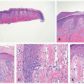
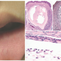
There is a broad clinical differential diagnosis for vitiligo. Hypopigmented lesions may sometimes be difficult to differentiate from depigmented lesions, and can be present in many conditions, including nevus depigmentosus/pigmentary mosaicism, pityriasis alba, and tinea versicolor (Figure 8-3). Postinflammatory hypo-and depigmentation should be preceded by inflammation, although a history of skin inflammation is not always elicited. Chemical leukoderma may present with a strikingly similar pattern to that of vitiligo and is often occupational in nature.

Conditions of congenital depigmentation, such as Waardenburg syndrome or piebaldism, should be considered for midline patches affecting the face and trunk. These patches may sometimes be present on the extremities as well. Syndromic vitiligo may be seen in Vogt-Koyanagi-Harada, Alezzandrini syndrome, and autoimmune polyendocrinopathy candidiasis-ectodermal dystrophy syndromes.

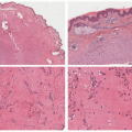
The histologic differential diagnosis includes chemical leukoderma, piebaldism, and nevus depigmentosus. These may be indistinguishable from vitiligo and require clinical history to differentiate. In inflammatory lesions, vitiligo may mimic lichenoid dermatoses such as lichen planus and its subtypes. These conditions lack clinical depigmentation, although hypopigmented lesions may appear similar to early lesions of vitiligo. Finally, patch-stage mycosis fungoides (MF) and inflammatory lesions of vitiligo may share clinical and histopathologic findings. MF, however, should have some degree of lymphocytic atypia, and Pautrier microabscesses may be present. In some cases, multiple biopsies may be necessary to distinguish the two entities.


FIGURE 8-3. Tinea versicolor. Widespread tinea versicolor mimicking vitiligo. Note the peripheral, scalloped border indicating coalescing macules. Clinically, the presence of fine scale and the retention of some pigment differentiates tinea versicolor from vitiligo, and the two can be easily distinguished by histology.


OCULOCUTANEOUS ALBINISM


Definition and Epidemiology

Oculocutaneous albinism (OCA) is a group of autosomal dominant, inherited disorders of melanin synthesis. Patients present at birth with variable hypopigmentation of skin, hair, and eyes. There are seven subtypes of nonsyndromic OCA, with types 1 to 4 accounting for the majority of reported cases.17,18

The incidence of OCA is estimated at 1 in 17 000 live births in the United States. Incidence in some worldwide populations is as high as 1 in 500 to 1 in 100 including in some areas of Africa and in native populations in North and South America (as high as 1 in 92).19 The most common form is OCA2, with prevalence estimated at 1 in 36 000 for Caucasian individuals and 1 in 3900 to 10 000 for those of African descent.20 OCA4 is the most common type in Japan, and OCA5-7 are rare, having been described in only one to a few families each.21,22,23,24,25


Etiology

Several genes-encoding proteins crucial for melanin synthesis have been identified as etiologic factors in OCA. Tyrosinase (TYR) is a key enzyme in the melanin production pathway, catalyzing the conversion of tyrosine to dopaquinone, a precursor of melanin. Absence or deficiency of TYR activity, as seen in OCA1a and OCA1b, results in diffuse depigmentation. P protein is a factor in the synthesis of melanosomes as well as typical processing and transport of TYR and Tyrosinase-related protein 1 (TYRP1). TYRP1 is a catalyst for the oxidation of 5,6-dihydroxyindole-2-carboxylic acid, another melanin precursor. TYRP1 and membrane-associated transporter protein are involved in the transport of TYR from the endoplasmic reticulum into melanosomes.


Clinical Presentation

Clinically, OCA is characterized by variable degrees of congenital pigmentary dilution of skin, eyes, and hair (Table 8-1). The lifetime development of pigmentation in affected patients is variable, with some patients devoid of pigment throughout life (as in OCA1a) and others able to create lentigines and melanocytic nevi. Ocular involvement includes characteristic translucent blue irides, hypopigmented retinal fundus, and nystagmus. In young adulthood, accelerated photoaging (telangiectasias, rhytids) are seen, in addition to actinic cheilitis, actinic keratoses, and skin malignancies.26 Squamous cell carcinoma and basal cell carcinoma are more common than melanoma, which, when present, is often amelanotic.26,27 OCA can be associated with significant psychosocial stress and, in some areas of the world, physical threat of assault or death caused by cultural conceptions surrounding albinism.28


TABLE 8-1. Causes and clinical features of oculocutaneous albinism.


Variant


Gene/Etiology


Clinical Manifestations


OCA1A26,27,29,30,31,32


TYR mutation—lack of protein


tyrosinase-negative OCA


Cutaneous: White skin and hair, blue irises. Hair may develop yellowish discoloration with age. Highest risk of premature photoaging and cutaneous malignancies.


Ocular: Photophobia, nystagmus, strabismus, and reduced visual acuity (often legally blind)


Associations: ADHD


OCA1B29,30


TYR—reduced activity of protein


tyrosinase-negative OCA


Cutaneous:


Yellow Mutant Variant: Hair turns yellow in first few years; gradually pigments to blonde/light brown.


Platinum Variant: Small amounts of pigment with metallic tinge in childhood


Minimal Pigment Variant: Pigment develops in eyes only.


OCA1—


Temperature Sensitive29,30


TYR—high temperaturedependent reduced activity of protein


Cutaneous: Born with white skin and hair, blue eyes. During the second decade of life, develop pigmentation at extremities and acral sites.


OCA233


P protein


Cutaneous:


Brown albinism: Slight to moderate pigmentary dilution in skin, hair, and eyes. Increased pigmentation including lentigines and nevi develop at sun-exposed sites. Lower risk of skin cancer.


Ocular: Less severe ocular manifestations


Associations: ˜1% of patients with Angelman or Prader-Willi syndromes due to contiguous deletions on chromosome 15


OCA3 (Rufous OCA)34,35


Tyrosinase-related protein 1 TRYP1


Red hair, reddish-bronze skin, and blue or brown irises.


OCA436


MATP


Similar to OCA2


OCA522


Unknown (mutation at 4q24)


Cutaneous: Golden hair and white skin


Ocular: Nystagmus, photophobia, foveal hypoplasia, and reduced visual acuity


OCA623,24


SLC24A5


Cutaneous: Blond hair that darkens with age, white skin


Ocular: Translucent light eyes (brown, green, or blue), nystagmus, photophobia, foveal hypoplasia


OCA725


c10orf11


Cutaneous: Light skin, pale blond to light brown hair


Ocular: Light blue or green eyes, nystagmus, mild photophobia


Abbreviations: ADHD, attention deficit disorder; MATP, membrane-associated transporter protein; OCA, oculocutaneous albinism; TYR, tyrosinase; TYRP1, tyrosinase-related protein 1.


Histologic Findings

Melanocytes are normal in number and morphology, but there is reduced or absent melanin, as can be seen with Fontana-Masson staining. Hair follicle melanin may be preserved in some forms of OCA.


Differential Diagnosis

The primary clinical differential diagnoses include syndromic causes of OCA, such as Hermansky-Pudlak,
Griscelli, and Chédiak-Higashi syndromes, which are discussed later in this chapter.

Histologically, other dermatoses with subtle findings of dyspigmentation may be considered. Vitiligo lacks melanocytes, and tinea versicolor has conspicuous fungal elements in the stratum corneum that should be seen on routine hematoxylin and eosin-stained sections.


HERMANSKY-PUDLAK SYNDROME


Definition and Epidemiology

Hermansky-Pudlak syndrome (HPS) is a group of autosomal recessive heritable disorders characterized by pigmentary dilution, platelet abnormalities, and accumulation of ceroid-lipofuscin within internal organs.37,38,39

HPS is common among Puerto Ricans, occurring in approximately 1 in 1800 to 1 in 400 people.37,39 There is also a high incidence among people of Dutch descent and East Indians from Madras. Males and females are equally affected.


TABLE 8-2. Hermansky-Pudlak syndrome.


Variant


Gene


Etiology


Unique Features


HPS-144


HPS1


Component of BLOC3


Nystagmus, reduced visual acuity, prolonged bleeding, pulmonary fibrosis, granulomatous colitis (up to 1/3 of patients)


HPS-2


AP3B1


Subunit of AP-3


Nystagmus, reduced visual acuity, prolonged bleeding, neutropenia, recurrent bacterial and viral infections, conductive hearing loss.


HPS-3


HPS3


Component of BLOC2


Nystagmus, reduced visual acuity, mild extraocular manifestation


HPS-4


HPS4


Component of BLOC3


HPS-542


HPS5


Component of BLOC2


Hypercholesterolemia and hypertriglyceridemia


HPS-6


HPS6


Component of BLOC2


HPS-741


DTNBP1


Subunits of BLOC1,


HPS-842


BLOC1S


Subunit of BLOC1


Nystagmus, reduced visual acuity, prolonged bleeding


HPS-947


HPS9/PLDN


Subunit of BLOC1


Nystagmus, immunodeficiency, no bleeding diathesis described to date.


HPS-10


AP3D1


Component of AP-3


Severe neurologic disease with developmental delay and refractory seizures, immunodeficiency.


Abbreviations: AP-3, adaptor protein; BLOC, biogenesis of lysosome-related organelles complex; DNTBP1, dystrobrevin binding protein 1; HPS, Hermansky-Pudlak syndrome; PLDN, Pallidin.


Etiology and Clinical Presentation

The genes involved in all subtypes of HPS encode proteins involved in the regulation of vesicular trafficking within lysosome-related organelles, including platelet dense granules and melanosomes.40,41,42,43 Table 8-2 lists gene associations and unique clinical features of the ten described variants of HPS. With the exceptions of HPS2 and 10, the involved genes are organized into functional structures of interacting proteins that participate in vesicular trafficking, known as BLOCs (biogenesis of lysosome-related organelles complex). Specific features of each HPS subtype depend on where each component is expressed: BLOC1 is present in melanosomes and platelet dense granules, BLOC2 is present in melanosomes alone and co-localizes with TYR and TYRP1, and biogenesis of lysosome-related organelles complex 3 (BLOC3) is present in melanosomes, dense granules, and lamellar bodies of type 2 pneumocytes. HPS2 is a subunit of AP-3, which mediates trafficking into lysosomal transport vesicles in multiple tissue types. HPS10 interacts with AP-3.

Patients with HPS exhibit variable pigmentary dilution of the skin and eyes, nystagmus, photophobia, and reduced visual acuity. Many experience systemic manifestations, the most common of which is pulmonary fibrosis, which may be life-threatening.

Hematologic manifestations include bleeding diathesis caused by defective dense granules in platelets.39 Patients may present with ecchymoses, epistaxis, and menometrorrhagia, but may not be apparent without the use of aspirin or prostaglandin inhibitors. Other rare complications include granulomatous colitis, cardiomyopathy, and renal failure.44,45,46 Life expectancy is 30 to 50 years.37

Only gold members can continue reading. Log In or Register to continue

Stay updated, free articles. Join our Telegram channel

May 8, 2019 | Posted by in Dermatology | Comments Off on Genetic Disorders of Pigmentation

Full access? Get Clinical Tree

Get Clinical Tree app for offline access