Genetic Disorders of Epidermal Maturation and Cornification


Genetic Disorders of Epidermal Maturation and Cornification


Jennifer A. Day

Adnan Mir


ICHTHYOSIS


Definition and Epidemiology

The ichthyoses are a large, heterogeneous group of heritable disorders characterized by abnormal epidermal differentiation and resulting in generalized scaling of the skin. They range from mild to severe and may occur in isolation or as a part of a broader syndrome. The nonsyndromic ichthyoses will be discussed in this chapter. These include ichthyosis vulgaris, X-linked recessive ichthyosis, the autosomal recessive congenital ichthyoses (ARCI, including lamellar ichthyosis, congenital ichthyosiform erythroderma, and Harlequin ichthyosis), and epidermolytic ichthyosis. The ichthyoses, the most common of which is ichthyosis vulgaris, are highly variable in incidence.


Etiology and Clinical Presentation

Table 7-1 highlights the genetic defects and clinical features of several common and uncommon ichthyoses. In general, the genetic defects in ARCI result in abnormal lipid metabolism, which in turn results in abnormalities of cornification. Ichthyosis vulgaris is caused by mutation in profilaggrin, which also results in abnormal formation of the stratum corneum. The epidermolytic ichthyosis is caused by mutations in keratins 1 and 10, leading to epidermolysis and superficial blister formation early in life.1

Several of these disorders present at birth with a collodion membrane—a taut, shiny membrane covering the infant’s entire body, derived from thickened stratum corneum. The tightness results in eclabium, ectropion, and in severe cases, restrictive pulmonary dysfunction. The membrane is shed over the first weeks of life, eventually revealing the lifelong phenotype.


Histologic Findings

Histopathologic changes are often subtle in ichthyotic conditions, except in certain disorders with characteristic findings (Table 7-2).8 Isolated hypogranulosis is suggestive of ichthyosis vulgaris, and epidermolytic hyperkeratosis is diagnostic of epidermolytic ichthyosis in the correct clinical context. This change can be seen in epidermal nevi that are caused by somatic mutations in KRT-1 and KRT-10. In general, a definitive diagnosis of the ichthyoses relies on molecular testing with multigene panels to determine a causative genetic mutation.

Collodion membranes, a shared finding in several ichthyotic disorders, demonstrate histologic findings of compact orthokeratotic hyperkeratosis of the stratum corneum.22


Differential Diagnosis

There is broad overlap in the clinical and histopathologic findings of the ichthyoses. They can be distinguished by the character and distribution of scale, as well as genetic evaluation. Orthokeratotic hyperkeratosis with a diminished granular layer is characteristic of ichthyosis vulgaris, but can also be seen in the syndromic ichthyoses Refsum syndrome, trichothiodystrophy, and Conradi-Hünermann-Happle Syndrome. Epidermolytic hyperkeratosis, present in epidermolytic ichthyoses, can also be found in epidermal nevi, ichthyosis bullosa of Siemens, or as an incidental finding.


TABLE 7-1. Selected nonsyndromic ichthyoses


Diagnosis


Epidemiology


Etiology


Clinical Presentation


Ichthyosis vulgaris2,3,4,5,6,7


(Figure 7-1)


1:100-1:250


Autosomal semidominant mutation in profilaggrin, resulting in the abnormal development of the cornified cell envelope


Cutaneous Manifestations: Fine, white scales on trunk and extremities; spares flexures; hyperlinear palms


Onset: Develops in infancy or early childhood, rarely present at birth with severe mutation, tends to improve in adolescence/adulthood


Associations: Atopic dermatitis and keratosis pilaris


X-linked recessive ichthyosis (steroid sulfatase deficiency)4,8,9


(Figure 7-2)


1:2000-1:9500 males


X-linked recessive mutation in steroid sulfatase, resulting in the accumulation of cholesterol metabolites in the epidermis and subsequent inhibition of transglutaminase 1 (TGM1)


Cutaneous Manifestation: Large, brown scales accentuated at neck; relative sparing of flexures


Other Manifestations: Asymptomatic corneal opacities, cryptorchidism, prolonged labor with affected fetuses due to insufficient cervical dilation


Associations: Mental retardation, Kallman syndrome, and/or chondrodysplasia punctata


Epidermolytic ichthyosis10,11


(Figure 7-3)


1:300 000


Usually autosomal dominant mutation in KRT1 or KRT10


Cutaneous Manifestations:


At birth: Superficial blistering


Childhood/adulthood: Progressive predominance of hyperkeratosis, classically described as corrugated, accentuated on flexure surfaces and over joints


ARCI—Lamellar ichthyosis11,12,13


1:300 000


Usually autosomal recessive


Transglutaminase 1 (TGM1)


Cutaneous Manifestations:


At birth: Collodion membrane


Childhood/adulthood: Large, plate-like scales in generalized distribution; ectropion, alopecia, nail dystrophy


ARCI—Congenital ichthyosiform erythroderma11,14,15,16,17


(Figures 7-4 and 7-5)


1:300 000


Autosomal recessive mutation in several genes:


TGM1 CYP4F22


ALOXE3 PNPLA1


ALOX12B LIPM


NIPAL4 CerS3


ABCA12


Cutaneous Manifestations:


At birth: Collodion membrane


Childhood/adulthood: Fine white scaling overlying generalized erythema, ectropion, alopecia


Other Manifestations: Neurologic abnormalities


ARCI—Harlequin ichthyosis18,19,20,21


(Figure 7-6)


Rare


Autosomal recessive mutation


ABCA12 (B61)—truncating mutations or deletions (B62)


Cutaneous Manifestations:


At birth: Harlequin fetus—thick, yellow/brown plates of scale interspersed with large, deep, bright red fissures; ectropion; eclabium; ear abnormalities; prematurity common


Childhood/adulthood: CIE (congenital ichthyosiform erythroderma)-like presentation


Mortality in the neonatal period is high, typically because of sepsis or respiratory failure


Abbreviations: ABCA12, ABC lipid transporter; ARCI, autosomal recessive congenital ichthyosis; FLG, profillagrin; KRT1/10, keratins 1/10; STS, steroid sulfatase; TGM1, transglutaminase 1.


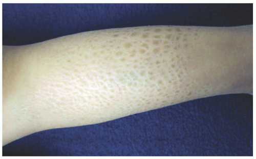
FIGURE 7-1. A child with ichthyosis vulgaris. The lower leg shows dry, polygonal, hyperpigmented scaling.


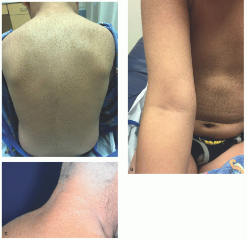
FIGURE 7-2. X-linked recessive ichthyosis with characteristic hyperpigmented scale on the back extending onto the neck (A and B) and conspicuous sparing of the antecubital fossa (C).


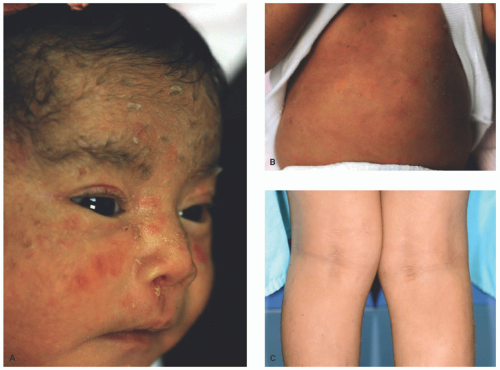
FIGURE 7-3. Epidermolytic ichthyosis. A baby showing blisters in the face, nose (A), and trunk (B). Progressive corrugated hyperkeratosis is seen in olden children, in association with a pattern of accentuation in flexural sites (C).


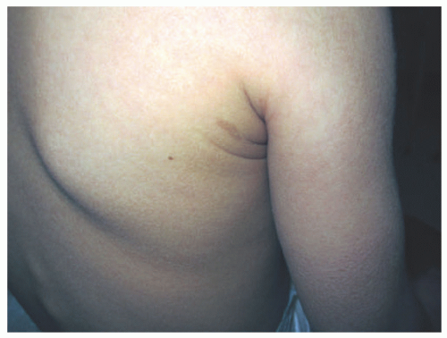
FIGURE 7-4. Autosomal recessive congenital ichthyosis with a congenital ichthyosiform erythroderma phenotype, showing fine, diffuse scale with mild underlying erythema.


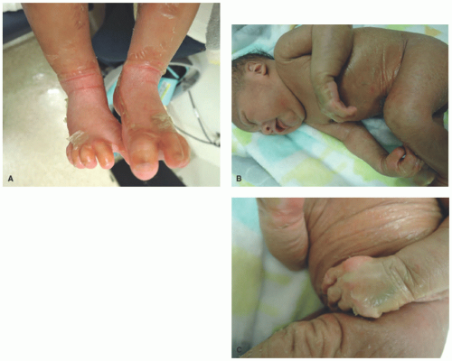
FIGURE 7-5. Collodion baby. This neonate is encased in a thick, yellowish, taut, glistening, parchment-like membrane that restricts its movements (A-C).


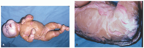
FIGURE 7-6. An infant with Harlequin ichthyosis with extremely thick, fissured scaled skin (A and B). Courtesy of Dr Kenneth Greer. Department of Dermatology. University of Virginia.


TABLE 7-2. Histopathologic findings for selected nonsyndromic ichthyoses


Diagnosis


Histopathology


Electron Microscopy


Other


Ichthyosis vulgaris11,23,24,25


(Figure 7-7)


SC: Mild hyperorthokeratosis


SG: ± Diminished


Absent or abnormal keratohyalin granules


Absent or diminished filaggrin staining by IHC


X-linked recessive ichthyosis (steroid sulfatase deficiency)11,23,25,26


(Figure 7-8)


SC: Ortho- or parakeratosis; melanosomes and desmosomes retained


SG: Normal or thickened ± Acanthosis


Hyperkeratosis of follicular and sweat duct orifices


Increased size and number of keratohyalin granules


Epidermolytic ichthyosis11,27,28


(Figure 7-9)


Epidermolytic hyperkeratosis:


SC: thick hyperorthokeratosis


SG: hypergranulosis


Acantholysis of suprabasal layers, leading to intraepidermal blisters


Mild perivascular lymphohistiocytic infiltrate


Clumped keratin intermediate filaments can be seen in keratinocytes


Clumped, fragmented keratin intermediate filaments in lower epidermis


Epidermolytic hyperkeratosis can also be seen in epidermolytic acanthomas (solitary papule) and epidermal nevi


ARCI— Congenital ichthyosiform erythroderma11,27,29


SC: Parakeratosis


SG: Thickened


Acanthosis


Increased number of lamellar bodies


Variable translucent lipid droplets in SC


Disorganized intercellular lamellae


Markedly increased epidermal proliferation


ARCI—Lamellar ichthyosis11,23,27,29,30


(Figure 7-10)


SC: Massive hyperorthokeratosis,


Acanthosis (can be psoriasiform or papillomatous)


Elongated cholesterol clefts


Variable translucent lipid droplets in SC


Thin or absent cornified envelope


Epidermal proliferation rate normal or mildly elevated.


Diminished staining of transglutaminase-1 on IHC


Diminished transglutaminase-1 activity in cultured keratinocytes


Other transglutaminase assays, not widely available.


ARCI—Harlequin ichthyosis31


SC: Massive hyperorthokeratosis ± parakeratosis


Hyperkeratotic plugging of follicles and sweat ducts


Hair follicles: concentric accumulation of hyperkeratotic material around hair shaft


Absent or abnormal lamellar bodies in SG


Absent extracellular lipid lamellae


Variable translucent lipid droplets in SC


Abbreviations: ARCI, autosomal recessive congenital ichthyosis; IHC, immunohistochemistry; SC, stratum corneum; SG, stratum granulosum.


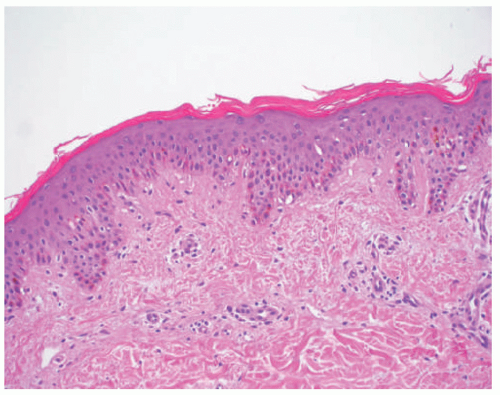
FIGURE 7-7. Ichthyosis vulgaris. Compact orthokeratosis and a diminished granular layer.


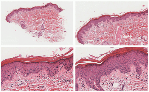
FIGURE 7-8. X-linked recessive ichthyosis. Mild epidermal acanthosis and hyperorthokeratosis. The granular cell layer is preserved (as opposed to ichthyosis vulgaris). There is mild hyperkeratosis around the sweat gland orifices. Digital slides courtesy of Path Presenter.com.


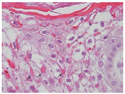
FIGURE 7-9. Epidermolytic hyperkeratosis with cytolysis of keratinocytes in the upper spinous layer and eosinophilic inclusions. Reprinted with permission from Bergman R, Khamaysi Z, Sprecher E. A Unique pattern of dyskeratosis characterizes epidermolytic hyperkeratosis and epidermolytic palmoplantar keratoderma. Am J Dermatopathol. 2008;30(2):101-105.


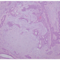
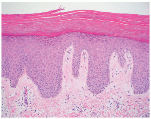
FIGURE 7-10. Lamellar ichthyosis. Thick hyperkeratosis with acanthosis and mild hypergranulosis.


NETHERTON SYNDROME


Definition and Epidemiology

Netherton syndrome (NS) is an autosomal recessive inherited disorder featuring congenital ichthyosiform erythroderma, characteristic hair abnormalities, severe atopic diathesis, and immune dysregulation. NS is thought to affect approximately 1:50 000 to 1:200 000 patients worldwide.32,33,34


Etiology

NS is caused by a mutation in the serine protease inhibitor Kazal type 5 (SPINK5) that encodes the protein lymphoepithelial Kazal-type-related inhibitor (LEKTI).33,34 LEKTI is primarily expressed in epidermal lamellar granules as well as lymphoid tissue. The causative mutation results in an overactivity of serine proteases, leading to disrupted regulation of lipid-processing enzymes and abnormal stratum corneum lipid envelope.35,36 Additionally, there is an enhanced degradation of desmoglein 1, contributing to impaired cellular cohesion in the stratum corneum.37,38


Clinical Presentation

Patients present at birth with congenital erythroderma and skin peeling.39 Within the neonatal period, potential complications include abnormal temperature regulation, electrolyte abnormalities, pneumonia, and sepsis.34,40,41 In childhood, patients tend to develop one of two phenotypes: ichthyosis linearis circumflexa (Figure 7-11) or congenital ichthyosiform erythroderma-like scale (Figure 7-12). The former is more common, characterized by migratory, circinate, scaly, erythematous plaques with a border of double-edged scale.42 In both presentations, pruritus and eczematous plaques are ubiquitous. Hair findings include trichorrhexis invaginata (Figure 7-13), giving the phenotype of dry, fragile, and poorly manageable hair.43 The distribution of affected hair within an individual is variable, but is often concentrated on lateral eyebrows. Thick, adherent scalp scale can also be seen.

Immune manifestations include severely elevated immunoglobulin E ([IgE] up to 10 000 IU/mL), impaired B- and NK-cell function, reduced response to vaccination, and increased levels of proinflammatory cytokines.44 Patients experience recurrent infections, particularly involving the respiratory tract and skin. The common microbes involved in skin infections include Staphylococcus aureus and human papillomavirus,45 the latter of which can predispose patients to squamous cell carcinoma.46 Food and environmental allergies are common.


Histologic Findings

The biopsy specimens of ichthyosis linearis circumflexa show hyperkeratosis, a normal granular cell layer, and
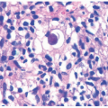
acanthosis. Focal parakeratosis, hypogranulosis, and exocytosis of neutrophils are variably present (Figure 7-14).47,48,49 In erythrodermic cases, parakeratosis can be confluent. A perivascular lymphohistiocytic infiltrate may be present in the superficial dermis.50 Periodic acid-Schiff staining demonstrates densely staining granules in upper epidermal layers.8 A microscopic evaluation of affected hair shafts reveals trichorrhexis invaginata, with a telescoping of the distal hair shaft into the proximal shaft.43 Other hair shaft findings, such as trichorrhexis nodosa, pili torti, and “matchstick” abnormality, can also be seen.


FIGURE 7-11. Ichthyosis linearis circumflexa in Netherton syndrome with the classic circinate, scaly, erythematous plaques with a border of double-edged scale. Courtesy of Dr Kenneth Greer. Department of Dermatology. University of Virginia.


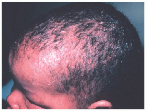
FIGURE 7-12. Congenital ichthyosiform erythroderma-like scale in Netherton syndrome. Diffuse scalp erythema and mild thin scaling is present. Courtesy of Dr Kenneth Greer. Department of Dermatology. University of Virginia.

The electron microscopic findings of premature secretion of lamellar bodies within the epidermis, electron-dense accumulations between corneocytes, and atypical splitting within the stratum corneum are fairly specific for NS.49


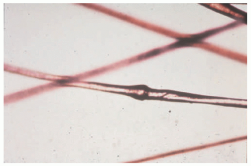
FIGURE 7-13. Trichorrhexis invaginata (Bamboo hair), typical of Netherton syndrome. Courtesy of Dr Kenneth Greer. Department of Dermatology. University of Virginia.


Differential Diagnosis

The clinical differential diagnosis of erythroderma in a neonate includes congenital ichthyosiform erythroderma, psoriasis, peeling skin syndrome, or another primary immunodeficiency. Features favoring NS include elevated serum IgE and characteristic trichorrhexis invaginata, although this often does not develop until after infancy. Immunodeficiency syndromes that may present with erythematous, eczematous eruption and high-serum IgE include autosomal dominant hyper IgE-syndrome, Wiskott Aldrich syndrome, Omenn syndrome, and IPEX (immune dysregulation, polyendocrinopathy, enteropathy, X-linked) syndrome. The main histopathologic differential diagnosis of ichthyosis linearis circumflexa is psoriasis, which may appear virtually identical. Differentiation is based on clinical history and physical findings. Additional considerations include other
syndromic and nonsyndromic ichthyoses, eczematous dermatitis, and dermatophytosis.


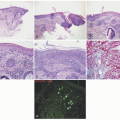
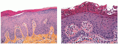
FIGURE 7-14. Skin biopsies from Netherton syndrome show psoriasiform hyperplasia and may have overlying parakeratosis (A) and neutrophils within the epidermis and stratum corneum (B). Differentiation from psoriasis must be made on a clinical basis. Reprinted with permission from Leclerc-Mercier S, Bodemer C, Furio L, et al. Skin biopsy in Netherton syndrome: a histological review of a large series and new findings. Am J Dermatopathol. 2016;38(2):83-91.


REFSUM DISEASE


Definition and Epidemiology

Refsum disease (RD) is an autosomal recessive inherited disorder caused by mutations in peroxisomal metabolism, leading to deficient oxidation of phytanic acid. Patients present with ichthyosis and progressive neurologic dysfunction. RD is rare, with fewer than 100 cases reported in the literature.51


Etiology

Causative mutations have been identified in phytanoyl-CoA hydroxylase (PHYH, Type 1) and peroxisomal targeting signal 2 receptor (PEX7, Type 2).52 Both genes are involved in the function of peroxisomes, and abnormalities lead to the inability to oxidize phytanic acid, resulting in aberrantly elevated serum levels.53 Phytanic acid, a branched, long-chain fatty acid, is derived from chlorophyll; thus, circulating levels in humans are delivered via dietary intake.54 Abnormally high levels interfere with cholesterol metabolism, leading to deficiencies in essential fatty acids, which in turn results in ichthyosis.


Clinical Presentation

Cutaneous findings include fine, white scaling on the trunk and extremities and hyperlinear palmoplantar markings, similar to those seen in ichthyosis vulgaris.53,54 This generally begins with or after neurologic symptoms.

The onset of neurologic abnormalities is typically in late childhood or adolescence. Prominent features include sensorineural deafness, anosmia, visual disturbance (reduced visual acuity and night blindness due to retinitis pigmentosa, with classic “salt and pepper” retinal pigmentation), and sensorimotor neuropathy leading to weakness and poor balance.51,53,54 Neurologic findings are typically insidious in onset and slowly progressive. Reduction in the dietary intake of sources of phytanic acid (green vegetables, dairy) may delay the progression of neurologic abnormalities. Additional manifestations developing over time include cataracts, renal tubular dysfunction, and cardiomyopathy.54,55


Histologic Findings

Skin biopsy specimens from patients with RD may show mild hyperorthokeratosis and a diminished granular layer. The accumulation of lipid droplets can be appreciated in specimens fixed in alcohol with lipid stains such as Sudan black or ultrastructural analysis with electron microscopy.55


Differential Diagnosis

The clinical differential diagnosis of the cutaneous manifestations of RD includes ichthyosis vulgaris and vitamin B deficiency. Mild ichthyosis in association with hearing loss can be seen in other peroxisomal deficiency disorders as well as neutral lipid storage disease. Prominent neurologic abnormalities are also seen in Sjögren-Larsson syndrome (SLS). The cutaneous histopathologic changes in RD are virtually identical to those seen in ichthyosis vulgaris. However, the accumulation of epidermal lipid is absent, and these patients lack neurologic symptoms.


SJÖGREN-LARSSON SYNDROME


Definition and Epidemiology

SLS is an autosomal recessive inherited disorder characterized by ichthyosis, progressive di- or tetraplegia, and intellectual disability. It is caused by mutation in the fatty aldehyde dehydrogenase ALDH3A2. SLS has an incidence of fewer than 1 in 100 000, with highest incidence in Sweden.56 It has been reported worldwide.


Etiology

Mutations in ALDH3A2, encoding fatty aldehyde dehydrogenase (FALDH), lead to a reduced oxidation of long-chain fatty acids. FALDH is normally involved in the recycling of cell membrane components, including ceramides in the epidermis and sphingolipids in the brain.57 Chronic pruritus is attributed to the accumulation of high levels of leukotriene B4.


Clinical Presentation

SLS presents at birth with variable degrees of erythema, scaling, and hyperkeratosis (Figure 7-15).58 Later, erythema fades and gives way to fine to plate-like scaling or nonscaling hyperkeratosis, predominantly on abdomen, neck, and flexures.58,59,60 The face tends to be spared. There is associated pruritus, accounting for commonly seen lichenification. Approximately 70% of affected patients have palmoplantar keratoderma.59,60 Hair and nails are normal.


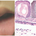
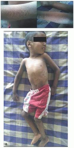
FIGURE 7-15. Marked ichthyosis in a patient with Sjögren-Larsson syndrome. A, Brown color diamond-shaped adherent scales on the upper limb and peeling of skin on the lower limb. B, Kyphoscoliosis of the trunk is present. Obtained with permission from Subramanian V, Hariharan P, Balaji J. Sjögren-Larsson syndrome: a rare neurocutaneous disorder. J Pediatr Neurosci. 2016;11(1):68-70, Figure 1 and 2.


Neurologic complications typically manifest by 1 year of age. Characteristic findings include delayed motor development, ultimately leading to gradually progressive spastic di- and tetraplegia, spasticity, and contractures.56,59 The other findings include intellectual disability, white matter abnormalities, and seizures.56,59,60

Ocular findings include perifoveal glistening white dots, a nearly pathognomonic feature. Photophobia is a common symptom.

May 8, 2019 | Posted by in Dermatology | Comments Off on Genetic Disorders of Epidermal Maturation and Cornification

Full access? Get Clinical Tree

Get Clinical Tree app for offline access