Genetic Disorders of Dermal Connective Tissue

Genetic Disorders of Dermal Connective Tissue
Adnan Mir
Kevin Y. Shi
EHLERS-DANLOS SYNDROME
Definition and Epidemiology
Ehlers-Danlos syndrome (EDS) is a heterogeneous group of diseases caused by hereditable anomalies of the extracellular matrix (ECM) resulting in skin hyperextensibility, fragility, and abnormal wound healing.1 EDS is currently classified into 13 subtypes on the basis of genetic defects and clinical features (Table 9-1).2
The incidence of EDS is approximately 1 in 5000 worldwide.3 The hypermobility and classical types account for 90% of the cases, whereas the vascular type accounts for another approximately 5%.4 Several subtypes have only been described in limited case reports. EDS may be inherited in autosomal dominant and/or recessive manner on the basis of subtype.
Etiology
The majority of EDS-associated mutations involve the biosynthesis of collagens I, III, and V.5 Because of the complex nature of collagen fibril assembly, assorted dominant negative, haploinsufficient, or biallelic null mutations account for the variable inheritance pattern of EDS.
EDS mutations other than those responsible for collagen assembly highlight the phenotypic convergence of heterogeneous genotypes. Classic-like and hypermobile EDS result from the loss of tenascin-X expression.1 Although its exact function is not well characterized, tenascin-X deficiency results in an aberrant organization of collagen subunits and morphologically irregular elastic fibers.6 Mutations in the C1r and C1s protease subunits of complement component 1 cause the periodontal type, which likely leads to abnormal intracellular protease activation.7 Perturbations in glycosaminoglycan biosynthesis (B4GALT7, B3GALT6), zinc homeostasis (SLC39A13), the transcription factors of ECM constituents (ZNF469 and PRDM5), and the catalysis of protein folding/chaperoning in the ER (FKBP14I) have all been linked to the subtypes of EDS.5,8,9
Clinical Presentation
EDS subtypes are characterized by varied clinical features, but they all present with some degree of skin hyperextensibility and fragility1 (Tables 9-1 and 9-2, Figure 9-1A). Patients also frequently exhibit wound expansion, dehiscence, and eventual formation of irregular, atrophic scars. Generalized joint flexibility is prominent in most types, except vascular and dermatosparaxis EDS (Figure 9-1B). Severe joint hyperflexibility is associated with chronic pain and poor functional status. Frequent bruising due to fragile small vessels is seen in all types of EDS. Benign cutaneous findings can be seen at the sites of frequent trauma such as spheroid nodules of calcified fat and molluscoid pseudotumor, which are nodules of skin and fibrous tissues found on the knees and elbows.
Histologic Findings
EDS is diagnosed on the basis of clinical and laboratory evaluation, without specific histopathologic findings.1,10 The epidermis is unremarkable, and loose dermal collagen bundles are seen in a few subtypes, but these findings are likely nonspecific. Transmission electron microscopy may show single collagen fibrils with irregular cross-sectional 
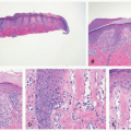
contours, significant variability in diameters, and disorganized packing into larger fibers.
TABLE 9-1. Categorization and features of Ehlers-Danlos syndrome

Type (Historic Category)

Genetic Mutations

Major Features

Inheritance Pattern

Salient Histologic/Ultrastructural Features

Classic (I, II)

COL5A1, COL5A2.

COL1A1

Skin hyperextensibility with atrophic scarring, GJH

AD

Flower-like collagen fibrils

Classic-like

TNXB

Skin hyperextensibility without atrophic scarring, easy bruising, GJH

AR

Cardiac valvular

COL1A2

Cardiac valvular disease, classic skin involvement, joint hypermobility

AR

Hypermobile (III)

Mostly unknown, partially due to TNXB

heterozygous

Joint hypermobility, less skin involvement, abdominal hernia, pelvic organ prolapse, aortic root dilatation

AD

Calcified deposits within an amorphous matrix of elastic fibers and clusters of hyaluronic acid

Vascular (IV)

COL3A1

Early arterial rupture, sigmoid colon perforation, gestational uterine rupture, peripartum perineum laceration, carotidcavernous sinus fistula, family history of vascular Ehlers-Danlos syndrome (vEDS)

AD

Thin dermis, irregular thickness of dermal-epidermal junction, fibroblasts with lysosome

Kyphoscoliotic (VIA)

PLOD1. FKBP14

Congenital hypotonia, congenital or earlyonset kyphoscoliosis, GJH

AR

Subtle irregularities in collagen fibril contour and spacing

Musculocontractural (VIB)

CHST14. DSE

Characteristic congenital contractures, characteristic facies, classic skin involvement with palmar wrinkling

AR

Dispersal of collagen fibrils

Arthrochalasia (VIIA, VIIB)

COL1A1, COL1A2

Congenital bilateral hip dislocation, severe

GJH, skin hyperextensibility

AD

Collagen fibrils with variable diameter and highly irregular contour

Dermatosparaxis (VIIC)

ADAMTS2

Extreme congenital skin fragility, characteristic facies, lax skin with palmar wrinkling, severe bruisability, umbilical hernia, growth retardation, short appendages, perinatal complications

AR

Hieroglyphic collagen fibrils

Periodontal (VIII)

C1R, C1S

Severe periodontitis, detached gingiva, pretibial plaques, family history of periodontal Ehlers-Danlos syndrome (pEDS)

AD

Spondylodysplastic

B4GALT7.

B3GALT6.

SLC39A13

Short stature, congenital hypotonia, limb bowing

AR

Brittle cornea syndrome

ZNF469. PRDM5

Thin cornea, keratoconus, keratoglobus, blue sclera

AR

Myopathic

COL12A1

Congenital hypotonia with/without atrophy, joint contractures, hypermobility of distal joints

AD or AR

Abbreviations: AD, autosomal dominant; AR, autosomal recessive; GJH, generalized joint hypermobility. Roman numerals in parentheses represent former numerical designations. Adapted from Malfait F, Francomano C, Byers P, et al. The 2017 international classification of the Ehlers-Danlos syndromes. Am J Med Genet C Semin Med Genet. 2017;175(1):8-26; Byers PH, Murray ML. Ehlers-Danlos syndrome: a showcase of conditions that lead to understanding matrix biology. Matrix Biol. 2014;33:10-15; Ong K-T, Plauchu H, Peyrol S, et al. Ultrastructural scoring of skin biopsies for diagnosis of vascular Ehlers-Danlos syndrome. Virchows Arch. 2012;460(6): 637-649; Rohrbach M, Vandersteen A, Yiş U, et al. Phenotypic variability of the kyphoscoliotic type of Ehlers-Danlos syndrome (EDS VIA): clinical, molecular and biochemical delineation. Orphanet J Rare Dis. 2011;6:46; Janecke AR, Li B, Boehm M, et al. The phenotype of the musculocontractural type of Ehlers-Danlos syndrome due to CHST14 mutations. Am J Med Genet A. 2016;170A(1):103-115.

TABLE 9-2. Pediatric findings in Ehlers-Danlos syndrome

Clinical Features

Ehlers-Danlos Syndrome Subtypes

Characteristic facies

Vascular, Plod1-associated dermatosparaxis, periodontal, kyphosclerotic, brittle cornea syndrome, spondylodysplastic, musculocontractural11

Congenital hearing loss

FKBP14-associated kyphoscoliotic

Severe neonatal skin tearing

Dermatosparaxis

Blue sclera

Brittle cornea syndrome, dermatosparaxis, kyphoscoliosis, spondylodysplastic, musculocontractural

Generalized hypotonia

Kyphoscoliotic, arthrochalasia, spondylodysplastic, myopathic

In utero and congenital hip dysplasia

Arthrochalasia12

Congenital or early-onset kyphoscoliosis

Kyphoscoliotic, arthrochalasia, B3GALT6-associated spondylodysplastic

Joint contractures

Musculocontractural, B3GALT6-associated spondylodysplastic

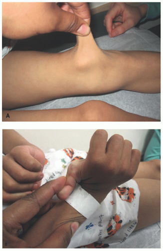
FIGURE 9-1. Ehlers-Danlos syndrome. Skin hyperextensibility (A) and joint hypermobility (B).
A few subtype-specific findings have been consistently reported. Significantly thinned dermis distinguishes vascular EDS. Gross fragmentation of elastic fibers has been seen in hypermobile EDS, which is associated with morphologic abnormalities of the amorphous elastin matrix. Distinctive cross-sectional contours of single collagen fibrils are seen in classic (flower like), vascular (hieroglyphic), and arthrochalasia (stellate) types.
Differential Diagnosis
The clinical differential diagnosis for EDS includes Marfan syndrome due to marfanoid habitus of kyphoscoliotic and periodontal subtypes. Cutis laxa features loose and doughy skin, which is seen in dermatosparaxis EDS. Loeys-Dietz syndrome (LDS; caused by TGF-β receptor mutations) presents with similar cutaneous and vascular complications to those found in vascular EDS, but presents with characteristic facies. Osteogenesis imperfecta type I can be distinguished by its significant bone pathology. Benign joint hypermobility syndrome should be easily excluded by the lack of cutaneous findings of hypermobile EDS and, if uncertain, by the strict application of diagnostic criteria.
MARFAN SYNDROME
Definition and Epidemiology
Marfan syndrome (MFS) is caused by mutations in FBN1, resulting in connective tissue defects in the skeletal, cardiovascular, ocular systems.13,14 Diagnosis is based on the 2010 revised Ghent nosology, which requires information on family history, syndromic features, and the identification of genetic mutations. The reported prevalence of MFS is approximately between 1/5000 and 1/15 000 without gender predilection.15
Etiology
Mutation in the FBN1 gene is identified in more than 90% of Marfan patients. Because of the gene’s extremely large size, more than 1800 different mutations have been identified to date in FBN1.16 Although most specific mutations do not predict organ system involvement, mutations located in exons 24 to 32 frequently result in neonatal MFS, the most severe form of the disease.17 About 25% of MFS-associated mutations are sporadic, but the majority are inherited in an autosomal dominant manner. Mutations in FBN1 cause reduced expression and misassembled microfibrils, leading to errors in elastic fiber formation and a compromise of the mechanical integrity of connective tissues.13 FBN1 has also been strongly implicated in the regulation of TGF-β signaling. Functional protein sequesters and inactivates TGF-β cytokine in the ECM. The loss of FBN1 results in an increase in TGB-β signaling, which likely accounts for some phenotypic elements (eg, cardiac valvular myxoma).
Clinical Presentation
The classic signs of MFS are reduced upper to lower segment ratio, severe pectus excavatum, scoliosis, ectopia lentis, dilatation and dissection of the ascending aorta, and lumbosacral dural ectasia.14 These features are not always present in childhood.18 Aortic root dilatation and ectopia lentis are the most constant features, and are, therefore, the most useful diagnostic criteria in the pediatric population.19 Patients present with a typical habitus characterized by tall, thin stature with long arms, legs, fingers, and toes (Figure 9-2).
Although the cutaneous findings of striae atrophicae and incisional hernias are found in MFS, they are not major diagnostic criteria.14 The prevalence of striae increases with age and is present in the vast majority of adult Marfan patients, appearing as atrophic, linear plaques ranging from erythematous to white in color.20,21 Presence in locations other than the thighs, buttocks, and hips occurs significantly more frequently in affected patients than in the general population. Infantile striae have been reported in the neonatal form of MFS.22 Abnormal wound healing is a common finding, with wide or atrophic scarring, dyspigmentation, and recurrent incisional hernias.
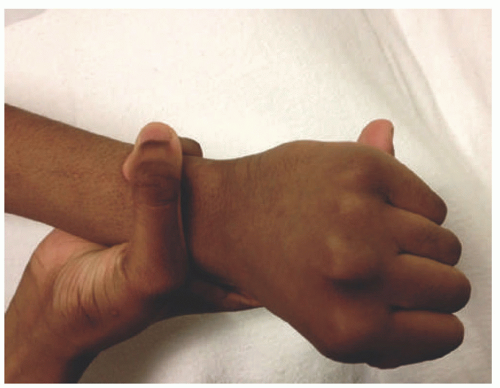
FIGURE 9-2. Marfan syndrome. Long fingers result in the thumb sign, in which the thumb protrudes from a closed fist, and the wrist sign, in which the thumb and fifth finger overlap when encircling the opposite wrist. Reprinted with permission from Bitterman AD, Sponseller PD. Marfan syndrome: a clinical update. J Am Acad Orthop Surg. 2017;25(9):603-609, Figure 2.
Histologic Findings
Clinically normal-appearing skin of patients with MFS may have characteristic histologic dermal findings. Elastic fibers appear fragmented with some scattering in orientation.23 Immunohistochemical evaluation for fibrillin protein shows discontinuous staining of the dermoepidermal junction, attenuation in the papillary dermis, and near-absence in the reticular dermis.24 The epidermis and dermal collagen appear normal. Ultrastructural studies show thinned elastic fibers that are randomly packed into large tortuous strands.23 Cross-sectional evaluation shows peripheral fragmentation of the elastin matrix, rendering a cobweb-like appearance.
As in striae from normal patients, biopsies from MFS patients show thinning of the epidermis with attenuation of the rete.25 The dermis is also thin, with hypertrophied collagen and elastic fibers run in parallel with the surface. Atrophic scars in a patient with incomplete MFS (ie, mitral valve prolapse, aortic enlargement, skin and skeletal findings syndrome [MASS]) show short, broken elastic fibers in the papillary dermis, which formed large aggregates in older lesions.26
Differential Diagnosis
LDS, a primary disorder of TGF-β signaling, and EDS share some clinical features with MFS.27 The distinct features of LDS include hypertelorism, bifid uvula or cleft palate, aortic aneurysm, and arterial tortuosity. Although some subtypes of EDS can have a marfanoid habitus, they are distinguished by skin hyperextensibility and atrophic wound healing in EDS, which are absent in MFS. Mutations in FBN2 cause congenital contractural arachnodactyly (CCA), which is characterized by contractures, arachnodactyly, scoliosis, and crumpled ears. These findings can be seen in neonatal MFS, but aortic root involvement and valvular disease is not typical of CCA.28 MFS-spectrum disorders (mitral valve prolapse syndrome, ectopia lentis syndrome, and MASS) can all be diagnosed under the current guidelines.14
TABLE 9-3. Causes and features of cutis laxa

Disorder

Inheritance

Gene

Distribution

Associated Features

ADCL type 1

ADCL type 2

AD

ELN

FBLN5

Generalized

Aortic dilatation, emphysema, GI diverticula.40

ARCL type 1A

ARCL type 1B

AR

FBLN5

FBLN4

Generalized

Aortic dilatation, supravalvular aortic stenosis, pulmonary artery stenosis, arterial tortuosity, emphysema, GI/GU diverticula, growth retardation, joint laxity, congenital hip dysplasia.30

ARCL type 1C (Urban-Rifkin-Davis syndrome)

AR

LTBP4

Generalized

Emphysema, pulmonary artery stenosis, valvular insufficiency, GI/GU diverticula, dysmorphic facial features

ARCL type 2A (Debré type)

AR

ATP6V0A2

Generalized. Improves in childhood41

  • Intrauterine growth restriction, joint laxity, congenital hip dysplasia, growth retardation, scoliosis, dysmorphic facial features,

  • De Barsy phenotype: mental retardation, athetosis, corneal clouding.42

  • Wrinkly skin syndrome is a phenotypic variant of ARCL 2A without GU involvement and milder facial and skin defects.

ARCL type 2B

De Barsy syndrome B (ARCL type 3B)

AR

PYCR1

Generalized. Dorsal acral and abdomen (2B).

ADCL type 343

AD

ALDH18A1

De Barsy syndrome A (ARCL type 3A)

AR

ARCL type 2C

ARCL type 2D

AR

ATP6V1E1

ATP6V1A

Generalized with sparse subcutaneous fat44

Hypotonia, cardiac abnormalities, aortic dilation, hip dysplasia, “mask-like” triangular face (2C), marfanoid habitus (2D), neurodevelopmental abnormalities (2D)

Geroderma osteodysplasticum

AR

GORAB

Cheeks, hands, feet, abdomen

Labial frenulum hypoplasia, malar hypoplasia, prognathism, oblique furrows on the lateral aspect of the face, osteoporosis, dwarfism

MACS

AR

RIN2

Generalized45

Macrocephaly, sparse hair, facial coarsening, gingival hypertrophy, scoliosis, osteoporosis, joint laxity

Occipital horn syndrome

X-linked recessive

ATP7A

Generalized

Occipital horn, coarse hair, arterial tortuosity, bladder diverticula, mental retardation, delayed motor development, joint laxity, osteoporosis, scoliosis. Allelic to Menke syndrome.

Abbreviations: AD, autosomal dominant; ADCL, autosomal dominant cutis laxa; AR, autosomal recessive; ARCL, autosomal recessive cutis laxa; GI, gastrointestinal; GU, genitourinary; MACS, Macrocephaly, Alopecia, Cutis Laxa, and Scoliosis.

CUTIS LAXA
Definition and Epidemiology
Cutis laxa (CL), or generalized elastolysis, is a group of disorders characterized by loose, hypoelastic skin folds caused by the disruption of dermal elastic fibers.29,30 Autosomal dominant, recessive, and X-linked inheritance patterns have all been reported with a number of implicated genes and associated systemic features. Acquired forms may be associated with inflammatory dermatoses and other systemic diseases. CL is rare, and there is no known predilection for specific populations.
Etiology
Heritable CL is subtyped on the basis of causal mutation that disrupts the synthesis and function of elastic fibers (Table 9-3).29 Mutation in ELN, which encodes for elastin, leads to an autosomal dominant version of CL. Recessive disease is caused by mutations in elastin support proteins (FBLN4 and FBLN5), the TGF-β pathway (LTBP4), vesicular ATPase (ATP6V0A2, ATP6V1E1, ATP6V1A), vesicular trafficking proteins (GORAB, RIN2), and mitochondrial components (PYCR1, ALDH18A1). Occipital horn syndrome, previously categorized as a subtype of EDS, is caused by an X-linked defect in cooper transport (ATP7A). Many other congenital syndromes also feature hypoelastic skin.
Postinflammatory reactions that cause the enzymatic degradation of the elastin network can cause acquired CL. It may be associated with conditions such as Sweet syndrome, infections, inflammatory connective tissue disease, plasma cell dyscrasias, and others.31 D-penicillamine, a cooper chelator that abrogates elastin cross-linking by inhibiting lysyl oxidase activity, is a cause of drug-induced CL.32
Clinical Presentation
The redundant skin in CL is hyperextensible but lacks the elastic recoil seen in EDS (with the exception of dermatosparaxis EDS). Pendulous skin folds around the cheeks, periorbital area, and neck create a progeroid appearance (Figure 9-3). Recessive forms of CL usually manifest in neonates and have a more aggressive phenotype, whereas autosomal and acquired forms can be adult-onset. Although skin findings are mostly generalized, more localized findings can be features of the autosomal recessive ARCL type 2B, geroderma osteodysplasticum, and acquired CL. Many recessive subtypes also have severe extracutaneous features such as neonatal emphysema, cardiovascular malformations, and developmental issues involving the neurologic, skeletal, gastrointestinal, and genitourinary systems. These associated findings overlap significantly between subtypes, and diagnosis can be difficult on clinical grounds alone.
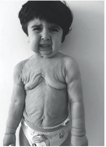
FIGURE 9-3. Cutis laxa. Pendulous skin folds confer a progeroid appearance. Reprinted with permission from Duz MB, Kirat E, Coucke PJ, et al. A novel case of autosomal dominant cutis laxa in a consanguineous family: report and literature review. Clin Dysmorphol. 2017;26(3):142-147.
Histologic Findings
For all forms of CL, elastic fibers found throughout the dermis can appear granular, clumped, or nearly absent.29 The epidermis appears normal. Dermal inflammation specific to particular dermatoses may be found in dermatosis-associated CL.33,34 Concurrent dermal immunoglobulin deposits can be detected in systemic diseases.35
Ultrastructural analysis demonstrates poorly organized clumps of elastin matrix around abnormal microfibrils.30 Immunoglobulin deposition along the edge of elastic fiber borders has been reported in patients with acquired CL.36,37
Differential Diagnosis
EDS is a disease of defective collagen and features hyperextensible skin that retains its elastic quality.2 In contrast to CL, joint hypermobility, skin fragility, and abnormal wound healing are dominant features of the disease. Elastic fiber loss and fragmentation are not seen on histology in EDS. Pseudoxanthoma elasticum (PXE) can lead to secondary CL, but primary papular lesions should precede the appearance of saggy skin. The presence of calcified elastic fibers in PXE is a distinct histologic finding.38 Mid-dermal elastolysis has the pathognomonic finding of a band-like loss of mid-dermal elastic fibers.39
APLASIA CUTIS CONGENITA
Definition and Epidemiology
Aplasia cutis congenita (ACC) is a neonatal finding of scarred or absent skin.46,47 Associated defects of the underlying bone and meninges are not uncommon. There is no single cause of ACC, which can be categorized on the basis of the number and location of lesions and associated clinical features (Table 9-4). The estimated incidence of ACC is approximately 3 in 10 000 births with a predominance in females.48,49
Etiology
ACC is a finding associated with a number of pathologic conditions.46 Developmental defects in skin morphogenesis are seen mostly in syndrome-associated ACC. For example, Adams-Oliver syndrome (AOS) features scalp ACC with frequent underlying skull defects, cutis marmorata telangiectatica congenita, transverse terminal limb defects, central nervous system (CNS) anomalies, and cardiac malformations.50 Loss-of-function mutations in Notch signaling and actin regulation pathways, both critical for general tissue morphogenesis, are known causes of AOS. ACC is also seen in focal dermal hypoplasia, a disorder of mesoectodermal development.51 A heterozygous missense mutation of BMS1 (a ribosomal GTPase) causes isolated, autosomal dominant ACC.52 BMS1 likely plays a role in cell cycle progression, critical for the rapidly proliferating cells of the developing scalp. In patients with epidermolysis bullosa, intrauterine trauma causes lesions localized to the extremities (Bart syndrome). Extrinsic factors that cause intrauterine insult, such as teratogens, infections, amniotic band sequence, and ischemic events, can also cause ACC.
TABLE 9-4. Causes of aplasia cutis congenita

Group

Clinical Features

Inheritance

Associated Anomalies

1. Scalp ACC, nonsyndromic

Vertex. Membranous or scar-like

AD (BMS1) or sporadic

Midline defects, tracheoesophageal fistula, patent ductus arteriosus, polycystic kidneys

2. Scalp ACC associated with Adams-Oliver syndrome

Scalp. Irregular borders and hemorrhagic to healing base

AD (ARHGAP31, RBPJ, NOTCH1, DLL4) or AR (DOCK6, EOGT)

Terminal transverse limb defect, cutis marmorata telangiectatica congenita, cardiac malformations, CNS abnormalities

3. Scalp ACC with skin/organoid nevi

Scalp. Membranous base

Sporadic

Epidermal nevi, congenital melanocytic nevi, ocular abnormalities, seizures.

4. ACC overlying embryologic malformations

Scalp, chest, abdomen, lumbosacral

Variable

Cranial malformations, spinal malformations, abdominal wall defect, sternal cleft, or combination

5. ACC with fetus papyraceus or placental infarcts

Scalp, trunk, extremity. Can be symmetric with irregular borders

Sporadic

Single umbilical artery, gastrointestinal atresia

6. ACC associated with epidermolysis bullosa

Trunk, extremity (Bart syndrome). Eroded or ulcerated

Varies on the basis of the type of epidermolysis bullosa (See Chapter 4)

Blistering of skin and/or mucous membranes, dystrophic nails, pyloric or duodenal atresia, abnormal ears and nose, ureteral stenosis, renal anomalies, amniotic bands

7. ACC localized to extremities without blistering

Extensor and dorsal surfaces of extremities

Autosomal dominant or recessive

Radial dysplasia

8. ACC caused by teratogens

Scalp (drugs), variable (infections)

Not inherited

Methimazole (with imperforate anus, urachal malformation), misoprostol low-molecularweight heparin, valproic acid, intrauterine infection with varicella, herpes simplex, rubella.

9. ACC associated with congenital syndromes

Variable

Variable

Trisomy 13, Wolf-Hirschhorn syndrome, focal dermal hypoplasia, focal facial dermal dysplasia, amniotic band sequence, microphthalmia with linear skin defects syndrome, Johanson-Blizzard syndrome, scalp-ear-nipple syndrome, oculocerebrocutaneous syndrome, oculoectodermal syndrome.

Abbreviations: ACC, aplasia cutis congenita; AD, autosomal dominant; AR, autosomal recessive; CNS, central nervous system Reprinted from Frieden IJ. Aplasia cutis congenita: a clinical review and proposal for classification. J Am Acad Dermatol. 1986;14(4):646-660. Copyright © 1986 Mosby, Inc. With permission. From Evers ME, Steijlen PM, Hamel BC. Aplasia cutis congenita and associated disorders: an update. Clin Genet. 1995;47(6):295-301. Copyright © 1995, John Wiley and Sons. Reprinted by permission of John Wiley & Sons, Inc.

Clinical Presentation
ACC predominantly occurs on the scalp (>85% of single lesions), usually on the vertex.47 The initial appearance of ACC is highly variable, with the typical end result being an atrophic or hypertrophic scar. The complete absence of skin presents as a translucent membrane overlying a punched out-appearing defect. A ring of coarse hair is sometimes present surrounding the membranous defect, the “hair collar sign,” and may indicate an underlying CNS malformation (Figure 9-4).53 Exposure and injury of the sagittal sinus has been reported for exceptionally deep lesions that involve the dura.54 Bullous ACC is a variant presenting with a serous fluid-filled vesicle with surface telangectasias.55 Patients may also present with lesions featuring ragged, stellate borders with a granulated or hemorrhagic base. This finding likely represents in utero wound healing and is usually secondary to intrauterine ischemic events, AOS, or the erosions of epidermolysis bullosa. Three cases of systemic ACC (>90% total body involvement) have been reported, in which cutaneous structures down to the subcutis were missing.56
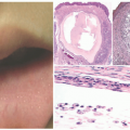
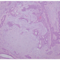
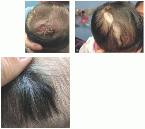
FIGURE 9-4. Aplasia cutis congenital. A, A crusted, healing erosion on the vertex of the scalp in a newborn infant. B, Extensive, well-healed aplasia cutis with resultant atrophic plaques. C, Aplasia cutis with hair collar sign—a rim of dark hair that may indicate underlying skull or central nervous system defects.
Histologic Features
The diagnosis of ACC is clinical, and histologic features most likely reflect various stages of tissue regeneration. Deeply ulcerated lesions may show a complete absence of cutaneous structures.47 As the lesion heals, the epidermis is flattened and may be remarkably thin at 1 to 2 cell layers thick.51 The underlying dermis takes on the appearance of granulation tissue or an atrophic scar.57,58 Adnexal structures are uniformly missing, and elastic fibers are sparse if present (Figure 9-5). For bullous ACC, the unremarkable 
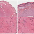
epidermis overlies an edematous dermis with very loose connective tissue.
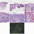
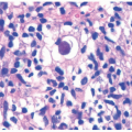
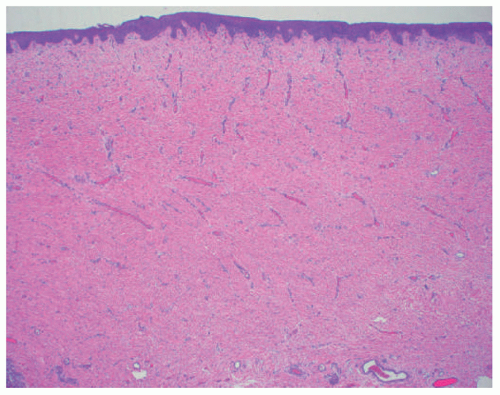
FIGURE 9-5. Aplasia cutis congenital. Thin epidermis overlies a dermal scar that lacks adnexal structures.

Only gold members can continue reading. Log In or Register to continue

Stay updated, free articles. Join our Telegram channel

May 8, 2019 | Posted by in Dermatology | Comments Off on Genetic Disorders of Dermal Connective Tissue

Full access? Get Clinical Tree

Get Clinical Tree app for offline access