Fig. 36.1
Generalized severe JEB presenting with generalized blistering and extensive areas of detachment of the epidermis
Secondary lesions following chronic, repeated (even intrauterine) tissue traumatization include atrophic scarring, webbing (intradermal scar formation between fingers and toes), contractures (typically in axillary vaults), and milia (keratin-filled cysts presenting as white papules).
Consecutive pigmentary abnormalities (hyper-, hypo- or demottled or mottled pigmentation) rarely also comprise EB nevi (quite common in JEB-generalized intermediate), i.e., large eruptive, asymmetrical, often irregularly pigmented, highly dynamic melanocytic lesions with sharply demarcated borders that frequently arise in areas of preceding blisters (Fig. 36.2). They may clinically (e.g., irregular lesions with explosive growth; frequent appearance of surrounding small satellite lesions) and dermoscopically (multicomponent pattern) mimic malignant melanoma [5].
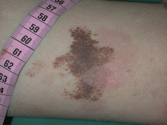
Fig. 36.2
EB nevus presenting as a large eruptive, asymmetrical, irregularly pigmented melanocytic lesions with sharply demarcated borders and small satellites arising in an area of preceding blister
Pseudosyndactyly due to repeated blistering on the hands and feet, initially presenting as partial interdigital fusion, webbing, or synechiae formation and followed by complete fusion of all of the digits to a keratinaceous cocoon-like structure, frequently causes marked functional disability, muscle atrophy, and bone absorption [3].
Exuberant granulation tissue (defined as moist, red, friable plaques with tendency to bleed), typically arising symmetrically around the mouth or nose as well as on the upper back, in axillary vaults, and around nail folds, is almost pathognomonic for JEB-gen sev [6]. Periorificial vegetation may cause complications such as total luminal occlusion of nares and may necessitate therapeutic intervention by laser and sharp dissection, or they may scar over and seal (Fig. 36.3). Differential diagnosis from squamous cell carcinoma is sometimes challenging and may be managed by continuous clinical and occasionally histopathological evaluation.
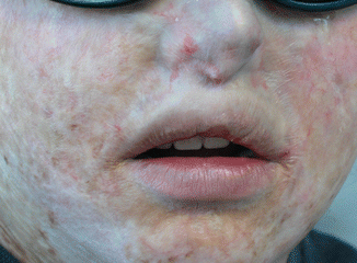
Fig. 36.3
Periluminal granulation has led to scarring over the nares (Courtesy: Dédée Murrell, MD)
Symptoms of involvement of the nail apparatus include peri- or subungual blistering with hemorrhages, paronychia-like lesions with nail bed erosions and development of granulation tissue soon after birth, as well as nail bed hyperkeratosis with onycholysis and onychomadesis (Fig. 36.4). Additionally, nail atrophy (very thin, brittle, short nail plate); onychodystrophy with thickened, yellowish, longitudinally grooved, eventually marked curved, and deformed nail plates (onychogryphosis); and, ultimately, absence (by shedding) of nails (anonychia) due to atrophy, scarring, and destruction of the nail bed and matrix by repetitive blistering are common findings in JEB-gen sev [7].
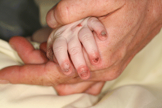
Fig. 36.4
Nail involvement with erosions and granulation tissue around the nail folds eventually leading to onycholysis
Despite being more prominent in generalized intermediate JEB, localized or more diffuse scarring alopecia can also be observed in the generalized severe JEB variant. Other, rather uncommon cutaneous manifestations of JEB-gen sev comprise palmoplantar keratoderma and congenital absence of skin (aplasia cutis congenita). The latter presents with ulcerated lesions and complete absence of all skin layers (triggered by intrauterine mechanical trauma?) [8]. In individuals with prolonged survival, denuded lesions can heal with a smooth epidermis (proliferation of fibroblasts in loose connective tissue stroma, capillary formation, absence of adnexal structures) presenting as red, angulated, flame-shaped, well-demarcated, depressed patches usually unilateral on the hands, feet, wrists, or ankles. Aplasia cutis congenita is not specific for JEB-gen sev and was also reported in EBS [9], DDEB, and RDEB [6].
36.3 Extracutaneous Involvement
Prototypical lesions of JEB-gen sev such as blisters and erosions, followed by strictures, contractures, and stenoses, also occur in many (epithelial) tissues outside the skin which express the laminin-332 protein, involving, e.g., the mucous membranes of the gastrointestinal, upper respiratory, and genitourinary tracts, the kidney, and external eye. This internal involvement makes JEB-gen sev a systemic, multidimensional disease and account for its considerably high morbidity and mortality.
Ophthalmic findings in the generalized severe JEB subtype include ocular surface (corneal, conjunctival) and eyelid abnormalities (blisters, erosions, and scarring), ectropion with exuberant granulation tissue, exposure keratitis, pannus formation, limbal broadening, symblepharon, and lacrimal duct obstruction [10].
Upper respiratory tract involvement is a frequent phenomenon in JEB-gen sev. Granulation tissue around nares and within nostrils can cause airway narrowing and repeated bleeding. The most common complications, i.e., chronic hoarseness, weak cry, or inspiratory stridor, are alarming findings seen in up to 50 % of all patients with JEB-gen sev [11]. They may signify a considerable respiratory affection with end-stage sequelae like laryngeal webs, stenosis, and (acute) airway obstruction due to occlusion by blisters, diffuse edema, progressive scar formation, and strictures or exuberant granulation tissue following blistering on the edges of the laryngeal/vocal cords. Airway injury arises spontaneously or follows episodes of coughing, crying, or upper respiratory tract infection. Precipitated or exacerbated by intubation or instrumentation as well as unrecognized or untreated gastroesophageal reflux disease, it may occur with rapid onset and life-threatening progression [12].
Partial or complete occlusion of upper airways and secondary aspiration pneumonia are the most severe otorhinolaryngeal complications in EB that are almost exclusively seen in the generalized subtypes of JEB within the first year of life [12]. The cumulative risk of acute airway obstruction in JEB-gen sev determined by analyses of the NEBR population is about 13 % by as early as age 1. In patients surviving beyond the neonatal period, it plateaus at a risk of nearly 40 % by age 6 and thereafter decreases most likely due to the age-related increase in the luminal diameter of airways. Consequently, early and regular surveillance and a comprehensive therapeutic management are mandatory.
Intraoral disease [13] is characterized by severe involvement of the oral soft and hard tissue, making both prophylactic and therapeutic approaches very challenging. Exuberant perioral granulation tissue frequently leads to a reduction of the oral opening (microstomia) and some loss of tissue mobility in the lips and perioral tissues [14].
Excessive caries results from a deficient oral hygiene due to painful peri- and intraoral blistering, erosions as well as scar formation with consecutive contractures (microstomia, ankyloglossia), and limited tongue mobility and food clearance. Moreover, the specific, extremely cariogenic EB diet, a generally higher risk for infections like candidiasis (reflecting the intrinsic barrier deficiency that stimulates colonization and invasiveness), and malnutrition (and consequent weakening of the immune system) are exacerbating factors that contribute to premature loss of teeth.
Related posts:
 Kindlin-1 and Its Role in Kindler Syndrome
 Cyclophosphamide in Autoimmune Blistering Diseases: Safety, Efficacy and Evidence Base
Management of Bullous Systemic Lupus Erythematosus
Cyclophosphamide in Autoimmune Blistering Diseases: Safety, Efficacy and Evidence Base
Management of Bullous Systemic Lupus Erythematosus
 Using Intravenous Immunoglobulins in Autoimmune Bullous Diseases
 Living with Epidermolysis Bullosa: Reviewing the Impact on Individuals’ Quality of Life
 How to Take a Skin Biopsy Correctly to Diagnose Epidermolysis Bullosa and Autoimmune Bullous Diseases
Stay updated, free articles. Join our Telegram channel

Full access? Get Clinical Tree


