General Considerations of Viral Diseases: Introduction
|
Cutaneous manifestations are a prominent feature in many viral infections and may arise from a direct result of viral replication in the epidermis or secondarily from viral replication in other organs. In the latter case, the cutaneous manifestations may be the presenting sign of a systemic infection requiring further evaluation. Although most viral infections with cutaneous involvement are mild and self-limited, occasionally severe or life-threatening complications can develop, especially in immunocompromised hosts. Developments in laboratory testing have improved diagnosis of viral infections in the skin. In addition antiviral medications have improved clinical management and outcomes for many of these viral illnesses. While specific diagnosis of the etiologic viral agent is not always feasible, laboratory diagnosis may also be important epidemiologically. Preventive vaccines are available for certain viruses and are currently the most effective strategy for decreasing morbidity and mortality associated with these diseases. Prophylactic vaccines led to the eradication of smallpox, and now include newer vaccines against herpes zoster virus and human papillomaviruses.
Definition
Viruses are unique and fascinating small subcellular agents that require host cells to replicate their genetic material.1 They contain genomic nucleic acid, a protein coat, and some viruses also have a lipid envelope. Lacking functional ribosomes or other organelles, viruses require host cellular machinery for replication of their genetic material.1,2 This dependence on the host cell for transcription and replication is why viruses are often referred to as obligate intracellular parasites. The intracellular location of the virus provides survival benefit to the virus against some of the host’s immune mechanisms.
Viruses are an important means of horizontal gene transfer and evolutionary genetic diversity.3,4 Historically, there have been many discussions regarding the etiology and categorization of viruses. Most experts currently view viruses as biological particles that can interact with cells, as opposed to biological organisms or life forms.3 Viruses have features of both of these groups, which accounts for some of their enigma, but lack some of the defining features of living organisms and are not classified taxonomically amongst the kingdoms of life.
The viral nucleic acid contains the genetic information necessary for directing the host cell to replicate the virus. The assembled virus is referred to as a virion, and contains a highly organized protein coat or capsid.5 The protein coat protects the viral nucleic acid from extracellular environmental insults such as nucleases and it facilitates virion attachment to the membrane of the host cell.6 After the virion has infected a cell, the now intracellular viral genetic information is usually found in a nonparticulate form. The various virion components are synthesized in separate pieces within the cell and assembled to form progeny particles. This assembly type of replication is characteristic of viruses.7
Classification
Viruses are grouped based upon their type of genomic nucleic acid, DNA or RNA, and by the number of strands of nucleic acid within each virion.2 Other traditional categorization has focused on viral size, morphology, and presence of an envelope, as shown in Table 191-1. A given family of viruses usually shares functional, genetic, biochemical, and immunologic features as well. Other features that may be evaluated in viral categorization are antigenic cross reactivity and host cell tropism. With the advent of more sophisticated techniques, recent viral classification has focused on relatedness of genome sequences.8
Group | Size (nm) | Shape | Envelope | Nucleic Acid | Capsid Assembly | Examples of Viruses |
|---|---|---|---|---|---|---|
Herpesvirus | 120–200 | Icosahedral | Yes | DNA | Nucleus | Herpes simplex virus (types 1 and 2), varicella zoster virus, cytomegalovirus, EBV, HHV-6, 7, and 8 |
Papillomavirus | 50–55 | Icosahedral | No | DNA | Nucleus | Papillomaviruses |
Polyomavirus | 45–50 | Icosahedral | No | DNA | Nucleus | SV40, JC virus, Merkel cell polyomavirus |
Poxvirus | 240 × 300 | Complex | Yes | DNA | Cytoplasm | Molluscum contagiosum, orf, milker’s nodules virus, variola virus, vaccinia virus |
Retrovirus | 80–120 | Spherical | Yes | RNA | Cytoplasm | HIV, HTLV |
Paramyxovirus | 150–300 | Helical | Yes | RNA | Cytoplasm | Measles virus, mumps virus |
Togavirus | 40–60 | Icosahedral | Yes | RNA | Cytoplasm | Rubella virus, Chikungunya virus, some arboviruses |
Parvovirus | 20 | Icosahedral | No | DNA | Nucleus | Parovirus B19 (agent of Erythema infectiosum) |
Hepadnavirus | 40–50 | Icosahedral | No | DNA | Nucleus | Hepatitis B virus |
Adenovirus | 70–80 | Icosahedral | No | DNA | Nucleus | Multiple human serotypes |
Picornavirus | 20–30 | Icosahedral | No | RNA | Cytoplasm | Enterovirus (coxsackievirus, echovirus, poliovirus), rhinovirus |
Bunyavirus | 90–100 | Helical | Yes | RNA | Cytoplasm | Some arboviruses |
Arenavirus | 85–120 | Spherical | Yes | RNA | Cytoplasm | Lassa virus |
Flavivirus | 40–50 | Spherical | Yes | RNA | Cytoplasm | Yellow fever virus, Dengue virus, hepatitis C virus, West Nile virus |
On a morphological basis viruses can be grouped as helical, icosahedral, or complex as determined by the type of capsomer and overall shape of the virus.9 In certain virus families (such as herpesviruses and retroviruses), the capsids are located inside an envelope (composed of lipid, protein, and carbohydrate), which is required for infectivity5 (see Table 191-1). The viral envelope is sensitive to drying, so that infectivity is lost with drying. The virions of some other viruses (e.g., papillomaviruses) do not possess an envelope, so their capsids are called naked. Because the capsid proteins are usually stable in dry environments, viruses whose virions lack an envelope typically remain infectious for long periods after drying.10
Figure 191-1 shows examples of virions from the three main families of viruses that multiply in the epidermis and appendageal structures (papillomaviruses, herpesviruses, and poxviruses). The viral genomes of these three families are composed of DNA. As noted earlier, papillomaviruses possess naked (nonenveloped) capsids, and herpesviruses have enveloped virions. Poxvirus virions are large, have a very complex structure, and are enveloped, but their envelope is not required for their virions to be infectious. Hence, poxviruses can remain infectious even after drying.11
Figure 191-1
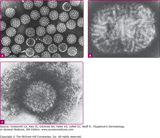
Electron micrographs of negatively stained virions (×200,000). A. Papillomavirus: multiple nonenveloped human papillomavirus (wart) virions showing capsid subunits (capsomeres). B. Poxvirus: single molluscum contagiosum virus virion, showing complex tubular structures. C. Herpesvirus: single varicella zoster virus virion showing capsid inside envelope. [Parts A and B used with permission from AF Howartson, JD Almeida, and MG Williams. Part C used with permission from Almeida JD et al: Morphology of varicella (chickenpox) virus. Virology 16:353, 1962.]
Viral Replication
Viral particles do not multiply through cell division because they are acellular. Instead, replication of viruses requires certain host cell genes, proteins, and organelles. The viral replication cycle involves several steps: adsorption, cellular uptake, uncoating, biosynthesis, virion assembly, and release.5
Adsorption proceeds with the attachment of virions to cells, involving a specific interaction between the viral capsid, or envelope, and host cell surface receptors. The specificity of this interaction between the viral protein and host receptor defines and limits the species and cell type that a particular virus can infect.6 Cells that lack the appropriate cell surface receptors for a particular virus are not susceptible to cellular viral entry and infection. For example, human immunodeficiency virus (HIV) enters cells via an interaction between viral envelope protein and two classes of receptors: cellular CD4 receptors and chemokine receptors.12 While the majority of people are known to be susceptible to HIV infection, rare genetic modifications of specific chemokine receptors have been identified which render hosts relatively resistant to HIV infection or the development of acquired immunodeficiency syndrome.13,14
After adsorption, the coat of enveloped viruses may fuse with the host cell membrane and release the virus nucleocapsid into the host cytoplasm. Alternately some viruses may enter the cell through invagination of the cell membrane forming vesicles in the cell cytoplasm.6 Uncoating subsequently occurs with release of the viral genome from its protective capsid, which begins the nonparticulate phase of the virus life cycle. The nucleic acid can now be transported within the cell.
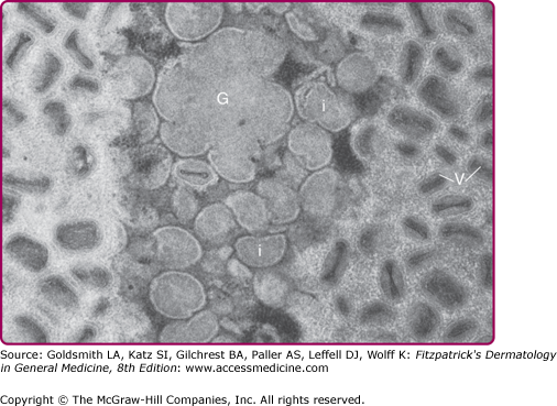
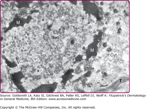
In the genomic activation or biosynthetic phase, messenger RNA (mRNA) is transcribed typically from viral DNA and translated by the host cell under regulation by the virus. DNA viruses, such as papillomaviruses and herpeviruses typically replicate in the nucleus. (Figs. 191-2 and 191-3) RNA viruses mainly replicate in the cytoplasm. However this is not absolute, as exemplified by poxviruses that contains DNA but replicate in the host cell’s cytoplasm15 (Fig. 191-4).
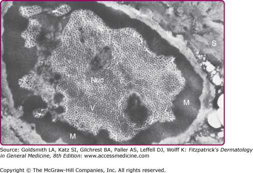
Figure 191-3
Cell infected with varicella zoster virus (a herpesvirus). Electron micrograph (×24,000). Portion of cell of the stratum spinosum. The nucleus (Nuc) contains varicella zoster virions (V). Chromatin is marginated at (M). Virions (V) and tonofilaments (T) are shown in the cytoplasm (Cyt).