Fig. 8.1
The tumor presents as a slowly growing, fleshy, sessile, or pedunculated lesion
Pathology
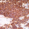
The tumor is characterized by a fenestrated pattern formed by thin anastomosing strands of basaloid and squamous cells, surrounded by abundant stroma (Fig. 8.2). The strands show connection with the epidermis or preexisting infundibula and terminate in nubbins of basaloid cells with peripheral palisading (Fig. 8.3). A cleft may be present between these nubbins and adjacent fibrotic stroma. The neoplastic cells are of two types, namely, basaloid or germinative cells and more fully differentiated squamous cells with abundant pink cytoplasm (Figs. 8.4, 8.5, and 8.6). The epithelial component is embedded in an abundant loose fibrovascular stroma. Follicular germs and rudimentary follicular papillae are sometimes observed. Even rarer, signs of a more advanced follicular differentiation in the form of cornification at the isthmus may be encountered (Fig. 8.5). Not uncommonly, a nodular type of BCC may be observed in continuity with a FEP.
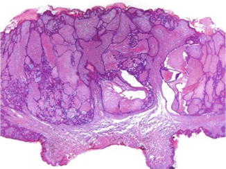
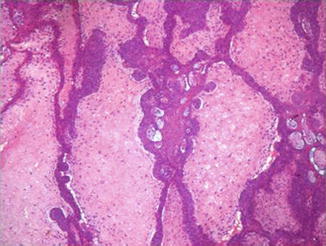
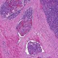
Fig. 8.2
This polypoid tumor is characterized by a fenestrated pattern formed by thin anastomosing strands of epithelial cells with multiple connections with the epidermis, surrounded by abundant stroma
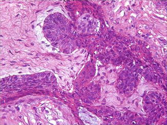
Fig. 8.3
The strands terminate in nubbins of basaloid cells with peripheral palisading that protrude into the surrounding stroma. Clefts are present between this nubbins and adjacent fibrotic stroma