Fat Injection to Correct Contour Deformities in the Reconstructed Breast
Scott L. Spear
Ali Al-Attar
Introduction
Breast reconstruction may be successfully accomplished using either prosthetic or autologous techniques or some combination of the two. Extensive experience with these techniques has led to progressive refinements that have raised the aesthetic standard for the reconstructed breast. Patients and surgeons alike are no longer satisfied with a simple “mound,” whether it is of prosthetic or autologous origin.
Although fat injection has been used since the mid-1980s to correct both congenital and iatrogenic contour deformities of the face, trunk, and extremities, its use in the breast is controversial and has only recently been addressed in the literature (1,2,3,4). Autologous fat has been reported to be used for cosmetic breast augmentation as well as for contour irregularities after reconstruction, but the current literature contains mainly case reports without well-conducted prospective trials that can guide clinical use (1,5,6,7).
Autologous fat augmentation is increasingly being used as a surgical soft tissue adjuvant in numerous parts of the body, and with clinical experience has come enhanced appreciation of its safety and efficacy. Nonetheless, its use in the breast raises the spectrum of diagnostic confusion due to the theoretical complication of lumps caused by fat necrosis and their potential to mimic the diagnosis of cancer recurrence. In addition, understanding of the science of autologous fat grafting is in its infancy, and numerous anecdotal reports suggest that it is a biologically active graft with potential immunologic and regenerative effects that may revolutionize the reconstructive surgical arsenal (3,8,9).
We have used fat injection since 1993 to correct contour deformities in breasts reconstructed by either prosthetic or autologous methods. More recently, we analyzed our 10-year experience because we were interested in determining the degree of improvement afforded by fat injection in the reconstructed breast, as well as any complications that resulted from using this technique, particularly given the controversy surrounding the procedure (1). Although fat injection in and around the reconstructed breast has limitations and may need to be repeated, our experience indicates that overall it is very safe and can improve or correct significant contour deformities that would otherwise be uncorrectable or require substantially more complicated or riskier procedures to improve.
History of Autologous Fat Grafting
Grafting of autologous fat is in fact an old concept, and its clinical history goes back to the nineteenth century. The allure of a reconstructive material that is autologous, soft, and plentiful has driven numerous surgeons to attempt this modality despite problems with reliability and resorption.
The first description of autologous fat grafting appeared in the 1890s in Europe, where macroscopic fat grafts were transferred to correct soft tissue deficits (10,11). The unpredictable resorption of fat grafts became immediately evident and was identified as the procedure’s critical blemish in the initial reports of its clinical applicability.
Careful analysis of the fate of transplanted autologous fat was conducted at the Mayo Clinic in a rodent model in the early 1900s (12). These investigators realized that tissue manipulation contributes to graft loss and suggested that careful handling can significantly reduce graft resorption. Lyndon Peer augmented the study of fat grafting with histologic analysis, and was the first investigator to establish that the survival of autologous fat grafts is essentially dependent on the survival of the individual grafted fat cells, unlike the model in some other autologous grafts (such as bone), where the donated tissue serves as a scaffold for replacement by recipient cells (13). This seminal work in the 1950s established the principles of autologous fat grafting: small graft sizes that maximize surface area to volume, permitting nutrition and oxygenation to the individual grafted adipocytes.
The use of clinical fat grafting accelerated in the late 1980s parallel to the development of liposuction, which provided abundant sources of donor tissue that is otherwise discarded. Furthermore, advances in microsurgery presented alternative techniques for soft tissue augmentation, which, combined with the advent of soft tissue fillers, raised the standards of performance that autologous fat grafting had to meet in order to be widely used.
During the last two decades plastic surgeons have led the investigation of autologous fat grafting in clinical trials and basic science research. Studies have yielded several tenets of clinical practice that have since been widely adopted: (a) grafting of freshly harvested fat, (b) gentle handling of tissues, (c) injection of fat using fine cannulae in thin strips, and (d) overcorrection to anticipate 20% to 50% resorption. Plastic surgeons now use fat grafting in multiple clinical applications, from cosmetic facial augmentation to craniofacial reconstruction and breast liporemodeling.
One major recent advance has come from Sidney Coleman’s findings that injected lipoaspirate appears to have a remedial effect on adjacent radiation-damaged skin. While the mechanism is unknown, Coleman and other surgeons have speculated that autologous fat transplantation might spark stem cell activity in the fat that can then modulate adjacent tissue. Although this mechanism is theoretical, a number of clinical and basic science studies have confirmed that transplanted fat can have positive modulatory effects in a paracrine fashion. The prospect of simultaneous cellular repair and soft tissue
augmentation makes autologous fat grafting a powerful instrument for solving difficult reconstructive problems.
augmentation makes autologous fat grafting a powerful instrument for solving difficult reconstructive problems.
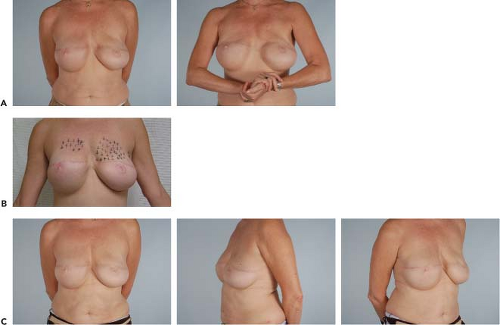
 Figure 76.1. A: A 57-year-old woman after bilateral mastectomy and reconstruction with tissue expanders and acellular dermal matrix, with later exchange to McGhan style 20 silicone implants. Note contour defects on the upper pole of both breasts, particularly on the left side. B: Preoperative markings performed with the patient standing; the areas marked with a plus sign indicate where soft tissue augmentation with fat injection will be performed. C: Postoperative photographs 2 months following autologous fat grafting, demonstrating improvement in upper pole fullness. Two hundred milliliters of autologous fat were injected into the left breast, and 85 mL were injected into the right breast.
Stay updated, free articles. Join our Telegram channel
Full access? Get Clinical Tree
 Get Clinical Tree app for offline access

|


