Introduction
The pathogenesis of facial aging may be explained on an anatomical basis. An anatomical approach to surgical rejuvenation of the face provides the way to obtaining a “natural” result that is lasting and with minimal morbidity. Fundamental to safety when operating in the face is a firm grounding in the principles on which the facial soft tissue layers are constructed. Understanding the relations of the facial nerve branches to these facial soft tissue layers, retaining ligaments, and soft tissue spaces is crucial in predicting and anticipating the location of the facial nerves. This is most important in addressing the overriding concern about the location and course of the facial nerve branches when performing surgical sub-SMAS (superficial musculoaponeurotic system) facelifts.
Five-Layered Anatomy, Facial Soft Tissue Spaces, and Retaining Ligaments of the Face
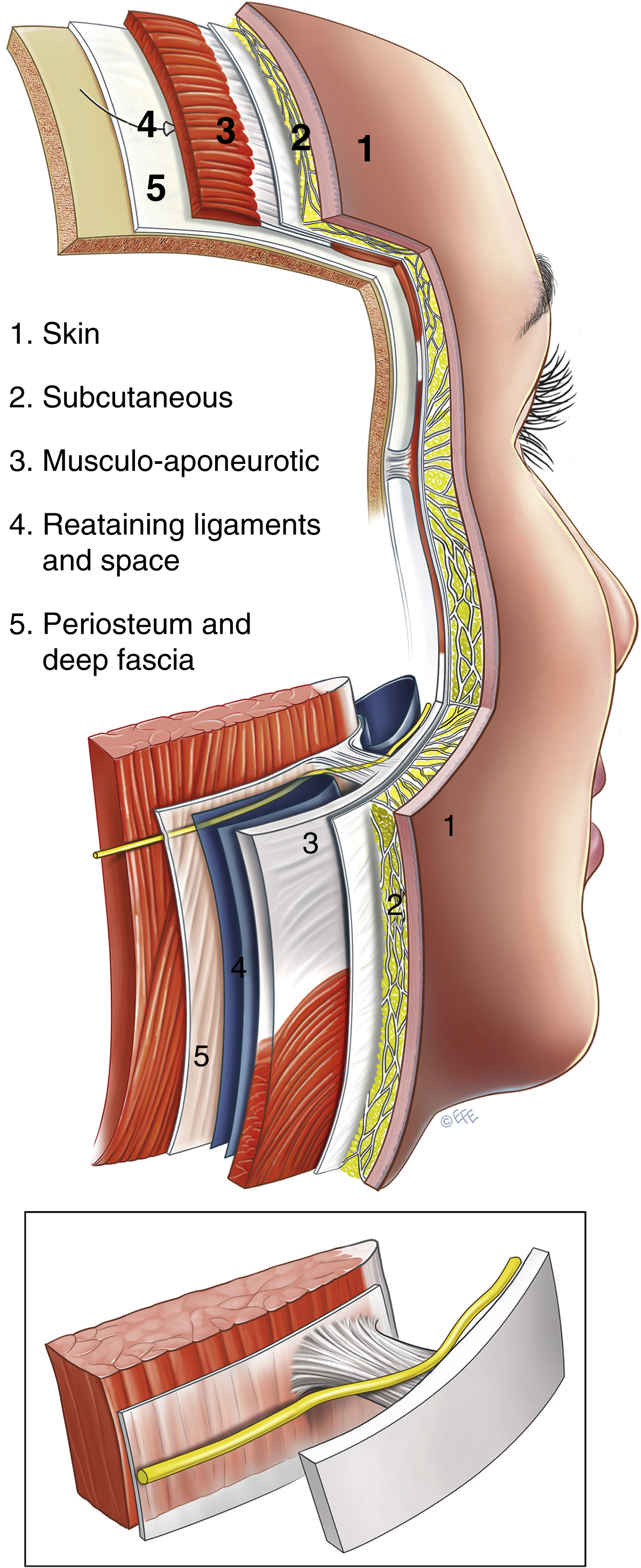
The five concentric layers of the face are: (1) skin; (2) subcutaneous tissue; (3) musculoaponeurotic layer; (4) loose areola tissue; (5) deep fascia. This five-layered arrangement is most clearly seen in the scalp and forehead (due to expansion of the soft tissues as a result of evolutionary expansion of the underlying cranial vault necessary to accommodate the highly developed frontal lobe in humans). Accordingly, the scalp is an excellent place to study the principles of the layered anatomy ( Fig. 62.1 ). Layer 4 (the loose areolar tissue) is the layer that allows the superficial fascia (defined as the composite flap of layers 1 through 3) to glide over the deep fascia (layer 5). This arrangement enables facial expressions by the frontalis and orbicularis oculi to function independent of the deeper muscle of mastication, the temporalis located deep to the deep fascia. The simplified anatomy over the scalp gives the basic prototype of layer 4. There are not any structures crossing this plane, which is essentially an avascular gliding space. At the boundaries of the scalp along the superior temporal line and across the supraorbital rim, the scalp and the forehead are firmly anchored by retaining ligaments. Vital structures such as nerves and vessels are always located in close proximity to the retaining ligaments when transitioning from deep to superficial in their course. In other parts of the face, the principles of this construction remain the same, albeit with considerably greater complexity. This is due to the compaction resulting from the reduction of forward projection of the mid and lower face (as occurs in other species) and the predominance of the orbital and oral cavities that limit the availability of a bony platform for attachment of retaining ligaments and muscles. To secure the mobile superficial layers to the facial skeleton, an elaborate system of retaining ligaments bind the dermis to the skeleton (or deep fascia where the facial skeleton is covered by skeletal muscles for mastication); the components of this system pass through all layers ( Figs. 62.2 and 62.3 ). ,
Layer 1 – Skin
The epidermis is a cell-rich layer composed mainly of differentiating keratinocytes and a smaller number of pigment-producing melanocytes and antigen-presenting Langerhans cells. A rich vascular plexus is an important component of the dermis. The thickness of the dermis relates to its function and tends to be inversely proportionate to its mobility. The dermis is thinnest in the eyelids and thickest over the forehead and the nasal tip.
Layer 2 – Subcutaneous Tissue
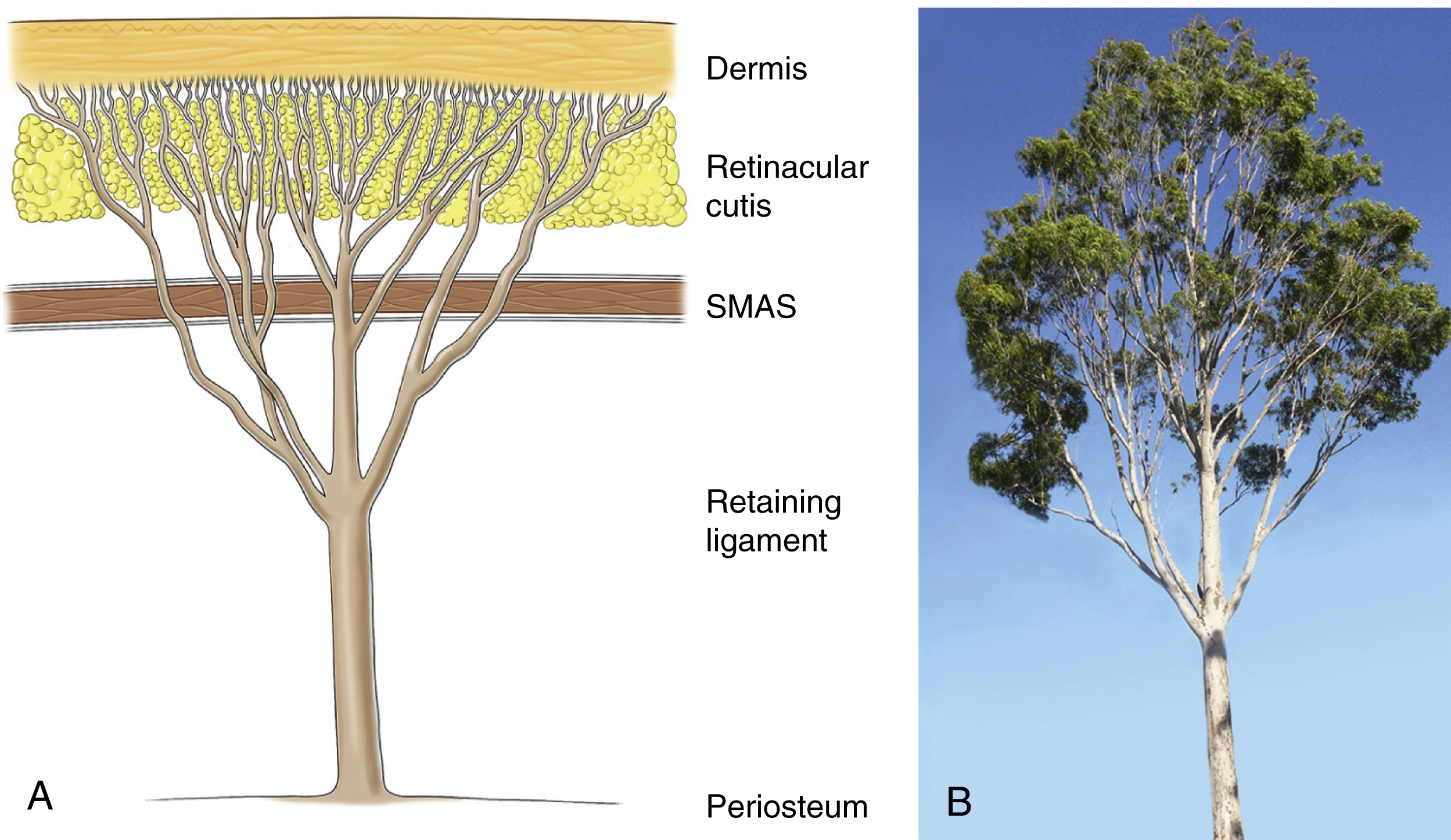
The subcutaneous layer has two components: the subcutaneous fat, which provides volume, and the fibrous retinacular cutis that binds the dermis to the underlying superficial musculoaponeurotic system (SMAS) of the face. Of note, the retinacular cutis is the name given to that portion of the retaining ligament that passes through the subcutaneous tissues. The amount, proportion, and arrangement of each component varies in different regions of the face. In the scalp, the subcutaneous layer has uniform thickness and consistency of fixation to the overlying dermis. In contrast, in the face proper, the subcutaneous layer has significant variation in thickness and attachments. In specialized areas such as the eyelids and lips, this layer is significantly compacted such that fat may appear non-existent. In other areas, such as the nasolabial segment, it is very thick. In areas with thick subcutaneous tissue, the retinacular cutis lengthens significantly, predisposing its fibers to weakening and distension with aging. Within the subcutaneous tissue, the overall attachment to the overlying dermis is stronger and denser than the attachment to the underlying SMAS. This is a result of the tree-like arrangement of the retinacular cutis fibers (see Fig. 62.2 ), with fewer but thicker fibers deep as its rises through the SMAS that progressively divide into multiple fine microligaments as they reach the dermis. This explains why it is easier to perform subcutaneous dissection in the deeper subcutaneous level (just on the surface of the underlying SMAS) than more superficially nearer the dermis, as there are fewer retinacular cutis fibers and the subcutaneous fat here does not attach directly to the outer surface of the underlying SMAS.
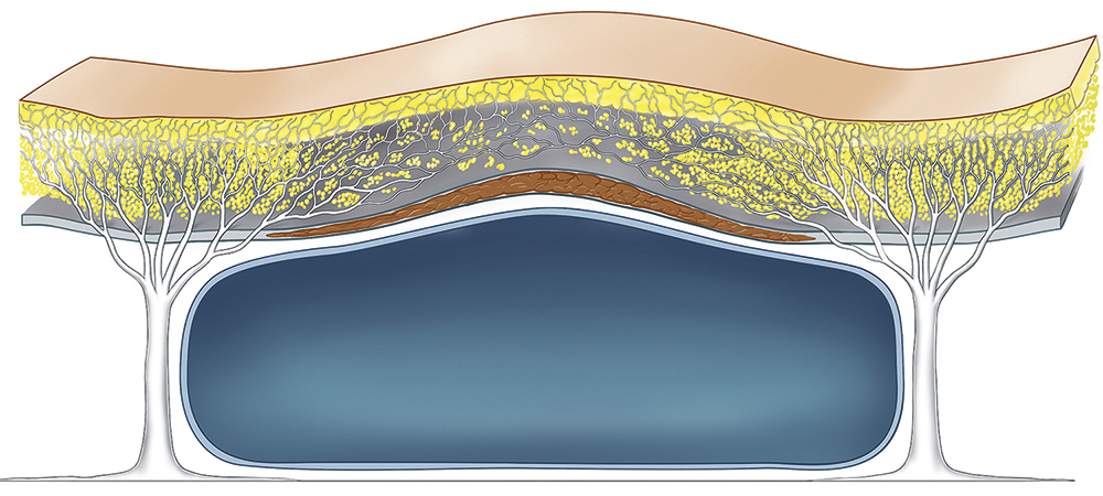
Furthermore, the retinacular cutis fibers are not uniform across the face, but vary in orientation and density according to the anatomy of the underlying deeper structures. As will become apparent when the anatomy of the underlying layer 4 is described, at the location of the retaining ligaments, the vertically orientated retinacular cutis fibers are the most dense and are the most effective in supporting the overlying soft tissues. In so doing, they form the boundaries that compartmentalize the subcutaneous fat. These areas, such as the so-called McGregor’s patch over the body of the zygoma, often require sharp release to mobilize. In between these retaining ligaments in layer 4 are located the soft tissue spaces of the face, that facilitate the mobility of the superficial fascia over the deep fascia. Where the subcutaneous fat overlies a layer 4 space, the retinacular fibers are less dense and orientated more horizontally, as a result of which, the tissues tend to separate with relative ease, often with just simple blunt finger dissection ( Fig. 62.4 ). This variation in the density and orientation of the retinacular cutis fibers in the subcutaneous fat is the anatomical basis for the so-called “superficial subcutaneous fat compartments” recently described. In these studies, injection of dyes into the subcutaneous fat results in staining of the fat in discrete compartments. While the relevance of superficial subcutaneous fat compartments remains debated, anatomically, they reflect the anatomy of layer 2, with diffusion of the injected dye being limited by the retaining ligaments at the boundaries of these compartments.
Layer 3 – Musculoaponeurotic Layer
The muscles of facial expression are unique and fundamentally different from skeletal muscles beneath the deep fascia because they are situated within the superficial fascia and they move the soft tissues of which they are a part. All muscles of facial expression have either all or the majority of their course within layer 3 and they are predominantly located over and around the orbital and oral cavities. In the prototype scalp, the occipital-frontalis moves the overlying soft tissue of the forehead, while its undersurface glides over the subgaleal aponeurotic space (layer 4). Layer 3 is continuous over the entire face, although for descriptive purposes, different names are given to certain parts according to the superficial muscle within. It is called the galea aponeurotica over the scalp, the temporoparietal (superficial temporal) fascia over the temple, the orbicularis fascia in the periorbital region, the superficial musculoaponeurotic system (SMAS) over the mid and lower face, and platysma in the neck.
Within layer 3, the facial muscles themselves have a layered configuration, with the broad, flat muscles forming the superficial layer that covers the anterior aspect of the face. The frontalis covers the upper, obicularis oculi the middle, and the platysma the lower thirds, respectively. The muscles of this superficial layer have minimal direct attachment to the bone, stabilized to the skeleton at their periphery indirectly by the vertically orientated retaining ligaments as noted earlier. The frontalis is fixed along the superior temporal line by the superior temporal septum, the obicularis oculi laterally by the lateral orbital thickening and the main zygomatic ligament at its inferolateral border and the platysma at its upper border by the lower key masseteric ligament. The deeper muscles in layer 3 provide greater functional control of the sphincters over the bony cavities. For the upper third, these are the corrugators and procerus and around the oral cavity, the elevators (zygomaticus major and minor, levator labii superioris, levator angulari oris) and levator labii superioris alaeque nasi, and the depressors (depressor angulari oris, depressor labii inferioris) around the oral sphincter and the mentalis.
Layer 4
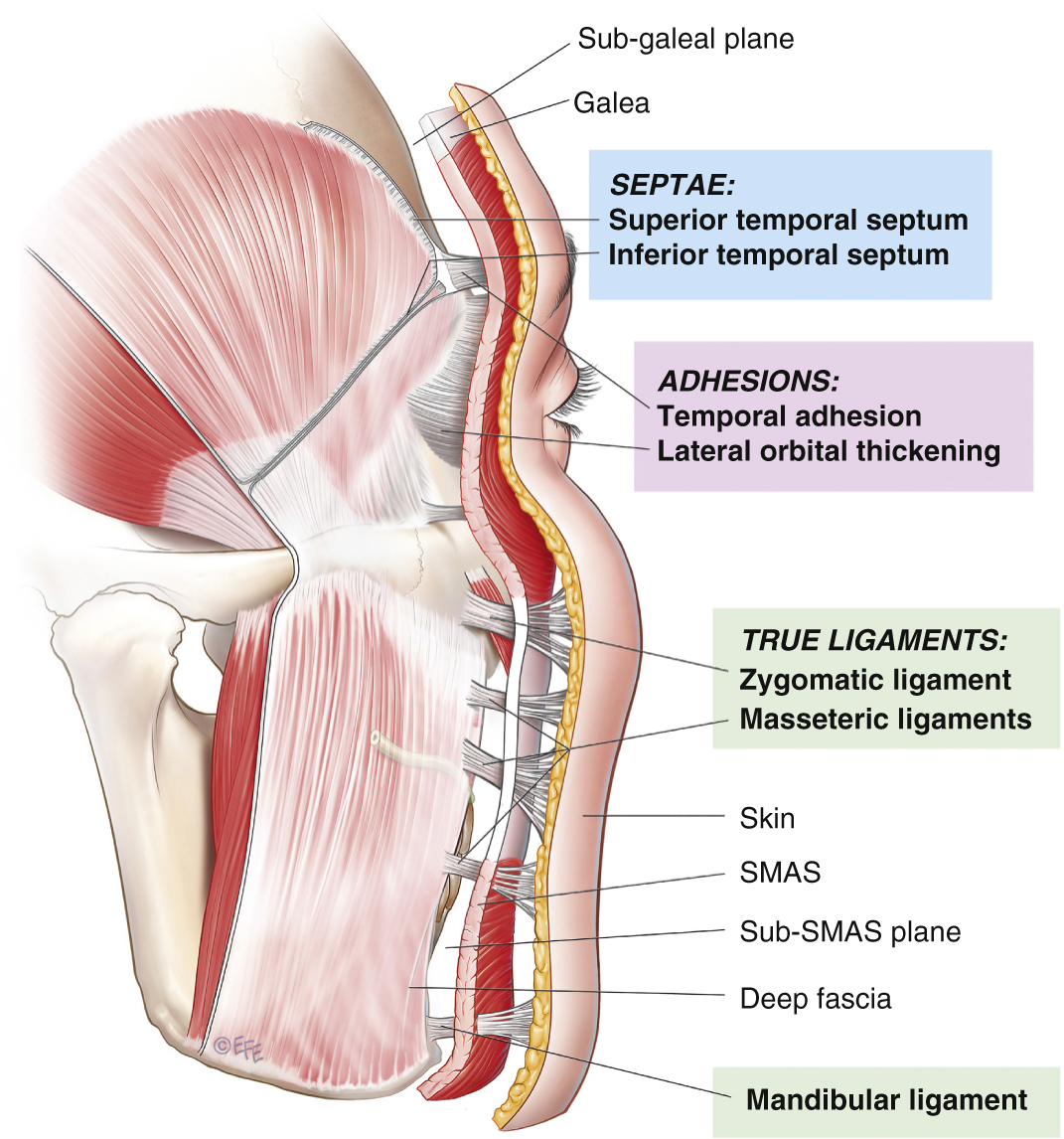
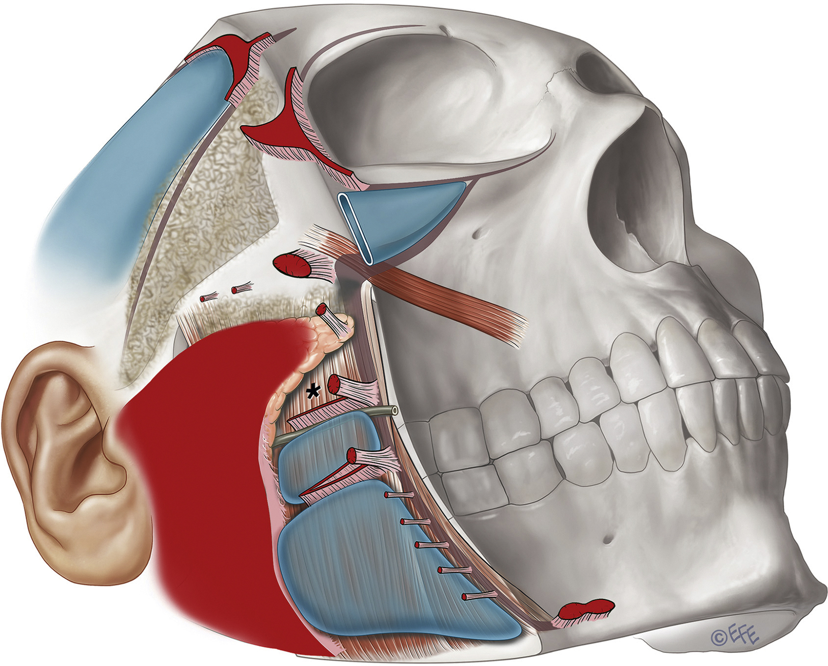
Layer 4 is the plane in which dissection is performed in sub-SMAS facelifts. It is an area of significant complexity and contains the following structures: (1) soft tissue spaces, (2) retaining ligaments, (3) deep part of the intrinsic muscles where passing from their bone attachment to their more superficial soft tissue origin, and (4) facial nerve branches, transitioning from deep to superficial. Functionally, a series of soft tissue spaces exist in layer 4 to allow independent movement of the periorbital and perioral muscle of facial expressions (located in the roof of the facial soft tissue spaces) over the deep fascia on the major muscles for mastication directly beneath (located beneath the floor of the facial soft tissue spaces). , The retaining ligaments of the face are strategically placed within the boundaries between the soft tissue spaces ( Fig. 62.5 ). In the lateral face, immediately in front of the ear, extending 25–30 mm forward of the ear cartilage to the posterior border of the platysma, is a diffuse area of ligamentous attachment, described by Furnas as the platysma auricular fascia (PAF). As no facial expression occurs here, the dermis, subcutaneous tissue, SMAS and the underlying parotid capsule (layers 1–5) are bound together as an area of diffuse retaining ligament. Layer 4 here is reduced to a fused layer, leaving it without a soft tissue space. There is no muscle in layer 3 here for mobility of this area of the superficial fascia. In contrast, in the anterior face where there is considerable movement over and around the bony cavities, the ligaments are significantly compacted and arranged around the edges of the bony cavities. These boundaries provide the last position where there is underlying deep fascia for the mobile shutters of the eyelids and lips to be supported. Importantly for the surgeon, the retaining ligaments also act as transition points for the facial nerve branches to pass from deep to superficial on their way to innervate their target muscles.
The soft tissue spaces of the face are in two forms: (1) those overlying bony cavities, such as the preseptal space of the eyelid over the orbit and the vestibule of the oral cavity under the lips and the lower nasolabial segment of the cheek and (2) those overlying bone, where the spaces allow the overlying superficial fascia to glide freely over the deeper layer 5.
Layer 5
The deep fascia, the deepest soft tissue layer of the face is formed by the periosteum where it overlies bone. Over the lateral face, where the muscles of mastication (temporalis and masseter) overlie the bone, the deep fascia is instead the fascial covering of the muscles, the deep temporal fascia and masseteric fascia, above and below the zygomatic arch respectively. The parotid fascia is also part of the deep fascia. The investing deep cervical fascia is the corresponding layer in the neck where it covers the supraomohyoid muscles and splits to form the submandibular space that contains the submandibular gland. The deep fascia, although thin, is tough and unyielding and gives attachment to the retaining ligaments of the face. In the mobile shutters over the bony cavities, the lids and lips, deep fascia is absent, being replaced by a mobile lining derived from the cavities, that of the conjunctiva or oral mucosa. The recently described “deep fat compartments” is fat located in layer 5, having more generally been called preperiosteal fat.
Facial Spaces
A large part of the sub-SMAS layer 4 consists of soft tissue “spaces.” These spaces have defined boundaries that are strategically reinforced by retaining ligaments. Significantly, from a surgical perspective, these areas are by definition anatomically “safe spaces” to dissect, as no structures cross within and all branches of the facial nerve are outside these spaces. As the roof of each space is the least supported part, laxity is more prone to develop here with aging, compared to their ligament-reinforced boundaries. This differential laxity accounts for much of the characteristic changes that occur with aging. Once a space has been surgically defined to its boundaries, the retaining ligaments in the boundary can then be precisely released under direct vision to achieve the desired mobilization while preserving the vital structures closely associated with the ligaments. A brief description of surgically significant facial soft tissue spaces is given below.
1
Upper Temporal Space
Overlying the temple are two compartments that are separated by the obliquely oriented inferior temporal septum ( Fig. 62.6 ). The upper compartment is a true space and the lower compartment is not a space, but an area containing important anatomy. Both compartments are interposed between the superficial temporal fascia (temporoparietal fascia) and the deep temporal fascia (temporalis muscle fascia). Anatomically, the upper compartment is an extension of the forehead anatomy down into the upper temple and the lower compartment is an upward extension of the upper cheek anatomy into the lower temple.
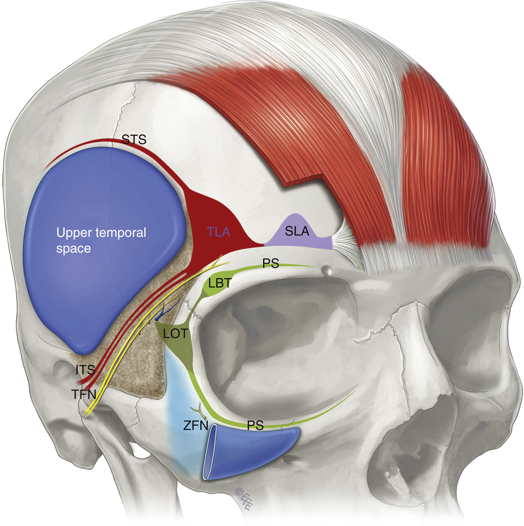
The upper temporal space is separated from the forehead by the superior temporal septum (STS) along the superior temporal line. Anteroinferiorly, the upper space is separated from the lower triangular temporal compartment, by the inferior temporal septum (ITS). These two septae merge at the triangular-shaped zone of adhesion called the temporal ligament. The upper temporal space provides safe surgical access to the lateral brow and upper midcheek. The space can be readily opened by blunt dissection to its boundaries. Once identified, the boundaries are then released by precise dissection. The superior temporal septum can be released sharply, taking care only to preserve the lateral (deep) branch of the supraorbital nerve, which runs parallel to the septum about 0.5 cm medial to it. The inferior temporal septum provides a marker to the important anatomy here as the temporal branches of the facial nerve are located parallel to and immediately inferior to this septum. To release the inferior temporal septum, the ceiling of the space is gently lifted off the deep temporal fascia floor, which three dimensionalises the septum in preparation for gentle release at the level of the floor, bearing in mind the frontal branches are located on the underside of the temporoparietal fascia in the ceiling, under the roof of the lower temporal area. Once released, the sentinel vein comes into view. The sentinel vein is not a good landmark for locating the temporal branches as they course cephalad to the vein, between it and the inferior temporal septum, where they travel in a layer of fat suspended on the underside of the temporoparietal fascia.
2
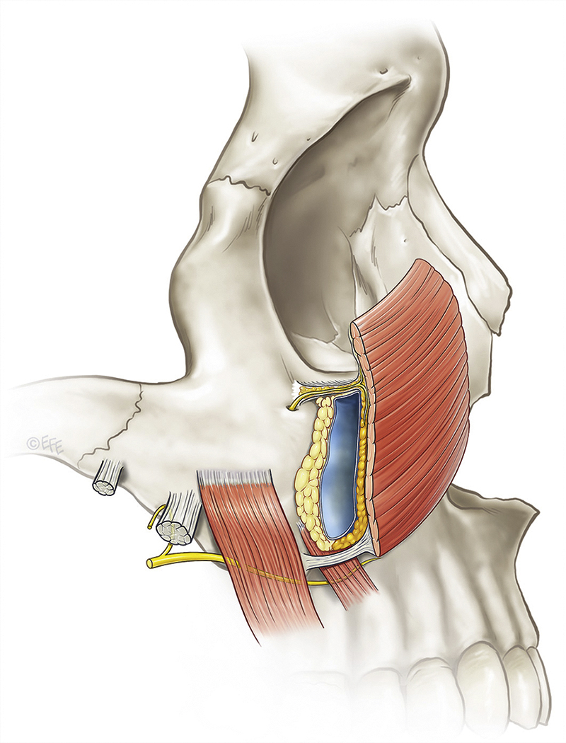
Prezygomatic Space
This triangular-shaped space overlies the body of the zygoma, its floor covering the origins of the zygomatic muscles. , The space allows independent movement of the obicularis oculi (pars orbitale) in its roof from the zygomatic muscles under the floor. Contraction of the overlying orbicularis elevates the prezygomatic soft tissues, which results in zygomatic smile lines (below the crow’s feet) ( Fig. 62.7 ). With aging laxity the roof of the space rests at a lower level. As a result, there is now a greater amplitude of movement on orbicularis contraction, which exaggerates the zygomatic lines with aging. This aging of the prezygomatic space, with bulging over its roof accentuated by its well-supported boundaries, is the anatomical basis for the clinical entity variously described as malar mounds, bags or malar crescent. This deformity indicates the presence of significant laxity and the treatment is directed to tightening the laxity of the roof.