Extremity burn reconstruction

19 Extremity burn reconstruction


Introduction


The upper and lower extremities are often involved in burn-related accidents. In particular, the arms and legs are the most exposed areas, as they constitute the first means of self-protection and are used to aid escape in accidents.


The majority of burn-related accidents involving the extremities, occur in the workplace and are mainly due to fire and electricity. In a domestic environment the majority of victims are babies, who suffer burns owing to contact with hot liquids.1,2


Extremity burns can be considered “difficult” burns, especially if they are extensive circumferential burns. Deep second- and third-degree burns, if not properly treated, can result in severe compartment syndromes, which can cause the loss of the extremity involved if not treated in a well-equipped burn-specialized facility.3,4


Knee and elbow joints are often prone to severe damage related to electrocution. The electric arc that is formed in this type of accident often finds its entry or exit point in one of these joints, causing the joint to explode.


Scarring often represents real challenges for the plastic surgeon. Some 50% of Z-plasties, as well as other surgical techniques such as scar debridement, involve the axillary cavity. In developing countries, the percentage of scar retraction causing disabilities is significant. This results in almost grotesque thoracic-brachial and brachio-antebrachial clinical picture and presents leg adhesions that are incompatible with the physiology of the structures involved.


While the patient is hospitalized, physio-kinesitherapy is specifically aimed at maintaining mobility of the major joints. Even if they may be only marginally involved in the accident, these parts are in fact often subject to arthritis, dysplasia, and joint calcification, which are not easy to treat.


These preliminary observations serve to point out that reconstruction of the extremities, which are a person’s main source of self-sufficiency, is not simply a matter of aesthetics but represents a real challenge for both the surgeon and the patient if functionality is to be restored. For the sake of clarity, the following analysis will consider the upper and the lower extremities separately.


History


The most important progress in limb burn wound therapy occurred during the Second World War, but many concepts such as early excision and grafting of small burns can be traced to Lustgarten in 1891. The major issues addressed during the Second World War era included early excision, skin grafting, splinting, and scheduled mobilization. The work of Allen and Koch in 1942 focused on the concept of closed dressings instead of the exposure technique in the care of burn patients.5 In the same period, many authors (e.g., McCorkle and Silvani) described for the first time the importance of early excision and grafting in order to prevent bacterial infections and to minimize hypertrophic scar sequelae in burns involving the upper limb and hands.6


Until the early 1960s, it was customary to maintain the eschar until demarcation, waiting for granulation to occur, and then perform skin grafting of the burn. Excisions failed because primary grafting was not performed immediately in same session.


Tangential excision preserving most of the viable tissue, with immediate skin grafting, was suggested initially by Janzekovic and colleagues in the early 1970s. They recommended performing this procedure on the 2nd to 5th day post-burn as the optimal time for best results, with less scarring and good preservation of function.7


There was no official protocol until 1987, when during an international Round Table discussion in Geneva, seven burn surgeons from around the world agreed that deep dermal and full-thickness burns of the hand and upper limb did better when excised and grafted early, with better functional results, as a result of earlier rehabilitation, and better aesthetic results because of the reduced development of thick hypertrophic scars. Their final results showed as much as a 50% decrease in hospitalization. We thus officially entered into cost-effectiveness considerations.


Early tangential excision of deep burns, followed by skin grafting, is probably the most significant breakthrough in recent years, leading to a reduction in the mortality rate in burns >40% TBSA.8,9


Basic science/disease process


Electrical burns


In cases of electrocution, the upper extremities are almost always involved, especially as entry or exit points of what is known as the electric arc.10


The electric arc mainly involves the wrist, elbow, and ankle joints with devastating effects on the joints themselves, the nervous structures, and the muscular masses.


Often the most serious damage is to the wrist and ankle. Since the hands and feet are the most frequent entry and exit points, the wrist and ankle are the nearest points of electrical resistance. The patient’s clinical condition on admission often looks less critical than it really is as the lesions may be misleading, with limited charring. In other cases, ample charred areas can be witnessed. These are the electric current’s entry and exit points. However, the underlying damage is always more extensive and may well involve the muscular fasciae. This is because nerves and vessels are very good conductors of electricity, and they may suffer damage a few days after the accident. Venous thrombosis or ischemia can be caused by internal destruction of the vessels catalyzed by the electric current. Peripheral neurological damage may occur either with total destruction of the nerve (neurotmesis), if close to the electricity entry and exit points, or with partial axonal damage (axonotmesis), which is generally resolved within a few months.11,12


In cases of high-voltage electrical burns (over 1000 volts), muscles, bones, and nerve structures can be totally destroyed and, as has been said, complete destruction of the major joints is a common observation.13


In such cases, reconstruction should be early, i.e., within 3–5 days of the accident, in order to save the affected joints and to remove destroyed muscular tissue as soon as possible. This is to prevent any serious acidosis related to massive quantities of myoglobin, which is a difficult condition to manage if a resuscitation procedure proves necessary.


Myoglobinuria is closely linked to muscular damage, since the destruction of muscle cell causes the release of myoglobin and in turn catalyses myoglobinemia. The first sign of this pathology is the dark pink color of the urine.14,15


However, it is important to note that an accurate picture of myoglobinuria can be provided only by serum differentiation between hemoglobin and myoglobin.


All patients with myoglobinuria should be treated with mannitol IV, bicarbonate IV, and Ringer’s lactate therapy in order to obtain alkaline osmotic diuresis. This can maximize the reduction of the precipitation of the pigment at the level of the renal tubules.


The damage caused by an electric current passing through the body, especially if of high voltage, depends not only on the characteristics of the electricity itself but also on the path it follows and on the characteristics of the tissues affected by the electricity. Even if adequate medical and surgical therapy is available, the risk of amputation of the extremity remains high.11


From a surgical point of view, debridement of necrotic tissue plays a fundamental role in the very first phase after the accident. As said, this can be very extensive compared to the skin lesion. Reconstruction in these cases rarely requires only skin graft coverage. It often also necessitates vascular reconstruction of the ulnar, radial, or tibial systems, nerve grafts, and the transposition of microvascular free flaps.


Vascular and neural reconstruction has to be carefully assessed, since electricity-related damage affecting the neurovascular structures can occur even some days after the surgical operation. This is due to irreversible endothelial damage, which affects small terminal vessels in particular. Hence it is preferable in such cases to postpone the more complex reconstruction procedures until secondary surgical procedures are performed.16 During the acute phase, it is therefore advisable to proceed by performing simple temporary skin coverage with a free skin graft and isolating and preparing the vascular and nervous structures that will be treated in a secondary surgical stage.17


Diagnosis/patient presentation


Deep burns in the entire upper extremities are statistically infrequent. The areas most often affected by burns are the hand and the forearm. The involvement of any part of the upper sleeve in any of its parts causes negative consequences below the burn, even though the burn itself does not affect that area.


Patients affected by upper extremity burns often suffer other associated traumas as a result of work-related accidents, which are the most frequent cause of this type of injury. An assessment of potential fractures, amputations, wounds, and blast and crush injuries is essential for planning subsequent reconstruction procedures. The functionality of the shoulder joints must be carefully examined.18 If possible, the nervous functionality of the median, ulnar, and radial nerves should also be evaluated.18


After this first check, and after calculating the extent of the burn surface area, it is of fundamental importance to assess whether there is any need of escharotomy. This surgical procedure, which has to be performed as an emergency, helps to prevent the compartment syndrome.


Fluid infusion and edema


Burn shock is characterized by specific hemodynamic modifications on both a hypovolemic and a cellular basis, and it can be defined as a nonhemorrhagic hypovolemic shock with loss of water, sodium, and plasma proteins but not of corpusculate elements. In the first 24–48 h fluid losses cannot be stopped but only replaced.


The objective of the treatment is to restore and preserve tissue perfusion and avoid ischemia. One of the biggest complications of burn shock is the increase of capillary permeability throughout the organism due to the effects of heat on the microcirculation. Edema reaches its maximum expression within 8–12 h of the accident in minor burns and within 12–24 h in major ones.


These phenomena are caused by endothelial damage and by hypoperfusion, which lead to the liberation of vasoactive substances and cytotoxic free radicals, which in turn are responsible for cellular edema and the generalized inflammatory reaction.12,14


When burns exceed 20–30% TBSA, even unburned areas develop edema owing to the increase of vascular permeability secondary to hypoperfusion and because of the plasma protein deficit.


The “emergency solution” of first choice for infusion in the first 8 h is Ringer’s lactate, in patients with extensive burns in whom it is not possible to achieve adequate tissue perfusion, even by doubling the infusion volume normally prescribed, it is possible to use hypertonic solutions (Ringer’s lactate + 50 mEq of NaHCO3 + 40 mEq Lactate). These solutions have to be used carefully, monitoring natremia (which must never exceed 160 mEq/dL) and plasma osmolarity. Note: hyperosmolar syndrome induces kidney failure.


The administration of proteins is not effective in the first 8 h post-burn; later on, it is possible to infuse solutions of albumin or fresh plasma. The quantity of proteins to infuse has to be defined: generally speaking, one can calculate 0.5–1 mL/kg/% TBSA in fresh plasma in the first 24 h, starting 8/10 h post-burn. Elderly patients and those with extensive burns (>50%) maintain better hemodynamic stability and develop less edema, thanks to the administration of proteins.


If no colloids have been used in the first hours after the trauma and oncotic pressure remains low because of the depletion of plasma proteins, it is necessary to reintegrate the losses: the 5% albumin demand during the second 24 h is 0.3–0.5 mL/kg/% TBSA. It is desirable to keep albumin blood levels above 2 g/dL to contrast peripherica edema.


The volume of fluids indicated in the resuscitation of burn patients depends on the gravity of the burn, the age and general condition of the patient, and the presence of associated lesions. In patients with burns in more than 15% TBSA, the quantity of fluid to infuse has to be calculated on the basis of the burned area and body dimensions (ABA Guidelines).3


Compartment syndrome


Deep second-degree and third-degree circumferential burns can trigger a serious compartment syndrome (Figs 19.119.6, Box 19.1), which if not adequately treated can easily result in the loss of the extremity involved.


Full circumferential burns, or burns involving at least three-quarters of the extremity’s circumference, are liable to the risk of the compartment syndrome. The clinical reaction that occurs when edema develops during the first hours post-burn triggers a pressure increase at the muscular level of the extremities. The extremities are blocked by the burned skin, which has no elasticity. After the initial restriction of venous outflow, if proper treatment is not given, there is the risk of a deficit in arterial flow, resulting in ischemic tissue downstream. It is important to perform invasive monitoring of compartment pressure. There are many external devices for measuring intramuscular pressure.


Main clinical signs of initial compartmental syndrome are failure of pulses in distal arteries, pallor and cold limb, initial intense pain which then decreases, high tension of the hand or the foot, pain at passive flexion-extension, paresthesia.


Screening with eco-color-Doppler could be useful during initial assessment, but this method cannot valuate the exact compartment pressure. If laboratory values give high levels of creatinine phosphokinase (CPK), it may indicate severe muscle damage, or ischemia.


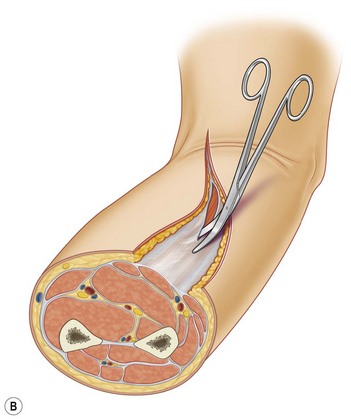
One of the most common devices which allow a precise measurement of compartment pressure is the Stryker Set®. This Device is assembled by a monitoring unit and a syringe with 3 mL of saline solution. After an accurate identification of the muscular compartment we want to check, we proceed with a simple injection of the 0.3 mL to re-equilibrate the pressure with interstitial fluids, and then we get the compartment pressure on the monitoring unit. For measurements exceeding 35–40 mmHg, it is necessary to adopt escharotomy-fasciotomy. In all these cases, it is necessary to perform full-thickness decompressive escharotomy in the very first hours in order to restore normal blood flow.19,20


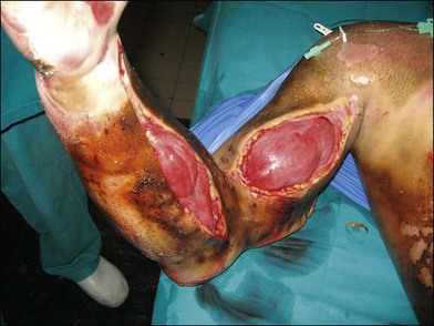
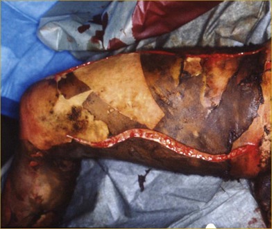
This procedure must involve all the burned tissues, skin, fat, and fasciae, until release of the compartment is observed. These escharotomies should follow the recommended procedures and be performed before the initial signs of either vascular deficit (frozen hand) or neurological deficits occur. Escharotomies can be performed with an electrosurgical knife or a scalpel, ensuring complete release of the muscles (Fig. 19.4B, 19.5).


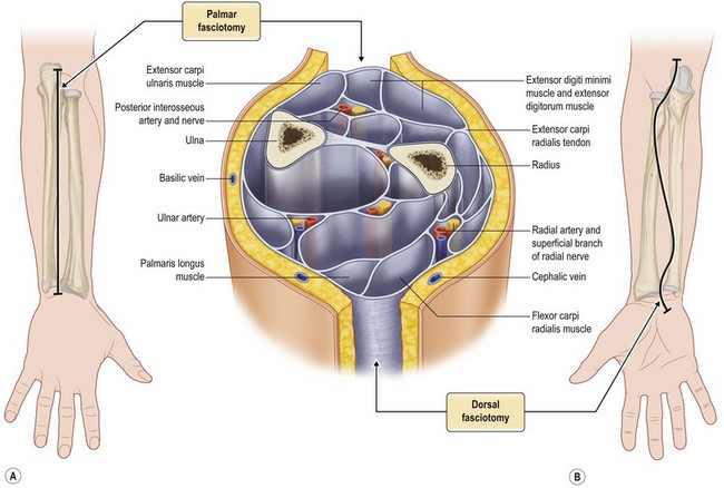
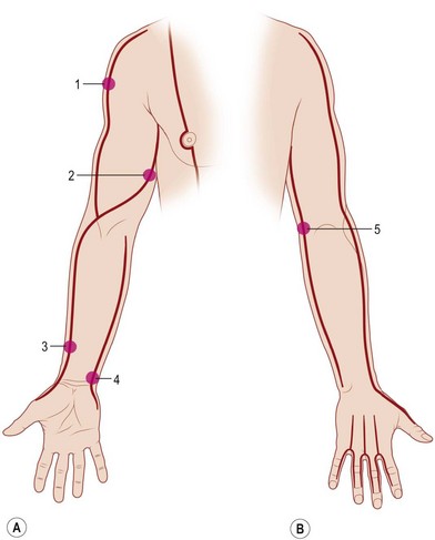
When treating the upper extremity, particular attention should be paid to the blood flow through the cephalic and basilic vein and superficial branch of the radial nerve at the level of the wrist. In the most serious cases, it may be necessary to perform complete decompression of the carpal tunnel (Figs 19.1, 19.2).


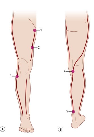
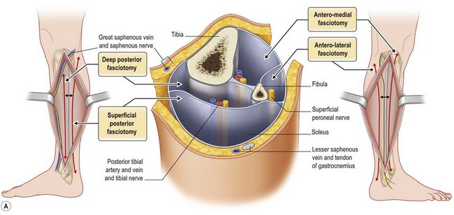
Escharotomy of the lower limb must be performed avoiding tibial anterior nerve, the medial saphenous vein and nerve, the tibial posterior nerve, the fibular nerve, and the external saphenous nerve (Figs 19.3, 19.4, 19.6).21


Medication of the extremities should be carried out after careful cleaning and debridement of the affected surface. Early mobilization of the joints is recommended unless otherwise specified. Nonadherent dressings such as Vaseline gauze are indicated for all burn-affected areas. For areas affected only superficially or at medium thickness, it is better to use medications such as Biobrane®, Mefix®, or Aquacel-Ag® to ensure good epithelialization. In areas that are deeply affected or clearly of third-degree, it is possible to proceed with occlusive dressings until the time comes for surgical procedures. Enzymatic debridement ointments or “debriders” can be useful for preparing the patient for the scheduled surgical operation. During hospitalization it is of fundamental importance to keep the upper extremities of the patient in an elevated position in order to control the formation of new edema and reduce existing edema.


Surgery of the burn areas should be performed early, since late recovery leads to a greater incidence of hypertrophic scars, weaker tissues, and delays in prompt mobilization of the extremities.22,23


Deep burned areas should be subjected to surgical treatment as soon as possible, while more superficial areas, if not healed within in 2 or 3 weeks, should also be considered for surgical treatment. Depending on the burn area, the extremities permit local anesthesia, but preferably only in patients with surgical indications but relatively limited burn areas. The preferred treatment remains debridement and autologous skin graft.


Related trauma


The trauma most frequently associated with burns is bone fracture. In this case, close collaboration between the plastic and the orthopedic surgeon is essential in order to choose the most appropriate surgical plan to follow. Stabilization of fractures contiguous to deeply burned areas should be performed early, preferably within the first 48 h after the accident, before bacterial colonization can hamper orthopedic action. It has frequently been reported in the literature that the early stabilization of burn patients drastically decreases the onset of complications related to the orthopedic intervention, thus reducing the risk of infective complications.


Various related trauma should be treated contextually or before their coverage with skin grafts, in order to avoid re-opening already treated burn areas.


In the deepest burn areas, especially as a result of electrocution or if accompanied by related trauma, the explosion of bones or tendons can occur, especially in the elbow, wrist, knee, and ankle areas. This condition imposes an additional priority in the planning of the surgical procedure to be performed. The exposed tendons are liable to rapid dehydration and consequent necrosis, and prompt coverage is therefore essential. In the preparatory phase it can be useful to use cadaver skin or biological medications, while in the surgical phase it is recommended to use dermis substitutes (such as Integra, Matriderm, etc.), which permit the formation of new tissue between the tendons and the grafted skin. Grafting directly on the exposed tendons is counter-indicated in terms of graft survival and of functional results.


The same considerations apply in the case of exposed bone, which should also be covered as early as possible, even though resistance to exposure is much higher and more easily resolved than in the case of tendons. Coverage can be performed at a second stage utilizing rotation or free flaps.24


Patient selection


Plastic surgeons possess all the necessary tools and scientific know-how to treat burn patients. From intensive care to the surgical phase, they should be able to take the best decisions for the patient. Plastic surgeon is in fact the only professional figures that have a clear and immediate understanding of patients’ overall surgical needs.


It is the plastic surgeon who decides the necessity of surgical procedures and in some cases, also decides to pursue a second-intention healing process. The preoperative care plan for scars is also crucial and should be prepared by the same medical team that treated the patient during the previous acute phase. The extremities are the most frequent sufferers from post-surgery scar contractures, often as a result of a lack of proper preventive follow-up measures (such as massage therapy, elastic compression, physio-kinesitherapy, and splinting) aimed at preventing pathological scarring. An assessment of the affected areas will suggest to the specialist whether to proceed with corrective measures or engage in additional surgical interventions.


The results of sequelae surgery can be extremely heterogeneous, ranging from scar contraction resolution at the joint level to vascular, nerve, and tendon reconstruction. The main objective of sequelae surgery in these localized areas is to re-establish the extremity’s proper functionality, considering that normal walking, physical autonomy, and self-dependence at work depend on the complete rehabilitation of the areas affected.25


Treatment/surgical technique


Burn surgery can be subdivided into acute-phase surgery and sequelae surgery. This distinction holds also as regards localized areas only.


Acute-phase extremity surgery is not fundamentally different from surgery in any other part of the body. Procedures such as fasciotomy, debridement, and coverage with autologous skin grafts, as previously described, follow the surgical guidelines of acute-phase general burns.


With regard to sequelae surgery, the extremities have some unique characteristics, e.g., the majority of scar retractions lead to disabilities, and localized burn-related joint blockages may occur.12


Acute-phase surgery: skin grafting


image Video1


Emergency procedures such as escharotomy and fasciotomy were largely dealt with in the foregoing section. In this section, we provide a preventative scheme concerning the incision lines to be used.


Focusing only on the extremities, the commonest surgical procedures continue to be surgical debridement and whole or meshed free-skin graft of autologous skin. These procedures should preferably be completed early, i.e., between the 5th and 7th day after the accident. As previously said, some ad hoc surgical variations may be necessary, depending on the type of damage affecting the extremities and the kind of structures involved. In order to limit bleeding and thus operate in a condition of controlled hemostasis, it is advisable to use a tourniquet. This procedure facilitates extremity surgery but at the same time, requires that greater attention should be paid to the assessment of vital tissue. In this way there is no microvascular bleeding.


Debridement should be performed up to the vital tissues, after careful removal of all necrotic skin, paying special attention to the flexion-extension areas and the axillary cavity areas. In these areas the necrotomy is more complex to perform. In deep or electric burns, it is advisable to reach up to the muscular fascia in order to avoid the risk of probable but undesirable liponecrosis. Unlike skin, the viable fat underneath is an unstable base for implanting free skin grafts.2629


During debridement of more superficially burned areas that do however require surgical treatment, instruments like VersaJet® provide useful support as they minimize the waste of vital tissue and also permit careful debridement in nonflat surfaces, as the extremities are.27


Skin grafts can adhere with mesher 1 : 1 or 1.5 : 1, and it is even possible to use 2 : 1 if the patient does not have donor areas. The elbow joint and the axillary and popliteal cavities are preferably grafted with medium-thickness healthy skin, which provides greater elasticity and resistance in areas subject to high mobility and traumatism. All skin grafts should be positioned considering the least relaxed skin tension lines in order to obtain the aesthetically best results. In the specific case of the arms, it is recommended to position the skin grafts longitudinally for the best aesthetic outcome.


Skin grafts must be checked after 4–5 days and treated until full recovery is completed.


Skin substitutes


image Video1


In the course of the last decade, the use of dermal substitutes in acute-phase burn surgery became more frequent. In particular, this technique is now used in acute-phase burn surgery in cases of extensive deep burns with exposure of bones and tendons.


The characteristics of an ideal skin substitute were defined in 1978 by Tavis, who stated that it should be adherent and impermeable to water, represent a barrier to electrolytes and proteins, be durable, and possess a bacteria barrier function. It should also show nontoxicity, nonantigenity, a hemostatic function, ease of application, and affordability. None of today’s skin substitutes fulfils all these criteria.


The currently available products (Integra®, Matriderm®, Allograft®, Terudermis®, etc.) have different characteristics but they all aim at the same objective: re-establishment of a layer of dermis under a free skin graft. There is an ongoing debate as to whether these newly formed tissues can be considered dermis or not.


A fundamental prerequisite for valid results is that the application should be onto vital tissue that is free of any contamination or necrotic residue.


In acute burns of the extremities the use of skin substitutes is extremely effective in the treatment of critical areas (armpit, elbow, popliteal cavity) and exposed bones and tendons.


These products provide immediate coverage of the burned area, resulting in reduced risk of infection or necrosis of the tissue. They also facilitate the regeneration of tissues between the deep structures and the skin graft.


Applying dermal substitutes is usually easy. However, when treating the extremities, some critical aspects should be considered, such as ensuring movement, the autonomy of the various segments, and perfect adherence between the product and the wound bed, since these products are mostly indicated for the treatment of critical points such as the elbow, armpit, and popliteal cavity. It has become increasingly common to use such products together with “negative pressure systems” during the post-surgical period. It is important to recall, however, that the commonest possible complication following their use in the acute phase is infection, which can lead to complete loss of the skin grafts applied in 30–40% of cases.


In the post-surgical period it is recommended that the surgeon should be guided by the instructions of the manufacturer of the product and by personal experience.2830

Only gold members can continue reading. Log In or Register to continue

Stay updated, free articles. Join our Telegram channel

Feb 21, 2016 | Posted by in General Surgery | Comments Off on Extremity burn reconstruction

Full access? Get Clinical Tree

Get Clinical Tree app for offline access