Excision of Soft Tissue Tumors of the Foot and Ankle
Raffi S. Avedian
Robert J. Steffner
Subhro K. Sen
DEFINITION
Soft tissue tumors of the ankle may be benign or malignant (soft tissue sarcoma) and exhibit a variable natural history ranging from latency to rapid growth.
Tumors located entirely above the muscle, fascia, or tendon are considered superficial, whereas tumors involving the fascia or located deep to it are considered deep.
ANATOMY
The ankle is a synovial joint involving the tibia, talus, and fibula. Tendons of the leg muscles travel around the ankle in discrete groups each contained in a retinaculum (FIG 1).
The anterior tibial artery and deep peroneal nerve travel anterior to the ankle joint just lateral to the anterior tibialis tendon.
The posterior tibial artery and tibial nerve travel along the medial aspect of the lower leg, in the fascial plane between the deep and superficial compartments. At the ankle, they course posterior to the posterior tibial tendon along the medial malleolus.
The tendons of the peroneus muscles travel along the posterior aspect of the lateral malleolus to their attachments in the foot.
PATHOGENESIS
The mechanism for soft tissue tumor formation is not known.
Risk factors for sarcoma development include radiation exposure, radiotherapy, pesticide exposure, and hereditary conditions including Li-Fraumeni syndrome and retinoblastoma gene mutation.
NATURAL HISTORY
All soft tissue sarcomas have the potential for local recurrence and metastasis.
Soft tissue sarcomas exhibit a spectrum of natural history from slow-growing low-grade tumors with low risk of metastasis to high-grade sarcoma that may grow rapidly and pose a high risk of metastasis.
Lungs are the most common site of metastasis. Lymph node involvement is rare.
Angiosarcoma, clear cell sarcoma, epithelioid sarcoma, rhabdomyosarcoma, myxofibrosarcoma, and synovial sarcoma are associated with increased risk of lymph node spread compared to other sarcomas.
Benign tumors by definition do not have metastatic potential but can grow to large sizes and cause symptoms.
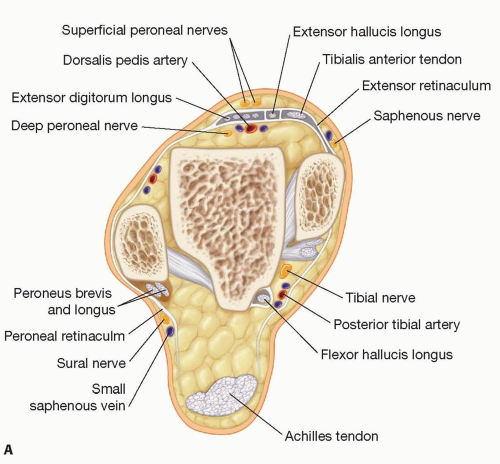 FIG 1 • A. Cross-sectional anatomy of the ankle. Notice that the tendons are contained within a retinacular layer. |
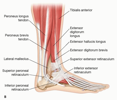 FIG 1 (Continued) • B. Oblique view of the ankle anatomy. |
PATIENT HISTORY AND PHYSICAL FINDINGS
Conducting a thorough history and examination is important to assess duration of symptoms, comorbidities, physical dysfunction, organ involvement, overall health, and patient expectations in order to best tailor treatment strategy for the individual patient.
Many sarcomas may be asymptomatic with the only patient complaint being the presence of a mass.
Neurovascular examination is mandatory for any extremity tumor.
IMAGING
Magnetic resonance imaging is the principal imaging modality used to characterize tumors, formulate differential diagnosis, define local tissue infiltration, and devise a surgical plan.
Plain radiographs are used if there is concern for bone involvement or to demonstrate mineralization within a tumor such as vascular malformations.
Staging for soft tissue sarcomas consists primarily of lung imaging.
DIFFERENTIAL DIAGNOSIS
The differential diagnosis for a soft tissue tumor includes benign tumors, sarcomas, lymphoma, infection, and inflammatory lesions (eg, rheumatoid nodules).
There are over 50 sarcoma subtypes. Common histologies include pleomorphic undifferentiated sarcoma, synovial sarcoma, leiomyosarcoma, malignant peripheral nerve sheath tumor, and liposarcoma.
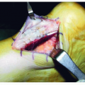
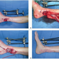
SURGICAL MANAGEMENT
Related posts:
Stay updated, free articles. Join our Telegram channel

Full access? Get Clinical Tree


