5Evidence for Efficacy of Migraine Surgery
Bahman Guyuron
Salient Points
• The first research on current migraine surgery was prompted by two patients who reported their migraine headaches (MHs) ceased after a forehead rejuvenation.
• A retrospective study on 314 patients who had undergone forehead rejuvenation demonstrated improvement or elimination of MH in 31 of 39 patients with MH.
• Twenty-one of the 22 patients in a prospective pilot study demonstrated either complete elimination (11 patients) or significant improvement (10 patients) of MH.
• Anatomical study on 20 fresh cadavers demonstrated passage of the greater occipital nerve through the muscle in all and emergence of the nerve from the muscle at a point about 3 cm caudal to the occipital tuberosity and 1.5 cm lateral to the midline.
• A study on 20 patients concluded that the main zygomaticotemporal branch of the trigeminal nerve emerges from the deep temporal fascia on average 17 mm lateral and 6 mm cephalad to the lateral palpebral commissure.
• In a level II randomized study, 82 of the 89 patients (92%) in the treatment group demonstrated at least 50% reduction in MH frequency, duration, or intensity compared to baseline; 31 (35%) reported elimination and 51 (57%) experienced improvement over a mean follow-up period of 396 days.
• In a level II 5-year follow-up study, 61 of 69 (88%) patients continued to have a positive response to the surgery (p < 0.0001). Twenty (29%) reported complete elimination of MH and 41 (59%) noticed a significant decrease in frequency, severity, and duration of MH (p < 0.0001).
• In a level I randomized study with sham surgery control, 41 of the 49 (83%) patients benefitted from the real surgery compared to 15 of 26 (56%) patients in the sham surgery group (p = 0.014). While 28 (57%) patients in the real surgery group observed complete elimination of MH, only 1 patient in the sham surgery group reported elimination (p < 0.0001).
• In a level III study, two independent departments at Case Western Reserve University (Center for Proteomics and Bioinformatics and Department of Neurosciences) compared the nerves of the patients with MH to those without and concluded that the nerves of patients with MH had myelin deficit.
• Through a level III study, it was demonstrated that the factors associated with migraine surgery failure include increased intraoperative bleeding and surgery on fewer trigger sites.
• In a level III study, it was discovered that the factors associated with migraine surgery success include older age of migraine onset, presence of visual symptoms, surgery at site I or II, and deactivation of all four common MH sites.
• A level III study demonstrated that there was no statistically significant difference between the injection of botulinum toxin type A and the use of a constellation of symptoms to identify the migraine trigger sites.
• A level III study demonstrated superior outcomes with supraorbital foraminotomy when indicated.
• A level III cost analysis study demonstrated total median cost reduction in migraine care of $3,949.70 per year postoperatively. The mean surgical cost was $8,378.
• Through a level III study, it was discovered that endoscopic nerve decompression was more successful in reducing or eliminating frontal MH than transpalpebral nerve decompression.
• -Another level III study verified that a positive botulinum toxin type A response is a significant predictor of migraine surgery success.
• A level III comparative study between those who were on narcotic analgesics and those who avoided these products demonstrated that the group that consumed narcotics had significantly lower rates of improvement from the surgery in all migraine indices.
• In a level III comparative study of surgery outcome on patients with episodic, chronic, and daily MHs, it was discovered that all cohorts, regardless of their frequency of MHs, achieved significant improvement in frequency, duration, severity, and MH index.
• We demonstrated that Doppler signal in the site of most intense pain can predictably confirm the presence of an artery in the most painful site and lead to precise location of the nerve irritation by a vessel and thus a successful removal of the offending artery.
• A level II prospective randomized study concluded that neurectomy and decompression of the zygomaticotemporal branch of the trigeminal nerve are both appropriate treatments for temporal MH.
• A level V retrospective review of an adolescent population has shown that migraine surgery may offer symptomatic improvement of MH frequency, duration, and severity in patients with identifiable anatomical trigger sites.
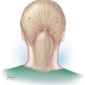
• In an anatomical study, the location of emergence of the lesser occipital nerve from the trapezius fascia was determined to be an area centered 65.4 ± 11.6 mm from midline and 53.3 ± 15.6 mm caudal to the line connecting the external auditory canals.
• The third occipital nerve was found 13.2 ± 5.3 mm from the midline and 62.0 ± 20.0 mm caudal to the line between the two external auditory canals.
• In another anatomical study, three patterns of interaction were found between the superficial temporal artery and auriculotemporal branch of the trigeminal: a single site where the artery is crossing over the nerve (62.5%), a helical intertwining relationship (18.8%), and nerve crossing over the artery (18.8%).
5.1 Introduction
While the initial reports from two patients indicating that their migraine headaches (MHs) had disappeared following cosmetic surgery were intriguing, one could not help being skeptical about the connection between cosmetic forehead procedures and migraine symptom disappearance. This onset rules out the possibility that the positive outcomes are related to a placebo effect, as claimed by the opponents of surgery, since neither the patients nor the surgeon was anticipating such a result.1–3 It would have been totally imprudent to ignore such a report by two independent patients. It was, therefore, sensible to design a study to investigate this connection. Thus, a retrospective study was conducted to determine whether there was an association between the removal of the corrugator supercilii muscle and the elimination or significant improvement of MH.4 Questionnaires were sent to 314 consecutive patients who had undergone corrugator supercilii muscle resection through endoscopic, transpalpebral, or open forehead rejuvenation procedures. The patients were queried as to whether they had a history of MH before the surgery and, if so, whether the headaches significantly improved or disappeared after surgery. If the answer was affirmative, then the patients were further questioned about the duration of the improvement or cessation of the headaches and the relationship to the timing of the surgery. After an initial evaluation of the completed questionnaires, a telephone interview was conducted to confirm the initial answers and to obtain further information necessary to ensure that the patients had a proper diagnosis of MH based on the International Headache Society criteria.
Of the 314 patients, 265 (84.4%) either responded to the questionnaire or were interviewed, or both. Of this group, 16 patients were excluded because of the provision of insufficient information to meet the International Headache Society criteria, the presence of organic problems, and other exclusions mandated by study design. Thirty-nine (15.7%) of the remaining 249 patients had MH that fulfilled the society criteria. Thirty-one of the 39 (79.5%) with preoperative migraine noted elimination or significant (>50%) improvement in MH immediately after surgery (p < 0.0001), and the benefits lasted over a mean follow-up period of 47 months. When the respondents with a positive history of MH were further divided, 16 patients (p < 0.0001; McNemar) noticed improvement over a mean follow-up period of 47 months, and 15 (p < 0.0001; McNemar) experienced total elimination of their MH over a mean follow-up period of 46.5 months. When divided by MH type, 29 patients (74%) had nonaura MH. Of these patients, the headaches disappeared in 11 patients, improved in 13 patients, and did not change in 5 patients (p < 0.0001). Ten patients experienced aura-type headaches, which disappeared or improved in 7 of the patients and did not change in 3 of the patients (p < 0.0001). This study suggested for the first time that there is indeed a strong correlation between forehead rejuvenation and potentially removal of the corrugator supercilii muscle and the elimination or significant improvement of MH. However, being a retrospective study, the results were open to criticism. This study offered additional compelling evidence against the placebo effect being the reason for the improvement or elimination of MH after surgery since these patients did not undergo surgery for treatment of MH nor were they aware of such a connection between their surgeries and MH.
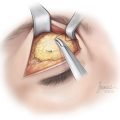
Intrigued by the findings from this study, we designed a prospective pilot study investigating the role of corrugator resection and removal of the zygomaticotemporal branch of the trigeminal nerve in MH on a small group of patients (n = 29 [24 women, and 5 men], with an average age of 44.9 years [range: 24–63 years]).5 The patients completed a comprehensive MH questionnaire. The research team neurologist evaluated the patients with moderate to severe MH to confirm the diagnosis using the criteria set forth by the International Headache Society. The team’s plastic surgeon injected 12.5 units of botulinum toxin A (BT-A) into each corrugator supercilii muscle group. The patients were asked to maintain an accurate log of their MH and to complete a monthly questionnaire documenting pertinent information related to their headaches. Patients who observed complete elimination of the MH following injection of BT-A underwent resection of the corrugator supercilii muscle group. Those who experienced only significant improvement and who had temple headaches underwent transection of the zygomaticotemporal branch of the trigeminal nerve with repositioning of the temple soft tissues, in addition to removal of the corrugator supercilii muscles. Once again, patients kept a detailed postoperative record of their headaches.
Twenty-four of 29 patients (82.8%; p < 0.001) reported a positive response to the injection of BT-A, 16 (55.2%; p < 0.001) observed complete elimination, 8 (27.6%; p < 0.04) experienced significant improvement (at least 50% reduction in intensity or severity), and 5 (17.2%) did not notice a significant change in their MH. Twenty-two of the 24 patients who had a favorable response to the injection of BT-A underwent surgery, and 21 (95.5%; p < 0.001) observed a postoperative improvement. Ten patients (45.5%; p < 0.01) reported elimination of MH and 11 patients (50.0%; p < 0.004) noted a considerable improvement. For the entire surgical group, the average intensity of the MH reduced from 8.9 to 4.1 on an analogue scale of 1 to 10, and the frequency of MH changed from an average of 5.2 per month to an average of 0.8 per month. For the group who only experienced an improvement, the intensity fell from 9.0 to 7.5 and the frequency was reduced from 5.6 to 1.0 per month. Only one patient (4.5%) did not notice any change. The follow-up ranged from 222 to 494 days, the average being 347 days.
This study confirmed the potential value of surgical treatment of MH since as many as 21 of 22 patients benefited significantly from the surgery. It was also evident that injection of BT-A is an extremely reliable predictor of surgical outcome. This study further confirmed our findings from the retrospective study. However, being a pilot study, it needed further validation since it did not include a control group. The study also raised the question as to why all of the patients did not respond to surgery if the surgery was indeed the answer. Furthermore, now that the connection was seemingly strong, it was time to start exploring the rationale behind the efficacy of the surgical treatment of MH.
It was about this time that research indicated the role of BT-A in treatment of MH.6 Based on the findings from the use of BT-A and preliminary surgical findings, we suspected that the elimination of the muscle function was the reason for these surprising positive findings. Additionally, after carefully listening to the patients who had undergone surgery, it became clear that most of these patients who had observed incomplete or partial elimination of MH had pain in the sites other than those that were the target of the surgery such as the occipital region and the retrobulbar area. Anatomical study of these sites seemed to be the next logical step.
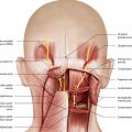
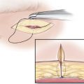
Our investigation related to the anatomy of the greater occipital nerve (GON) revealed some enthralling findings.7 The objective of this study was to determine the GON course, conceivable anatomic variations, and potential sites of entrapment or compression. Twenty fresh cadaver heads were dissected to trace the course of the GON from where the occipital nerve penetrates the semispinalis muscle. It was discovered that the nerve travels through the thickness of the muscle on every specimen. Standardized measurements were used on 14 specimens to determine the location of the emergence of the nerve using the midline and occipital protuberance as landmarks. The point of emergence of the GON from the semispinalis muscle was determined to be centered approximately 3 cm below the occipital protuberance and 1.5 cm lateral to the midline.
As we were completing this anatomical study, a patient who had undergone septorhinoplasty reported that his MH had disappeared. Searching for a potential connection in the literature, we came across an article from Switzerland demonstrating that indeed this surgery had been performed for MH and improved or eliminated MH on many patients.8 In this study, 299 patients with various types of headaches, including migraine, cluster, and so-called idiopathic headaches, were operated on between 1973 and 1991. Septal correction, resection of the middle concha, ethmoidectomy, and sphenoidectomy on the corresponding headache side or occasionally on both sides were carried out. Most patients (235; 78.5%) were totally asymptomatic postoperatively; 34 (11.5%) had a sensation of pressure in the head on rare occasions but no further migraines, and 30 (11%) continued to experience headaches that occurred only rarely and were mild and of short duration.
Up to this point, the prevailing theory behind MH pathogenesis included four basic concepts: (1) neuronal hyperexcitability during the interictal phase; (2) cortical spreading depression (CSD) as the basis of aura; (3) trigeminal nerve activation at a peripheral and central origin that accounts for the headache; and (4) the provocative concept that progressive central sensitization is possibly related to periaqueductal gray matter damage. Of these four concepts, our findings supported peripheral activation of the trigeminal nerve. MH is postulated to be caused by dilatation of large vessels innervated by the trigeminal nerve. Vasodilatation is the consequence of release of calcitonin gene-related peptide, substance P, and neurokinin A found in the cell bodies of trigeminal neurons. What prompts the release of these peptides remains unclear. We proposed that it may be the mechanical stimulation of the potentially hyperexcited, peripheral sensory nerves that instigates the cascade of events. In fact, in 3 out of 4 trigger sites that we studied, the sensory nerves traverse the muscles. As to the fourth site, contact between the turbinates and the deviated septum may cause MH in some patients.
More knowledge about the anatomy of the zygomaticotemporal branch of the trigeminal nerve seemed in order at this time. A study was conducted to determine the site of emergence of this nerve from the deep temporal fascia and to identify the number of its accessory branches and their locations. An initial pilot study, conducted on 20 patients, concluded that the main zygomaticotemporal branch emerges from the deep temporal fascia at a point on average 17 mm lateral and 6 mm cephalad to the lateral palpebral commissure, commonly referred to as the lateral canthus. These measurements, however, were obtained after dissection of the temporal area, rendering the findings less reliable. Another study included 20 consecutive patients, 19 women and 1 man, between the ages of 26 and 85 years, with an average age of 47.6 years.9 Those who had a history of previous trauma or surgery in the temple area were excluded. Before the start of the endoscopic forehead procedure, the likely topographic site of the zygomaticotemporal branch was marked 17 mm lateral and 6 mm cephalad to the lateral orbital commissure on the basis of the information extrapolated from the pilot study. The surface mark was then transferred to the deeper layers using a 25-gauge needle stained with brilliant green. After endoscopic exposure of the marked site, the distance between the main branch of the trigeminal nerve or its accessory branches and the tattoo mark was measured in posterolateral and cephalocaudal directions. In addition, the number and locations of the accessory branches of the trigeminal nerve were recorded.
On the left side, the average distance of the emergence site of the main zygomaticotemporal branch of the trigeminal nerve from the palpebral fissure was 16.8 mm (range: 12–31 mm) in the posterolateral direction and an average of 6.4 mm (range: 4–11 mm) in the cephalad direction. On the right side, the average measurements for the main branch were 17.1 mm (range: 15–21 mm) in the posterolateral direction and 6.65 mm (range: 5–11 mm) in the cephalic direction. Three types of accessory branches were found in relation to the main branch: (1) accessory branch cephalad, (2) accessory branch lateral, and (3) accessory branches in the immediate vicinity of the main branch. This anatomical information has proven colossally helpful in injection of BT-A in the temporalis muscle to eliminate the trigger sites in the parietotemporal region and surgical management of MHs triggered from this zone.
Armed with the anatomical information and having an understanding that there are at least four common trigger sites, we designed a prospective randomized study.10
Related posts:
 An Overview of Migraine Headaches
 Detection of Migraine Headache Trigger Sites
 Surgical Anatomy of the Frontal and Occipital Trigger Sites
 Surgical Treatment of Auriculotemporal Migraine Headaches (Site V)
 Surgical Treatment of Frontal Migraine Headaches (Site I)
 Surgical Treatment of Lesser Occipital Migraine Headaches (Site VI)
Stay updated, free articles. Join our Telegram channel

Full access? Get Clinical Tree


