Vesicobullous and inflammatory diseases produce vulvar erosions that may exhibit nonspecific morphology and represent a diagnostic challenge. An approach to arriving at the correct diagnosis is presented. Most common etiologies are reviewed.
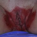
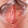
Erosive diseases in the vulvovaginal area are often chronic, painful, progressive, and debilitating conditions that frequently result in considerable patient anxiety and frustration. These conditions, which represent a mixture of inflammatory, infectious, and neoplastic processes, are often difficult to diagnose and treat. The morphology of erosive disease is often nonspecific and therefore a thorough knowledge of the differential diagnosis is important ( Box 1 ). This article provides an overview of the clinical features, causes/associations, histology, and treatment of some of the more commonly encountered conditions causing erosive vulvar disorders. Particular emphasis is given to lichen planus (LP), which is the most common cause of erosive vulvitis.
Inflammatory dermatoses with possible erosion
Lichen planus
Lichen sclerosus
Fixed drug eruption
Stevens-Johnson syndrome/toxic epidermal necrolysis
Irritant/allergic contact dermatitis
Plasma cell (Zoon) vulvitis
Lichen simplex chronicus
Inflammatory vesiculobullous diseases
Subepidermal blistering diseases
Bullous pemphigoid
Cicatricial pemphigoid
Linear IgA disease
Bullous systemic lupus erythematosus
Epidermolysis bullosa acquisita
Intraepidermal blistering diseases
Pemphigus vulgaris
Benign familial pemphigus (Hailey-Hailey disease)
Nutritional
Vitamin B 2 deficiency
Acrodermatitis enteropathica
Infections
Candidiasis
Impetigo
Herpes simplex
Herpes zoster
Malignancies
Vulvar intraepithelial neoplasia
Squamous cell carcinoma
Basal cell carcinoma
Extramammary Paget disease
Langerhans cell histiocytosis
Paraneoplastic diseases
Necrolytic migratory erythema
Paraneoplastic pemphigus
General approach to the evaluation of erosive vulvar disease
In approaching the patient with erosive vulvar disease, a spectrum of disease processes must be considered. Consequently, the clinician must be attuned to a range of details both in history and physical examination to arrive at the correct diagnosis ( Box 2 ). Occam’s razor often does not apply to genital skin; multiple problems may coexist. For example, a patient with lichen sclerosus (LS) may also have an irritant dermatitis from urinary incontinence as well as fissures from a yeast infection. In addition, when the patient continues to complain of pain despite a normal examination, the diagnosis of vulvodynia should be entertained.
History
- •
Itching versus burning
- •
Hygiene practices
- •
Use of sanitary pads
- •
Estrogen status (eg, posttotal hysterectomy, postmenopausal, postpartum)
- •
Incontinence problems
- •
Underlying immunosuppressive disease
- •
Related symptoms: abnormal vaginal discharge, vaginal bleeding, dyspareunia/apareunia, dysuria, oral symptoms, ocular symptoms, dysphagia, hearing difficulties
- •
History of abnormal Pap smear
- •
Previous abdominal surgery
- •
Medications
- •
Review of all topical agents (especially benzocaine-containing products) that come into contact with genital skin
- •
Review all prescription and over-the-counter oral medications
- •
Review of systems
- •
Screen for symptoms associated with autoimmune disorders such as hypothyroidism, diabetes, connective tissue disease
- •
Screen for depression and anxiety
- •
Physical examination
- •
Full cutaneous examination with attention to oral, ocular, and perianal skin
- •
Vulvar examination: make a checklist for presence or absence of all normal structures, examine for evidence of scarring
- •
Vaginal examination with wet mount of secretions
- •
Related posts:

Stay updated, free articles. Join our Telegram channel

Full access? Get Clinical Tree


