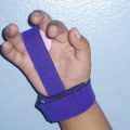
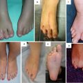
Fig. 25.1
Example of pseudosyndactyly
History
EB was first described by Austrian dermatologist Von Hebra in 1870 [4]. In 1879, Tilbury Fox, a British dermatologist, described the inheritance of EB in two cases involving a 6-year-old girl who presented with hand blisters and her sister, a 2-year-old with blisters on multiple areas and ulcerations on the tongue [5]. In 1886, this condition was named epidermolysis bullosa by Heinrich Koebner.
EB Types
There are four major types of EB based on the Report of the Third International Consensus Meeting on Diagnosis and Classification of EB [6]. EB is classified according to the level at which cleavage occurs: (1) EB simplex (intraepidermal separation), (2) junctional EB (intra-lamina lucida separation), (3) dystrophic EB (sub-basal lamina separation), and (4) Kindler syndrome (mixed cleavage points).
EB simplex (EBS) is the most common type that follows an autosomal dominant inheritance pattern with several subtypes having an autosomal recessive pattern of inheritance. It is characterized by localized or widespread blisters depending on subtype and typically presents at birth or early infancy. Blister formation occurs within the epidermis and blisters heal without scar formation. Most patients with the localized subtype will have a normal life expectancy [2, 7]. Nail dystrophy, milia, and mucosal involvement are rare in all types of EBS compared to the other major types [1].
Junctional EB (JEB) is inherited in an autosomal recessive fashion and can vary from mild to severe disease. Cleavage occurs at the dermal–epidermal junction within the lamina lucida. The hallmark clinical feature of all forms of JEB is enamel hypoplasia that has an appearance of pitting on tooth surfaces [1]. There are two general subtypes of JEB, which include the JEB-Herlitz and the more common generalized atrophic benign EB [1, 2]. The junctional EB Herlitz variant is the most severe form that is present at birth and carries a high risk of death within the first 2 years of life. Blisters heal with atrophic scars. Manifestations can occur in the eyes, and gastrointestinal and genitourinary systems. Death is related to complications from malnutrition, infection, and respiratory failure [2, 7]. In the milder form, generalized atrophic benign EB, patients can have a normal lifespan. They may have scalp involvement, which can lead to hair loss.
Dystrophic EB is characterized by cleavage below the basal lamina and has both autosomal dominant and recessive inheritance patterns [1, 2, 7]. The autosomal dominate subtype (DDEB) has a milder presentation with blistering occurring primarily in the upper and lower extremities. In contrast, the recessive type (RDEB) manifests with widespread blistering with involvement of the eyes, gastrointestinal tract, genitourinary tract, kidney, and heart. Blisters heal with severe scar formation that can result in nail dystrophy, joint flexion contractures, and pseudosyndactyly of the fingers and toes. This ongoing process of blister and scar formation can result in significant hand dysfunction. There is also an increased risk of the development of squamous cell carcinoma in areas of chronic non-healing ulcers. Because there is mucosal involvement, scar formation and erosions can occur in the esophagus, cornea, and genitourinary and gastrointestinal tracts. Death typically ensues in early adult life secondary to malnutrition, infection, and cutaneous carcinomas.
Kindler syndrome is an autosomal recessive form of EB with cleavage occurring at multiple sites including intraepidermal, junctional, and sublamina densa [1, 2]. Blistering presents at birth and typically occurs in acral locations. Blister healing occurs with atrophic scar. Patients generally have a normal lifespan with blister formation decreasing with time. This form EB is associated with photosensitivity, skin atrophy, and dyspigmentation and can also have mucosal involvement [2].
Diagnosis
EB commonly presents at birth with skin blisters that must be distinguished from other skin lesions occurring during the neonatal period. The diagnosis of EB begins with a thorough history and physical examination including a family history to rule out inheritable causes of skin blisters and erosions [8]. The differential diagnosis of a newborn with skin blisters is quite extensive as described by Nischler et al. Some notable conditions include inheritable causes, traumatic blisters, infectious causes such as herpes simplex, staphylococcal scalded skin syndrome, bullous impetigo, and immunobullous disorders including bullous pemphigoid, and epidermolysis bullosa acquisita [8]. If there is a high suspicion for EB, a skin biopsy of a blister is required for a definitive diagnosis. Immunofluorescence mapping (IFM) of a skin biopsy is the primary diagnostic tool used in the diagnosis of EB and can identify the EB subtype in addition to the level of skin separation. If IFM is inconclusive, transmission electron microscopy and genetic testing can be performed to aid in the diagnosis [2, 6, 7].
Hand Contractures
Hand deformities can occur with all major types of EB; however, they more commonly occur with recessive dystrophic EB subtypes. Le Touze et al. described four characteristic lesions seen in EB affecting the hand: (1) complete loss of nails, (2) flexion contracture of fingers and palm, (3) thumb adduction contracture, and (4) pseudosyndactyly. Deformities develop from repeated blister healing that forms dense scar tissue causing partial fusion between digital web spaces. As this process continues, fusion of entire digits can result and scar tissue can eventually encase the entire hand, a condition referred to as a mitten hand (Fig. 25.2). This mitten hand restricts movement of fingers, causing finger contractures, adduction contracture of the thumb and proximal muscle atrophy, and even joint destruction in cases with severe long-standing deformities [9]. This renders the hand non-functional, making it difficult for patients to carry out even simple tasks. Fine et al. reviewed the National Epidermolysis Bullosa Registry to determine the frequency and risk of developing hand and foot deformities in the major and subtypes of EB. This database contains information on 3,280 patients with EB that enrolled between the period of 1986 and 2002. There were 2,748 patients identified who had sufficient data to allow classification into 10 different subtypes of EB. The authors concluded that mitten hand deformities occur less commonly in EBS, JEB, and DDEB than in RDEB. The frequency of mitten hand deformities observed in EBS and JEB ranged from 0.0 to 4.39 % and 0.53 to 6.82 % depending on subtype. This is in comparison to 41.18 % and 51.13 % seen with non-Hallopeau-Siemens RDEB and RDEB-inversa, respectively. The highest frequency was observed in the Hallopeau-Siemens RDEB subtype at 95 %. The frequency of mitten hand deformities was only 2.35 % in the DDEB subtype.
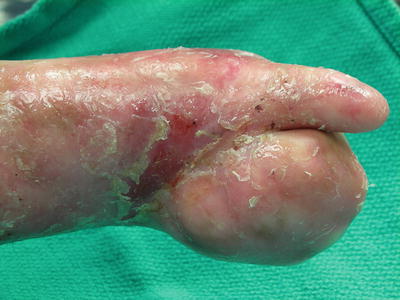
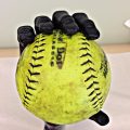
Fig. 25.2
Mitten hand
Management
Nonoperative
Treatment of EB is best delivered in a multidisciplinary fashion with involvement of the patient’s family, primary care physician, dermatologist, dentist, nutritionist, and physical and occupational therapists. Involvement of subspecialty services such as gastroenterology, urology, ophthalmology, pain management, plastic surgery, and hand surgery is determined based on need. Because there is currently no cure available for EB, nonoperative treatment is aimed at preventing blister formation by minimizing skin trauma, providing adequate nutrition, wound care, and preventing infection [2].
Operative Treatment Indications
The indications for operative treatment of the hand in patients with EB include progressive loss of hand function as demonstrated by decreased grasp or pinch, formation of pseudosyndactyly, loss of finger independence, flexion contractures, and formation of a mitten hand. The goals of surgery are to improve overall hand function by reestablishing pinch and grasp function, and to delay recurrence [2, 10, 11]. The timing of surgery will depend on surgeon preference. Although some patients will present with severe contractures and a mitten hand deformity, we prefer early surgical intervention when patients maintain some hand function with only mild to moderate contractures. Early surgical intervention is advocated in order to avoid interruptions in a developing child [3, 12]. Surgery is considered when parents and patients can cooperate with the postoperative regimen [3, 11].
Preoperative Considerations
It is important to recognize that EB can affect many areas of the body, and extreme care must be exercised when treating the patient. An air mattress should be utilized when possible. All bony prominences should be well-padded and sequential compression devices are preferentially not utilized as they can further damage fragile skin. No rubbing of skin should be allowed and the use of adhesive tape is contraindicated. A blood pressure cuff should only be applied to a well-padded extremity [12]. Electrocardiographic leads are placed over petroleum non-adhesive dressing as shown in Fig. 25.3 [1]. An intravenous line is placed after the patient has been adequately sedated and is sutured into place and secured with a cotton wrap and Coban (3 M Corp., St. Paul, MN) (Fig. 25.4). It is important that all members participating in the care of the patient are aware of these precautions.
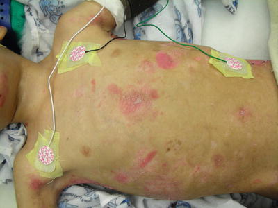
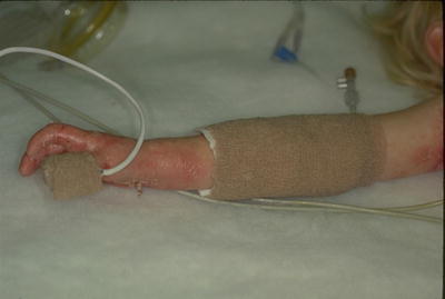
Fig. 25.3
Electrocardiographic leads placed over non-adhesive dressings
Fig. 25.4
Intravenous line secured with Coban
Surgical Technique
The operative technique performed by the senior author is similar to that described by several other surgeons [3, 12–15]. However, our technique differs such that no tourniquet is used, full thickness skin grafts are used instead of split thickness, and pinning of the thumb is not performed after release of its adduction contracture [11]. After carrying out the preoperative precautions described previously, the patient is placed on synthetic sheepskin overlying the operative table. Although some surgeons prefer the use of tourniquet [12–15], we elect not to use a tourniquet to avoid skin trauma at the site of tourniquet placement. The patient is given one dose of appropriately dosed cefazolin intraoperatively if there are no allergic contraindications. Bleeding generally consists of slow ooze that can be controlled with Hemostatic collagen (Avitene, Alcon Puerto Rico Inc., Humacao, Puerto Rico) or thrombin-soaked cellulose. There is also a constant serous ooze from the surgical sites that must be monitored carefully in order to adequately provide fluid resuscitation intraoperatively. A median nerve wrist block of dose appropriate 0.25 % bupivacaine with epinephrine 1:200,000 is administered. This block decreases anesthetic requirements and also helps with postoperative pain (Fig. 25.5). The abdomen and extremity are prepped without mechanical scrubbing, pouring dilute chlorhexidine soap over the proposed operative sites.
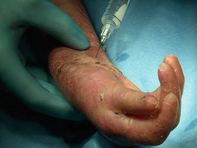
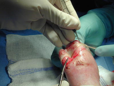
Fig. 25.5
Median nerve block
Surgery begins with epidermal degloving of the hand by scoring only the epidermis with a scalpel, then gently teasing away the epidermal cocoon using fine forceps, a Freer elevator, and selective separation with Littler scissors (Figs. 25.6 and 25.7). The pseudosyndactyly that forms between the fingers is released with the elevator, advanced with selective cuts, and gently separated with opposing traction on the digits. When applying traction, a single layer of gauze padding over each digit absorbs the ooze, which can create slippery surfaces. Degloving alone has proven to be ineffective and is associated with early recurrence [16]. The first web space contracture usually requires release of the adductor fascia, and only occasionally full thickness skin grafting. A four flap Z-plasty is not used to release the first web space contracture as the skin non-pliable.
 Anesthesia Concerns in Congenital Anomalies of the Upper Extremity
General Skeletal Disorders
Anesthesia Concerns in Congenital Anomalies of the Upper Extremity
General Skeletal Disorders
 Therapy Management of Children with Congenital Anomalies of the Upper Extremity
 Physical Medicine and Rehabilitation Management of Children with Congenital Anomalies of the Upper Extremity
 Ulnar Polydactyly and Ulnar Dimelia
 Macrodactyly


Related posts:
Anesthesia Concerns in Congenital Anomalies of the Upper Extremity
General Skeletal Disorders
Therapy Management of Children with Congenital Anomalies of the Upper Extremity
Physical Medicine and Rehabilitation Management of Children with Congenital Anomalies of the Upper Extremity
Ulnar Polydactyly and Ulnar Dimelia
Macrodactyly
Stay updated, free articles. Join our Telegram channel
Full access? Get Clinical Tree
