Epidermal Nevi, Neoplasms, and Cysts


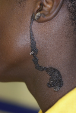
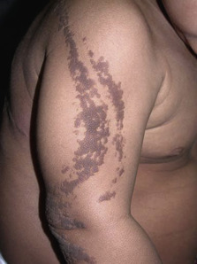
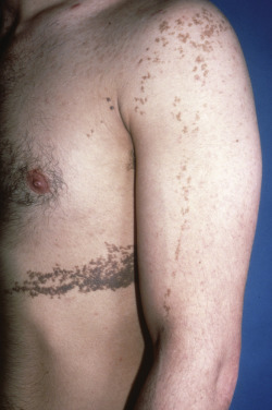
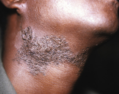
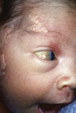
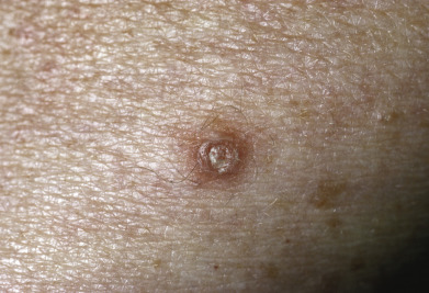
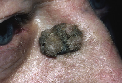
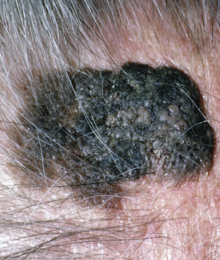
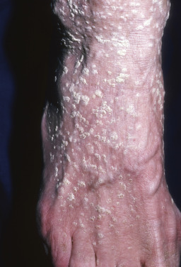
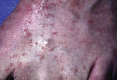
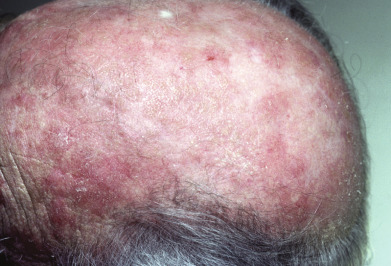
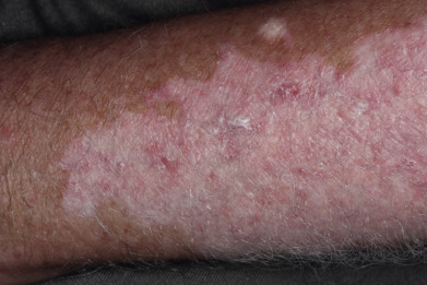
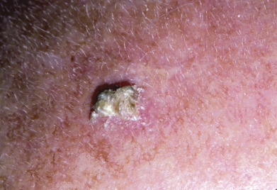
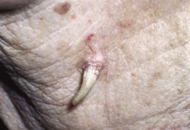
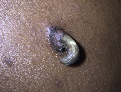
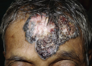
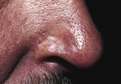
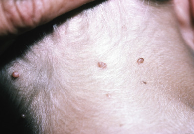
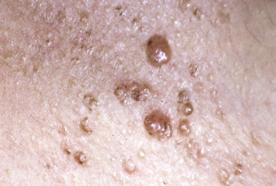
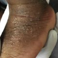
Epidermal neoplasms commonly present with hyperkeratosis, acanthosis, or papillomatosis. These may manifest clinically as a cutaneous horn, scale, palpable induration, a velvety or filiform appearance, or a smooth lesion raised above the surrounding skin surface. As an example, epidermal nevi tend to be raised and linear in configuration, following Blaschko lines. They may be hyperpigmented, hypopigmented, fleshy, or keratotic in appearance.


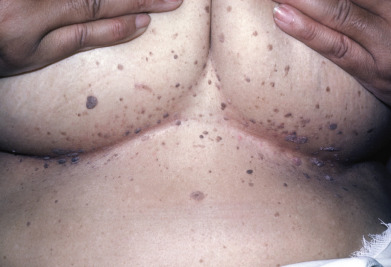
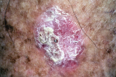
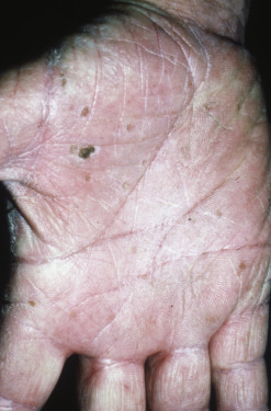
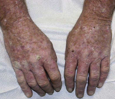
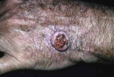
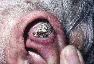
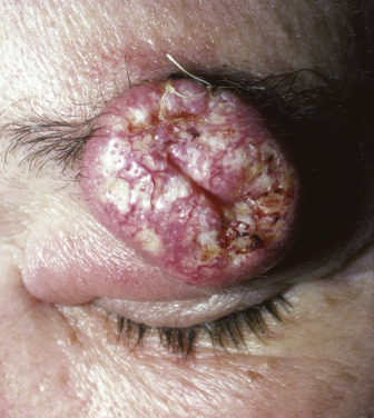
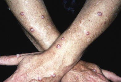
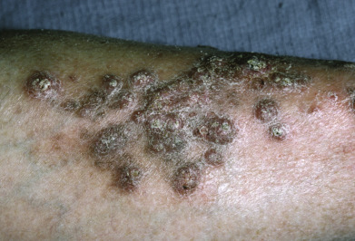
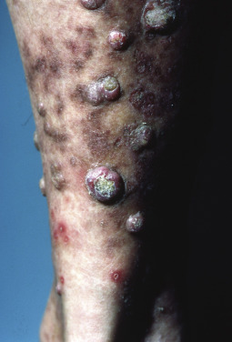
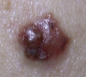
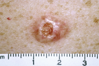
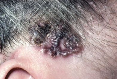
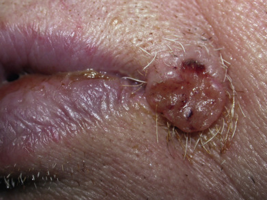
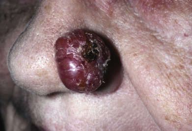
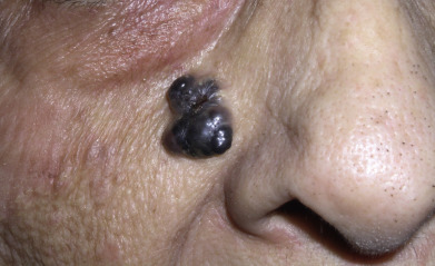
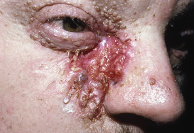
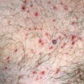
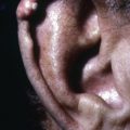
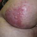
Cysts and dermal epithelial neoplasms displace the overlying skin and may produce overlying atrophy, erythema, or telangiectasia. The presence of tumor-associated vascularity lends a red or blue appearance, depending on the speed of blood flow and oxygen saturation of the blood. The presence of cytoplasm lends a yellow appearance due to carotenoids dissolved in the aqueous phase of cytoplasm. A brown appearance most commonly relates to melanin within the epithelial cells and underlying dermis, but can also be a result of dermal hemosiderin or lipofuscin deposition. Lipofuscin dissolved in apocrine sweat often lends a blue appearance to portions of sweat gland tumors as a result of diffraction of light (the Tyndall effect). Sebaceous elements lend a yellow or orange appearance. An appreciation of the color, morphology, and distribution of the lesions will help the physician narrow the differential diagnosis.


Fig. 29.1


Epidermal nevus.


Fig. 29.2


Epidermal nevus.


Fig. 29.3


Epidermal nevus.


Fig. 29.4


CLOVE syndrome.


Fig. 29.5


Nevus comedonicus.


Fig. 29.6


Schimmelpenning syndrome with a lipodermoid of the conjunctiva.


Fig. 29.7


Inflammatory linear verrucous epidermal nevus.


Fig. 29.8


Clear cell acanthoma.

Courtesy Ken Greer, MD.


Fig. 29.9


Multiple clear cell acanthomas.


Fig. 29.10


Warty dyskeratomas.


Fig. 29.11


Seborrheic keratosis.

Courtesy Steven Binnick, MD.


Fig. 29.12


Seborrheic keratosis.

Courtesy Steven Binnick, MD.


Fig. 29.13


Seborrheic keratosis.

Courtesy Steven Binnick, MD.


Fig. 29.14


Seborrheic keratosis.


Fig. 29.15


Seborrheic keratosis.


Fig. 29.16


Dermatosis papulosa nigra.

Courtesy Steven Binnick, MD.


Fig. 29.17


Dermatosis papulosa nigra.


Fig. 29.18


Stucco keratosis.


Fig. 29.19


Flegel disease.


Fig. 29.20


Benign lichenoid keratoses.


Fig. 29.21


Arsenical keratoses.


Fig. 29.22


Actinic keratoses.


Fig. 29.23


Actinic keratoses.


Fig. 29.24


Actinic keratoses in vitiliginous skin.


Fig. 29.25


Hypertrophic actinic keratosis.


Fig. 29.26


Cutaneous horn.

Courtesy Steven Binnick, MD.


Fig. 29.27


Cutaneous horn.

Courtesy Debabrata Bandyopadhyay.


Fig. 29.28


Keratoacanthoma.

Courtesy Curt Samlaska, MD.


Fig. 29.29


Keratoacanthoma.

Courtesy Steven Binnick, MD.


Fig. 29.30


Keratoacanthoma.


Fig. 29.31


Multiple keratoacanthomas.


Fig. 29.32


Eruptive keratoacanthoma.


Fig. 29.33


Eruptive keratoacanthoma.


Fig. 29.34


Basal cell carcinoma.


Fig. 29.35


Basal cell carcinoma.


Fig. 29.36


Basal cell carcinoma.


Fig. 29.37


Basal cell carcinoma.


Fig. 29.38


Basal cell carcinoma.

Courtesy Steven Binnick, MD.


Fig. 29.39


Superficial basal cell carcinoma.


Fig. 29.40


Pigmented basal cell carcinoma.

Courtesy Curt Samlaska, MD.


Fig. 29.41


Pigmented basal cell carcinoma.

Courtesy Debabrata Bandyopadhyay, MD.


Fig. 29.42


Morpheaform basal cell carcinoma.

Courtesy Steven Binnick, MD.


Fig. 29.43


Basal cell carcinoma.


Fig. 29.44


Large basal cell carcinoma.


Fig. 29.45


Aggressive basal cell carcinoma.


Fig. 29.46


Multiple basal cell carcinomas in a radiation site.

Courtesy Steven Binnick, MD.


Fig. 29.47


Basal cell nevus syndrome with skin tag–like basal cell carcinomas.

Courtesy Ken Greer, MD.


Fig. 29.48


Basal cell nevus syndrome.

Courtesy Steven Binnick, MD.


Sep 3, 2019 | Posted by in Dermatology | Comments Off on Epidermal Nevi, Neoplasms, and Cysts

Full access? Get Clinical Tree

Get Clinical Tree app for offline access