Endoscopic browplasty


Table 22.1

Indications for surgery


Forehead rhytids
Moderate brow ptosis
Temporal hooding
Facial nerve palsy with brow ptosis


Table 22.2

Preoperative evaluation


Position of brow
Age and gender of patient
Magnitude of brow ptosis
Location of hairline and quantity of follicles
Prior eyelid, facial surgery or trauma
Qualitative and quantitative assessment of tear film


Introduction


The endoscopic forehead lift is primarily a cosmetic operation and is useful for moderate brow ptosis. It can smoothen out forehead rhytids, weaken brow depressors, and elevate the tail of the brow. For mild brow ptosis, an internal brow elevation can be performed ( Chapter 21 ). This procedure is most effective in preventing post-upper blepharoplasty brow descent but provides minimal brow elevation. For moderate brow ptosis where function takes precedence over form, a direct browplasty can be performed to elevate a ptotic brow ( Chapter 20 ). For more severe brow ptosis in select patients, pretrichial browplasty can be performed ( Chapter 23 ).


Critics of the endoscopic technique claim that this procedure is not as effective in lifting the brow as an open procedure and that the effects are not long lasting. However, we have found the procedure to be useful in mild-to-moderate brow ptosis with good long-term results. Furthermore, the endoscopic procedure has the advantage of resulting in less hair loss, hypesthesia, and skin scarring compared to open brow approaches.


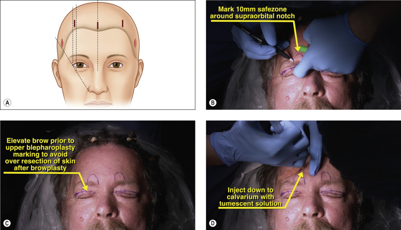
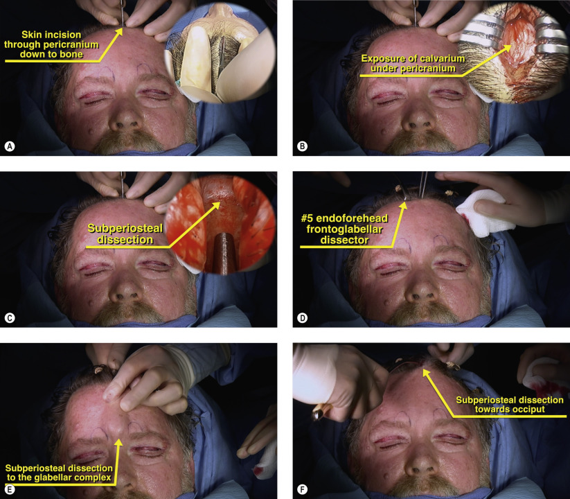
The procedure involves the creation of three optical cavities through five incisions: one central, two paramedial, and two temporal. All pockets are communicated into one larger cavity. After release of the periosteum and weakening of the brow depressors, the forehead flap is elevated and fixated. Options for central fixation include an absorbable Endotine Forehead implant (MicroAire, Charlotesville, VA) removable screw, and surgeon-drilled bone tunnels. Our preferred fixation method is use of bone tunnels. Temporally, the superficial temporalis fascia is advanced and secured to the deep temporalis fascia. Skin incisions are then closed with staples or sutures.


Surgical Technique


Figures 22.1A–D


Marking of incision sites

Five incision sites are typically made for endoscopic browplasty ( Figure 22.1A ). A vertically oriented central incision is made 3–4 mm posterior to the hairline. Paired paramedial marks are made in line with the peak of the brow which corresponds to the lateral corneal limbus. The two temporal marks are made at a location bisected by a plane parallel to the preauricular crease and a line extrapolated from the nasal ala through the lateral canthus. Prior to the start of surgery, the hair is parted with multiple rubber ties. The supraorbital notches are palpated and a 10-mm safe zone is marked ( Figure 22.1B ). When performing concomitant upper blepharoplasty, the brow should be manually lifted while marking the skin for excision ( Figure 22.1C ). This prevents over-resection of skin after browplasty. The incision sites are injected with 2% lidocaine and 1 : 100,000 epinephrine at least 10 minutes prior to incision for maximal hemostasis. The forehead and temporal region down to the brow and zygomatic arch are infiltrated with 50 to 100cc of dilute 2% lidocaine with epinephrine 1 : 100,000 and saline diluted down to a 1 : 5 ratio ( Figure 22.1D ). At least 10 minutes should elapse for maximal vasconstrictive effect. A full face sterile preparation is performed to include the hair. A plastic bag is placed under the head rest to capture any bleeding or saline rinses during and after surgery.


Figures 22.2A–F


Incision of central and paramedial sites

The central incision and paramedial incisions are performed with a #15 or #10 blade down through the pericranium to expose bone ( Figure 22.2A ). With adequate local anesthetic, bleeding from the incision should be minimal and excessive cautery should be avoided to prevent the development of localized alopecia. Point bleeders can be controlled with conservative bipolar cautery. Senn retractors are used for horizontal exposure to identify the calvarium ( Figure 22.2B ). A Freer or periosteal elevator is then used to perform a subperiosteal dissection inferiorly towards the brow ( Figure 22.2C ). Once the periosteum near the incision is elevated, #5 endoforehead frontoglabellar dissector is introduced in the subperiosteal plane ( Figure 22.2D ). The dissection is continued inferiorly towards the glabellar complex to gently elevate the procerus muscle ( Figure 22.2E ) Aggressive dissection lateral to the midline should be avoided to minimize trauma to the supraorbital and supratrochlear nerves as well as to prevent damage to the corrugator and depressor supercilii which may result in lateral splaying of the brow. Finally, the dissection is continued posteriorly towards the occiput ( Figure 22.2F ). These portions of the dissection can be performed safely without endoscopic visualization as long as the dissectors remain on the calvarium in the subperiosteal plane.

Only gold members can continue reading. Log In or Register to continue

Stay updated, free articles. Join our Telegram channel

May 16, 2019 | Posted by in Reconstructive surgery | Comments Off on Endoscopic browplasty

Full access? Get Clinical Tree

Get Clinical Tree app for offline access