Ductal Carcinoma in Situ: An Oncoplastic Treatment Approach
Melvin J. Silverstein
Introduction
Ductal carcinoma in situ (DCIS) of the breast is a heterogeneous group of lesions with diverse malignant potential and a range of treatment options. It is the most rapidly growing subgroup in the breast cancer family of disease, with more than 67,000 new cases diagnosed in the United States during 2008 (27% of all new cases of breast cancer) (1). Most new cases (>90%) are nonpalpable and discovered mammographically.
It is now well appreciated that DCIS is a stage in the neoplastic continuum in which most of the molecular changes that characterize invasive breast cancer are already present (2). All that remains on the way to invasion are quantitative changes in the expression of genes that have already been altered. Genes that might play a role in invasion control a number of functions, including angiogenesis, adhesion, cell motility, the composition of extracellular matrix, and more. To date, genes that are uniquely associated with invasion have not been identified. DCIS is clearly the precursor lesion for most invasive breast cancers, but not all DCIS lesions have the time or the genetic ability to progress to become invasive breast cancer (3,4,5).
Therapy for DCIS ranges from simple excision to various forms of wider excision (segmental resection, quadrant resection, oncoplastic resection, etc.), all of which may or may not be followed by radiation therapy. When breast preservation is not feasible, total mastectomy, with or without immediate reconstruction, is generally performed.
Since DCIS is a heterogeneous group of lesions rather than a single entity (6,7) and because patients have a wide range of personal needs that must be considered during treatment selection, it is clear that no single approach will be appropriate for all forms of the disease or for all patients. At the current time, treatment decisions are based upon a variety of measurable parameters (tumor extent, margin width, nuclear grade, the presence or absence of comedonecrosis, age, etc.), physician experience and bias, and randomized trial data, which suggest that all conservatively treated patients should be managed with postexcisional radiation therapy.
The Changing Nature of Ductal Carcinoma in Situ
There have been dramatic changes in the last 20 years that have affected DCIS. Before mammography was common, DCIS was rare, representing less than 1% of all breast cancer (8). Today, DCIS is common, representing 27% of all newly diagnosed cases and as much as 30% to 50% of cases of breast cancer diagnosed by mammography (1,115,116,117,118,119).
Previously, most patients with DCIS presented with clinical symptoms, such as breast mass, bloody nipple discharge, or Paget’s disease (9,10). Today, most lesions are nonpalpable and generally detected by mammography alone.
Until approximately 20 years ago, the treatment for most patients with DCIS was mastectomy. Today, almost 75% of newly diagnosed patients with DCIS are treated with breast preservation (11). In the past, when mastectomy was common, reconstruction was uncommon; if it was performed, it was generally done as a delayed procedure. Today, reconstruction for patients with DCIS treated by mastectomy is common; when it is performed, it is generally done immediately, at the time of mastectomy. In the past, when a mastectomy was performed, large amounts of skin were discarded. Today, it is considered perfectly safe to perform a skin-sparing mastectomy for DCIS and, in some instances, nipple-areola sparing-mastectomy. In the past, there was little confusion. All breast cancers were considered essentially the same, and mastectomy was the only treatment. Today, all breast cancers are different, and there is a range of acceptable treatments for every lesion. For those that choose breast conservation, there continues to be a debate as to whether radiation therapy is necessary in every case. These changes were brought about by a number of factors. Most important were increased mammographic utilization and the acceptance of breast conservation therapy for invasive breast cancer.
The widespread use of mammography changed the way DCIS was detected. In addition, it changed the nature of the disease detected by allowing us to enter the neoplastic continuum at an earlier time. It is interesting to note the impact that mammography had on the Breast Center in Van Nuys, California, in terms of the number of DCIS cases diagnosed and the way they were diagnosed (12).
From 1979 to 1981, the Van Nuys group treated a total of only 15 patients with DCIS, 5 per year. Only 2 lesions (13%) were nonpalpable and detected by mammography. In other words, 13 patients (87%) presented with clinically apparent disease. Two state-of-the-art mammography units and a full-time experienced radiologist were added in 1982, and the number of new DCIS cases dramatically increased to more than 30 per year, most of them nonpalpable. When a third machine was added in 1987, the number of new cases increased to 40 per year. In 1994, the Van Nuys group added a fourth mammography machine and a prone stereotactic biopsy unit. In 1998, I moved to the Norris Comprehensive Cancer Center at the University of Southern California (USC) and continued to accrue DCIS patients. In March 2008, I became director of the Breast Program at Hoag Memorial Hospital Presbyterian in Newport Beach, California, and the DCIS series continued.
Analysis of the entire series of 1,411 patients through December 2008 shows that 1,242 lesions (88%) were nonpalpable (subclinical). If we look at only those diagnosed during the last 5 years that I was at the USC/Norris Cancer Center, 95% were nonpalpable.
The second factor that changed how we think about DCIS was the acceptance of breast conservation therapy (lumpectomy, axillary node dissection, and radiation therapy) for patients with invasive breast cancer. Until 1981, the treatment for most patients with any form of breast cancer was generally mastectomy. Since that time, numerous prospective randomized trials have shown an equivalent rate of survival for selected patients with invasive breast cancer treated with breast conservation therapy (13,14,15,16,17,18). Based on these results, it made little sense to continue treating less aggressive DCIS with mastectomy while treating more aggressive invasive breast cancer with breast preservation.
Current data suggest that many patients with DCIS can be successfully treated with breast preservation, with or without radiation therapy. This chapter will show how easily available data can be used to help in the complex treatment selection process.
Pathology
Classification
Although there is no universally accepted histopathologic classification, most pathologists divide DCIS into five major architectural subtypes (papillary, micropapillary, cribriform, solid, and comedo), often comparing the first four (noncomedo) with comedo (6,19,20). Comedo DCIS is frequently associated with high-nuclear-grade (6,19,20,21), aneuploidy, a higher proliferation rate (22), HER2/neu gene amplification or protein overexpression (23,24,25,26,27), and clinically more aggressive behavior (28,29,30,31). Noncomedo lesions tend to be just the opposite.
The division by architecture alone, comedo versus noncomedo, is an oversimplification and does not work if the purpose of the division is to sort the patients into those with a high risk of local recurrence versus those with a low risk. It is not uncommon for high-nuclear-grade noncomedo lesions to express markers similar to those of high-grade comedo lesions and to have a risk of local recurrence similar to comedo lesions. Adding to the confusion is the fact that mixtures of various architectural subtypes within a single biopsy specimen are common. In my series, more than 70% of all lesions had significant amounts of two or more architectural subtypes, making division into a predominant architectural subtype problematic.
Regarding comedo DCIS, there is no uniform agreement among pathologists of exactly how much comedo DCIS needs to be present to consider the lesion a comedo DCIS. Although it is clear that lesions exhibiting a predominant high-grade comedo DCIS pattern are generally more aggressive and more likely to recur if treated conservatively than are low-grade noncomedo lesions, architectural subtyping does not reflect current biologic thinking. Rather, it is the concept of nuclear grading that has assumed importance. Nuclear grade is a better biologic predictor than architecture, and therefore it has emerged as a key histopathologic factor for identifying aggressive behavior (28,31,32,33,34,35). In 1995, the Van Nuys group introduced a new pathologic DCIS classification (36) based on the presence or absence of high-nuclear-grade and comedo-type necrosis (the Van Nuys classification).
The Van Nuys group chose high nuclear grade as the most important factor in their classification because there was general agreement that patients with high-nuclear-grade lesions were more likely to recur at a higher rate and in a shorter time period after breast conservation than patients with low-nuclear-grade lesions (28,31,34,37,38,39). Comedo-type necrosis was chosen because its presence also suggests a poorer prognosis (40,41) and it is easy to recognize (42).
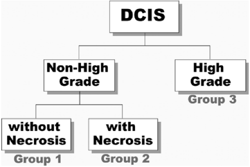
The pathologist, using standardized criteria as noted below, first determines whether the lesion is high nuclear grade (nuclear grade 3) or non–high nuclear grade (nuclear grade 1 or 2). Then, the presence or absence of necrosis is assessed in the non–high-grade lesions. This results in three groups (Fig. 7.1).
Nuclear grade is scored by previously described methods (36). Essentially, low-grade nuclei (grade 1) are defined as nuclei 1 to 1.5 red blood cells in diameter with diffuse chromatin and unapparent nucleoli. Intermediate nuclei (grade 2) are defined as nuclei 1 to 2 red blood cells in diameter with coarse chromatin and infrequent nucleoli. High-grade nuclei (grade 3) are defined as nuclei with a diameter greater than 2 red blood cells, with vesicular chromatin, and one or more nucleoli.
In the Van Nuys classification, no requirement is made for a minimum or specific amount of high-nuclear-grade DCIS, nor is any requirement made for a minimum amount of comedo-type necrosis. Occasional desquamated or individually necrotic cells are ignored and are not scored as comedo-type necrosis.
The most difficult part of most classifications is nuclear grading, particularly the intermediate-grade lesions. The subtleties of the intermediate-grade lesion are not important to the Van Nuys classification; only nuclear grade 3 need be recognized. The cells must be large and pleomorphic, lack architectural differentiation and polarity, have prominent nucleoli and coarse, clumped chromatin, and generally show mitoses (36,40).
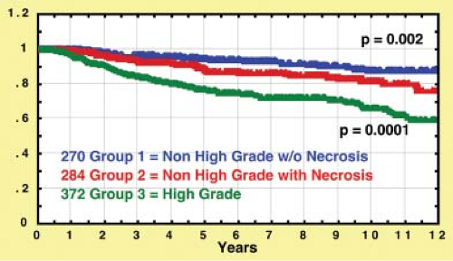
The Van Nuys classification is useful because it divides DCIS into three different biologic groups with different risks of local recurrence after breast conservation therapy (Fig. 7.2). This pathologic classification, when combined with tumor size, age, and margin status, is an integral part of the USC/Van Nuys Prognostic Index (USC/VNPI), a system that will be discussed in detail.
Progression to Invasive Breast Cancer
Which DCIS lesions will become invasive, and when will that happen? These are the most important questions in the DCIS field. There is intense molecular genetic study and knowledge
regarding the progression of normal breast epithelium through hyperplastic and atypical hyperplastic changes to DCIS and then to invasive breast cancer. Most of the genetic and epigenetic changes present in invasive breast cancer are already present in DCIS. No genes uniquely associated with invasive cancer have been identified (2,11). As DCIS progresses to invasive breast cancer, quantitative changes in the expression of genes related to angiogenesis, adhesion, cell motility, and the composition of the extracellular matrix may occur (2,11). Using gene-array technology, researchers are attempting to identify high-risk patterns, which will require quicker and more aggressive treatment.
regarding the progression of normal breast epithelium through hyperplastic and atypical hyperplastic changes to DCIS and then to invasive breast cancer. Most of the genetic and epigenetic changes present in invasive breast cancer are already present in DCIS. No genes uniquely associated with invasive cancer have been identified (2,11). As DCIS progresses to invasive breast cancer, quantitative changes in the expression of genes related to angiogenesis, adhesion, cell motility, and the composition of the extracellular matrix may occur (2,11). Using gene-array technology, researchers are attempting to identify high-risk patterns, which will require quicker and more aggressive treatment.
 Figure 7.2. Probability of local recurrence–free survival for 926 breast conservation patients using the Van Nuys ductal carcinoma in situ pathologic classification. |
Because most patients with DCIS have been treated with mastectomy, knowledge of the natural history of this disease is relatively scant. In a study of 110 consecutive, medicolegal autopsies of young and middle-aged women between the ages of 20 and 54 years, 14% were found to have DCIS (43), suggesting that the subclinical prevalence of DCIS is significantly higher than the clinical expression of the disease.
The studies of Page et al. (44,45) and Rosen et al. (46) shed light on the nontreatment of DCIS. In these studies, patients with noncomedo DCIS were initially misdiagnosed as having benign lesions and therefore went untreated. Subsequently, approximately 25% to 35% of these patients developed invasive breast cancer, generally within 10 years (44,45). Had the lesions been high-grade comedo DCIS, the invasive breast cancer rate likely would have been higher than 35% and the time to invasive recurrence shorter. With few exceptions, in both of these studies, the invasive breast carcinoma was of the ductal type and was located at the site of the original DCIS. These findings and the fact that autopsy series have shown up to a 14% incidence of DCIS suggest that not all DCIS lesions progress to invasive breast cancer or become clinically significant (43,47). There is far more microscopic DCIS than clinically apparent DCIS.
Page and associates recently updated their series (44,45,48). Of 28 women with low-grade DCIS misdiagnosed with benign lesions and treated with biopsy between 1950 and 1968, 11 patients recurred locally with invasive breast cancer (39%). Eight patients developed recurrence within the first 12 years. The remaining 3 were diagnosed over 23 to 42 years. Five patients developed metastatic breast cancer (18%) and died from the disease within 7 years of developing invasive breast cancer. These recurrence and mortality rates, at first glance, seem alarmingly high. However, they are only slightly worse than what can be expected with long-term follow-up of patients with lobular carcinoma in situ, a disease that most clinicians are willing to treat with careful clinical follow-up. In addition, these patients were treated with biopsy only. No attempt was made to excise these lesions with a clear surgical margin. The natural history of low-grade DCIS can extend over 40 years and is markedly different from that of high-grade DCIS.
Microinvasion
The incidence of microinvasion is difficult to quantitate because until recently there was no formal and universally accepted definition of exactly what constitutes microinvasion. The 1997 edition of AJCC Cancer Staging Manual (5th ed.) carried the first official definition of what is now classified as pT1mic and read as follows (114):
Microinvasion is the extension of cancer cells beyond the basement membrane into adjacent tissues with no focus more than 0.1 cm in greatest dimension. When there are multiple foci of microinvasion the size of only the largest focus is used to classify the microinvasion (do not use the sum of all individual foci). The presence of multiple foci of microinvasion should be noted, as it is with multiple larger invasive carcinomas.
The reported incidence of occult invasion (invasive disease at mastectomy in patients with a biopsy diagnosis of DCIS) varies greatly, ranging from as little as 2% to as much as 21% (49). This problem was addressed in the investigations of Lagios et al. (28).
Lagios et al. performed a meticulous serial subgross dissection correlated with specimen radiography. Occult invasion was found in 13 of 111 mastectomy specimens from patients who had initially undergone excisional biopsy of DCIS. All occult invasive cancers were associated with DCIS greater than 45 mm in diameter; the incidence of occult invasion approached 50% for DCIS greater than 55 mm. In the study of Gump et al. (50), foci of occult invasion were found in 11% of patients with palpable DCIS but in no patients with clinically occult DCIS. These results suggest a correlation between the size of the DCIS lesion and the incidence of occult invasion. Clearly, as the size of the DCIS lesion increases, microinvasion and occult invasion become more likely.
If even the smallest amount of invasion is found, the lesion should not be classified as DCIS. It is a T1mic (if the largest invasive component is 1 mm or less) with an extensive intraductal component (EIC). If the invasive component is 1.1 to 5 mm, it is a T1a lesion with EIC. If there is only a single focus of invasion, these patients do quite well. When there are many tiny foci of invasion, these patients have a poorer prognosis than expected (51). Unfortunately, the TNM staging system does not have a T category that fully reflects the malignant potential of lesions with multiple foci of invasion since they are all classified by their largest single focus of invasion.
Multicentricity and Multifocality of Ductal Carcinoma in Situ
Multicentricity is generally defined as DCIS in a quadrant other than the quadrant in which the original DCIS (index quadrant) was diagnosed. There must be normal breast tissue separating the two foci. However, definitions of multicentricity vary among investigators. Hence, the reported incidence of multicentricity also varies. Rates from 0% to 78% (7,46,52,53),
averaging about 30%, have been reported. Twenty years ago, the 30% average rate of multicentricity was used by surgeons as the rationale for mastectomy in patients with DCIS.
averaging about 30%, have been reported. Twenty years ago, the 30% average rate of multicentricity was used by surgeons as the rationale for mastectomy in patients with DCIS.
Holland et al. (54) evaluated 82 mastectomy specimens by taking a whole-organ section every 5 mm. Each section was radiographed. Paraffin blocks were made from every radiographically suspicious spot. In addition, an average of 25 blocks were taken from the quadrant containing the index cancer; random samples were taken from all other quadrants, the central subareolar area, and the nipple. The microscopic extension of each lesion was verified on the radiographs. This technique permitted a three-dimensional reconstruction of each lesion. This study demonstrated that most DCIS lesions were larger than expected (50% were greater than 50 mm), involved more than one quadrant by continuous extension (23%), but most important, were unicentric (98.8%). Only one of 82 mastectomy specimens (1.2%) had “true” multicentric distribution with a separate lesion in a different quadrant. This study suggests that complete excision of a DCIS lesion is possible due to unicentricity but may be extremely difficult due to larger than expected size. In a recent update, Holland and Faverly reported whole-organ studies in 119 patients, 118 of whom had unicentric disease (55). This information, when combined with the fact that most local recurrences are at or near the original DCIS, suggests that the problem of multicentricity per se is not important in the DCIS treatment decision-making process.
Multifocality is defined as separate foci of DCIS within the same ductal system. The studies of Holland et al. (54,55) and Noguchi et al. (56) suggest that a great deal of multifocality may be artifactual, resulting from looking at a three-dimensional arborizing entity in two dimensions on a glass slide. It would be analogous to saying that the branches of a tree were not connected if the branches were cut at one plane, placed separately on a slide, and viewed in cross section (44). Multifocality may be due to small gaps of DCIS or skip areas within ducts as described by Faverly et al. (39).
Detection and Diagnosis
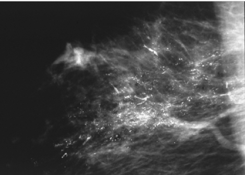
The importance of quality mammography cannot be overemphasized. Currently, most patients with DCIS (more than 90%) present with nonpalpable lesions. A few percent are detected as random findings during a biopsy for a breast thickening or some other benign fibrocystic change; most lesions, however, are detected by mammography. The most common mammographic findings are microcalcifications, frequently clustered and generally without an associated soft-tissue abnormality. More than 80% of DCIS patients exhibit microcalcifications on preoperative mammography. The patterns of these microcalcifications may be focal, diffuse, or ductal, with variable size and shape. Patients with comedo DCIS tend to have “casting calcifications.” These are linear, branching, and bizarre and are almost pathognomonic for comedo DCIS (57) (Fig. 7.3). Almost all comedo lesions have calcifications that can be visualized on mammography.
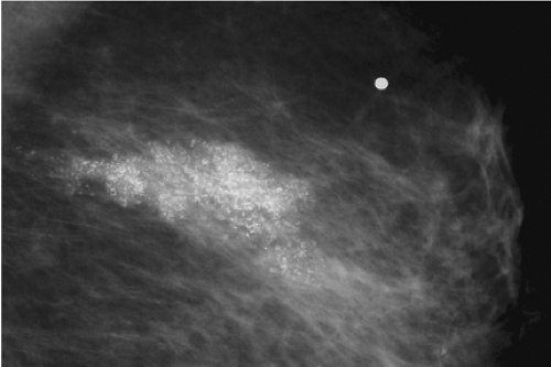
Thirty-two percent of noncomedo lesions in my series did not have mammographic calcifications, making them more difficult to find and the patients more difficult to follow, if treated conservatively. When noncomedo lesions are calcified, they tend to have fine, granular, powdery calcifications or crushed stone–like calcifications (Fig. 7.4).
 Figure 7.3. Mediolateral mammography in a 43-year-old woman showing irregular branching calcifications. Histopathology showed high-grade comedo ductal carcinoma in situ, Van Nuys group 3. |
A major problem confronting surgeons relates to the fact that calcifications do not always map out the entire DCIS lesion, particularly those of the noncomedo type. Even though all the calcifications are removed, in some cases, noncalcified DCIS may be left behind. Conversely, in some patients, the majority of the calcifications are benign and map out a lesion bigger than the true DCIS lesion. In other words, the DCIS lesion may be smaller than, larger than, or the same size as the calcifications that lead to its identification. Calcifications more accurately approximate the size of high-grade comedo lesions than low-grade noncomedo lesions (54).
Before mammography was common or of good quality, most DCIS was usually clinically apparent, diagnosed by palpation or inspection; it was gross disease. Gump et al. (50) divided DCIS by method of diagnosis into gross and microscopic disease. Similarly, Schwartz et al. (29) divided DCIS into two groups: clinical and subclinical. Both groups of researchers thought patients presenting with a palpable mass, a nipple discharge, or Paget’s disease of the nipple required more aggressive treatment. Schwartz believed that palpable DCIS should be treated as though it were an invasive lesion. He suggested that the pathologist simply has not found the area of invasion. Although
it makes perfect sense to believe that the change from nonpalpable to palpable disease is a poor prognostic sign, our group has not been able to demonstrate this for DCIS. In our series, when equivalent patients (by size and nuclear grade) with palpable and nonpalpable DCIS were compared, they did not differ in the rate of local recurrence or mortality.
it makes perfect sense to believe that the change from nonpalpable to palpable disease is a poor prognostic sign, our group has not been able to demonstrate this for DCIS. In our series, when equivalent patients (by size and nuclear grade) with palpable and nonpalpable DCIS were compared, they did not differ in the rate of local recurrence or mortality.
If a patient’s mammogram shows an abnormality, most likely it will be microcalcifications, but it could be a nonpalpable mass or a subtle architectural distortion. At this point, additional radiologic workup needs to be performed. This may include compression mammography, magnification views, or ultrasonography. Magnetic resonance imaging (MRI) has become increasingly popular to map out the size and shape of biopsy-proven DCIS lesions or invasive breast cancers. I obtain a preoperative MRI on every patient with a diagnosis of breast cancer.
Biopsy and Tissue Handling
If radiologic workup shows an occult lesion that requires biopsy, there are multiple approaches: fine-needle aspiration biopsy (FNAB), core biopsy (with various types and sizes of needles), and directed surgical biopsy using guide wires or radioactivity. FNA is generally of little help for nonpalpable DCIS. With FNA, it is possible to obtain cancer cells, but because there is insufficient tissue, there is no architecture. Thus, although the cytopathologist can say that malignant cells are present, the cytopathologist generally cannot say whether the lesion is invasive.
Stereotactic core biopsy became widely available in the early 1990s, and it is now widely used. Dedicated digital tables make this a precise tool in experienced hands. Currently, large-gauge (11 gauge or larger) vacuum-assisted needles are the tools of choice for diagnosing DCIS. Ultrasound-guided biopsy also became very popular in the 1990s but is of less value for DCIS since most DCIS lesions do not present with a mass that can be visualized by ultrasound. All suspicious microcalcifications should be evaluated by ultrasound since a mass will be found in 5% to 15% of cases (58).
Open surgical biopsy should only be used if the lesion cannot be biopsied using minimally invasive techniques. This should be a rare event with current image-guided biopsy techniques (58,59) and occur in less than 5% of cases. Needle localization segmental resection should be a critical part of the treatment not the diagnosis.
Whenever needle localization excision is performed, whether for diagnosis or treatment, intraoperative specimen radiography and correlation with the preoperative mammogram should be performed. Margins should be inked or dyed, and specimens should be serially sectioned at 3- to 4-mm intervals. The tissue sections should be arranged and processed in sequence. Pathologic reporting should include a description of all architectural subtypes, a determination of nuclear grade, an assessment of the presence or absence of necrosis, the measured size or extent of the lesion, and the margin status with measurement of the closest margin.
Tumor size should be determined by direct measurement or ocular micrometry from stained slides for smaller lesions. For larger lesions, a combination of direct measurement and estimation, based on the distribution of the lesion in a sequential series of slides, should be used. The proximity of DCIS to an inked margin should be determined by direct measurement or ocular micrometry. The closest single distance between any involved duct containing DCIS and an inked margin should be reported.
If the lesion is large and the diagnosis unproven, either stereotactic or ultrasound-guided vacuum-assisted biopsy should be the first step. If the patient is motivated for breast conservation, a multiple-wire–directed oncoplastic excision can be planned. This will give the patient her best chance at two opposing goals: clear margins and good cosmesis. The best chance at completely removing a large lesion is with a large initial excision. The best chance at good cosmesis is with a small initial excision. It is the surgeon’s job to optimize these opposing goals. A large-quadrant resection should not be performed unless there is histologic proof of malignancy. This type of resection may lead to breast deformity, and should the diagnosis prove to be benign, the patient will be unhappy.
Removal of nonpalpable lesions is best performed by an integrated team of surgeon, radiologist, and pathologist. The radiologist who places the wires and interprets the specimen radiograph must be experienced, as must the surgeon who removes the lesion and the pathologist who processes the tissue.
Treatment
For most patients with DCIS, there will be no single correct treatment. There will generally be a choice. The choices, although seemingly simple, are not. As the choices increase and become more complicated, frustration increases for both the patient and her physician (60,61).
Counseling the Patient with Biopsy-Proven Ductal Carcinoma in Situ
It is never easy to tell a patient that she has breast cancer. But is DCIS really cancer? From a biologic point of view, DCIS is unequivocally cancer. When we think of cancer, however, we generally think of a disease that, if untreated, runs an inexorable course toward death. That is certainly not the case with DCIS (45). We must emphasize to the patient that she has a borderline cancerous lesion, a preinvasive lesion, which at this time is not a threat to her life. In our series of 1,411 patients with DCIS, the breast cancer–specific mortality rate is 0.5%. Numerous other DCIS series (62,63,64,65,66,67) confirm an extremely low mortality rate.
Patients often ask why there is any mortality rate at all if DCIS is truly a noninvasive lesion. If DCIS recurs as an invasive lesion and the patient goes on to die from metastatic breast cancer, the source of the metastases is clear. But what about the patient who undergoes mastectomy and sometime later develops metastatic disease, or a patient who is treated with breast preservation who never develops a local invasive recurrence but still dies of metastatic breast cancer? These latter patients probably had an invasive focus with established metastases at the time of their original treatment, but the invasive focus was missed during routine histopathologic evaluation. No matter how carefully and thoroughly a specimen is examined, it is still a sampling process, and a 1- to 2-mm focus of invasion can be missed.
One of the most frequent concerns expressed by patients once a diagnosis of cancer has been made is the fear that the cancer has spread. We are able to assure patients with DCIS that no invasion was seen microscopically and the likelihood of systemic spread is minimal.
The patient needs to be educated that the term “breast cancer” encompasses a multitude of lesions of varying degrees of aggressiveness and lethal potential. The patient with DCIS needs to be reassured that she has a minimal lesion and that she is likely going to need some additional treatment, which may include surgery, radiation therapy, an antiestrogen, or some combination. She needs reassurance that she will not need chemotherapy, that her hair will not fall out, and that it is highly unlikely that she will die from this lesion. She will, of course, need careful clinical follow-up.
Endpoints for Patients with Ductal Carcinoma in Situ
When evaluating the results of treatment for patients with breast cancer, a variety of endpoints must be considered. Important endpoints include local recurrence (both invasive and DCIS), regional recurrence (such as the axilla), distant recurrence, breast cancer–specific survival, overall survival, and quality of life. The importance of each endpoint varies depending on whether the patient has DCIS or invasive breast cancer
When treating invasive cancer, the most important endpoints are distant recurrence and breast cancer–specific survival, in other words, living with or dying from breast cancer. For invasive breast cancer, a variety of different systemic treatments have been shown to significantly improve survival. These include a wide range of chemotherapeutic regimens and endocrine treatments. Variations in local treatment were incorrectly thought not to affect survival (18,68). They do, however, affect local recurrence. Recently the literature has shown that for every four local recurrences prevented, one breast cancer death is prevented (69).
DCIS is similar to invasive breast cancer in that variations in local treatment affect local recurrence, but no study has shown a significant difference in distant disease-free or breast cancer–specific survival, regardless of any treatment (systemic or local), and no study is likely to show a difference since there are so few breast cancer deaths in patients with pure DCIS. The most important outcome measure, breast cancer–specific survival, is essentially the same no matter what local or systemic treatment is given. Consequently, local recurrence has become the most commonly used endpoint when evaluating treatment for patients with DCIS.
A meta-analysis of four randomized DCIS trials comparing excision plus radiation therapy versus excision alone was published in 2007. It contained 3,665 patients. Radiation therapy decreased local control by a statistically significant 60%, but overall survival was slightly worse in the radiotherapy group, with a relative risk of 1.08 (66). These data are dissimilar to those of the Early Breast Cancer Trialists’ Collaborative Group and deserve further analysis (69). Half of the recurrences in the meta-analysis were DCIS and could not possibly affect survival. Of the remaining invasive recurrences, 80% to 90% were cured by early detection and treatment. This should result in a slight trend toward a lower survival for the excision-alone group, but exactly the opposite was seen, a nonsignificant trend toward a better survival. The authors of the meta-analysis felt that with longer follow-up, the higher local recurrence rate for excision alone will likely result in a lower overall survival at some point in time. For the time being, however, that has not happened, and a detrimental effect secondary to radiation therapy must be considered a possibility.
Local recurrences are clearly important to prevent in patients treated with DCIS. They are demoralizing. They often lead to mastectomy and, theoretically, if they are invasive, they increase the stage of the patient and are a threat to life. However, protecting DCIS patients from local recurrence must be balanced against the potential detrimental effects of the treatments given.
Following treatment for DCIS, 40% to 50% of all local recurrences are invasive. About 10% to 20% of DCIS patients who develop local invasive recurrences develop distant metastases and die from breast cancer (70,71). Long term, this could translate into a mortality rate of about 0% to 0.5% for patients treated with mastectomy, 1% to 2% for conservatively treated patients who receive radiation therapy (if there is no mortality associated with radiation therapy), and 2% to 3% for patients treated with excision alone. In order to save their breasts, many patients are willing to accept this theoretical, and as of now statistically unproven, small absolute risk associated with breast conservation therapy.
Treatment Options
Mastectomy
Mastectomy is, by far, the most effective treatment available for DCIS if our goal is simply to prevent local recurrence. Most mastectomy series reveal local recurrence rates of approximately 1%, with mortality rates close to zero (72). In my series, I have had only 1 breast cancer death among 485 patients treated with mastectomy (0.2%).
However, mastectomy is an aggressive form of treatment for patients with DCIS. It clearly provides a local recurrence benefit but only a theoretical survival benefit. It is, therefore, often difficult to justify mastectomy, particularly for otherwise healthy women with screen-detected DCIS, during an era of increasing utilization of breast conservation for invasive breast carcinoma. Mastectomy is indicated in cases of true multicentricity (multiquadrant disease) and when a unicentric DCIS lesion is too large to excise with clear margins and an acceptable cosmetic result.
Genetic positivity to one of the breast cancer–associated genes (BRCA1, BRCA2) is not an absolute contraindication to breast preservation, but many patients who are genetically positive and who develop DCIS seriously consider bilateral mastectomy and oophorectomy.
Breast Conservation
The most recent Surveillance Epidemiology and End Results (SEER) data reveal that 74% of patients with DCIS are treated with breast conservation. While breast conservation is now widely accepted as the treatment of choice for DCIS, not all patients are good candidates. Certainly, there are patients with DCIS whose local recurrence rate with breast preservation is so high (based on factors that will be discussed later in this chapter) that mastectomy is clearly a more appropriate treatment. However, the majority of women with DCIS diagnosed currently are candidates for breast conservation. Clinical trials have shown that local excision and radiation therapy in patients with negative margins can provide excellent rates of local control (62,65,66,67,73,74,75,76). However, even radiation therapy may be overly aggressive since many cases of DCIS may not
recur or progress to invasive carcinoma when treated by excision alone (28,45,77,78,79,80).
recur or progress to invasive carcinoma when treated by excision alone (28,45,77,78,79,80).
Reasons to Consider Excision Alone
There are a number of lines of reasoning that suggest that excision alone may be an acceptable treatment for selected patients with DCIS.
The prevalent use of excision. Excision alone is already common in spite of the randomized data that suggest that all conservatively treated patients benefit from radiation therapy. The 2003 SEER data indicated that excision alone was being used as complete treatment for DCIS in 35% of all DCIS patients. American doctors and patients have embraced the concept of excision alone.
Anatomic considerations. Evaluation of mastectomy specimens using the serial subgross tissue processing technique reveals that most DCIS is unicentric (involves a single breast segment and is radial in its distribution) (35,39,54,55,81,82). This means that in many cases, it is possible to excise the entire lesion with a segment or quadrant resection. Since DCIS, by definition, is not invasive and has not metastasized, it can be thought of in Halstedian terms. Complete excision should cure the patient without any additional therapy. Faverly et al. showed that if 10-mm margins are achieved in all directions, the likelihood of residual DCIS is less than 10% (39).
Stay updated, free articles. Join our Telegram channel

Full access? Get Clinical Tree


