Chapter 7
Drug Rashes
OVERVIEW
- Drug reactions in the skin are common. Cutaneous adverse reactions account for a third of all adverse reactions to drugs.
- Drugs can cause adverse reactions in several ways: by changing normal skin function, by exacerbating an existing dermatosis, by causing an idiopathic dermatosis such as urticaria, by causing a specific drug eruption (lichenoid drug rashes) or by precipitating a severe drug reaction (toxic epidermal necrolysis).
- Identifying the culprit drug requires careful history-taking and knowledge of the notoriety of certain drugs in the causation of certain reactions.
- Careful skin examination is required to identify the morphology of the rash and the correct classification of the drug reaction.
- The most important step in management of drug rashes is identification and withdrawal of the culprit medication.
- Some drug reactions are mild and resolve quickly (maculopapular exanthems); others are more severe and carry considerable morbidity and mortality (Stevens–Johnson syndrome and toxic epidermal necrolysis).
Introduction
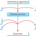
Adverse reactions in the skin to medications are very common and are an important cause of iatrogenic illness. Drug rashes are usually self-limiting and resolve completely upon withdrawal of the culprit medication, but a small number (<2%) can cause serious morbidity and mortality. This may not only have medicolegal and economic implications but may undermine the patient’s confidence in the prescriber and affect future adherence. Diagnosis of drug-induced skin disease may be difficult for a number of reasons:
- Almost any drug can cause any rash.
- Unrelated drugs can cause similar reactions.
- The same medication can cause a different rash in different individuals.
- Patients may not volunteer information about medicines that they have taken, which they deem not to be relevant (over-the-counter (OTC) preparations and complementary medicines).
History
It is imperative to take a thorough history from patients in whom a drug reaction is suspected. Eliciting the temporal association of the ingestion of the drug and the onset of the eruption is key (Table 7.2). Apart from noting any medications taken for the first time in the 3 months prior to the appearance of the rash, patients should be specifically asked about any recent changes to brand, dosing or preparation of long-term medications. Patients may not volunteer information about drugs they have taken that they assume are not relevant, such as paracetamol taken for a headache, or an antihistamine taken for hay fever. Direct questioning about OTC preparations should always form part of a thorough drug history. Medications recommended by alternative/complementary health practitioners may not be revealed spontaneously and should be asked about directly. Both generic and brand names of all drugs should be recorded, and the patient should be asked about any history of sensitivity to medications. Knowledge of whether the patient has been previously exposed to suspected culprit drugs is also relevant.
Examination
The patient should be exposed fully to allow complete examination of the skin. The morphology of the rash should be described—for example, lichenoid, urticated, vasculitic, maculopapular or bullous. The distribution of the rash should be noted: Is it widespread? Limited? Acral (hands and feet)? Photo-distributed? These features will help to classify the eruption and may give a pointer as to the causative drug. Special attention should be paid to the mucosal sites—eyes, mouth, genitalia—as involvement at these sites can indicate one of the severe cutaneous adverse reaction (SCAR) syndromes. Early diagnosis of these syndromes is crucial as patients may become unwell and deteriorate quickly. Careful examination of the appendageal structures such as hair, nails and teeth should also be carried out as these can be affected by certain medications.
Investigations
In most cases, careful history and examination will provide all the necessary clues to make a confident diagnosis of a drug rash. A skin biopsy can be helpful to confirm the diagnosis, but the result of this is likely to be delayed following an acute presentation, and so action based on clinical assessment will usually precede this. Exclusion of differential diagnoses such as infection may require other investigations such as blood tests (white cell counts and CRP levels). There are no consistently reproducible diagnostic tests which confirm specific drug allergy in the convalescent period; however, certain investigations such as measurement of specific IgE, patch testing, intradermal testing and in vitro tests such as lymphocyte transformation tests and cytokine release assays may be helpful. However, these investigations should be carried out by experts as their interpretation is highly specialised.
Classification of drug reactions in the skin
Cutaneous reactions to medications are extremely varied. They may be classified in a number of ways. Pathogenetically, drug reactions in the skin may be classified as immune-mediated or non-immune mediated. Immune-mediated rashes are the most common and include hypersensitivity reactions from types I to IV. Type I reactions (immediate reactions, usually mediated by IgE or drug-specific receptors bound to mast cells and other immune cell membranes) tend to manifest in the skin as urticaria or angio-oedema. Type II reactions (cytotoxic reactions) result in cutaneous purpura. Type III (immune complex-mediated) reactions lead to cutaneous vasculitis. Type IV delayed hypersensitivity reactions are by far the most common group of drug rashes resulting in generalized exanthems, phototoxic rashes and severe drug reactions such as toxic epidermal necrolysis (TEN).
Non-immune-mediated rashes include accumulation of medications in the skin (causing pigment changes), instability of mast cells (causing histamine release), slow acetylators (metabolism of drugs affected) and photosensitivity reactions (increased susceptibility to UV light).
However, drug reactions in the skin may also be classified clinically, and this is the approach adopted here. Drug reactions in the skin are discussed under the following headings:
- Drugs which alter normal skin function.
- Drugs which exacerbate an existing dermatosis.
- Common drug-induced rashes—maculopapular exanthem, urticaria, vasculitis, lichenoid drug reaction and fixed drug eruption.
- Severe drug-induced rashes—Stevens–Johnson syndrome (SJS) and TEN, acute generalised exanthematous pustulosis (AGEP) and drug reaction with eosinophilia and systemic symptoms (DRESS).
Drugs which alter normal skin function
Photosensitivity
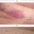
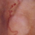
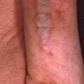
Drugs may cause excessive sensitivity to light in two ways: phototoxic reactions and photoallergic reactions. Phototoxic reactions (Figure 7.1a and b) are the more common, and resemble sunburn and may blister. The reaction is confined to light-exposed sites and may be characterised by a sharp demarcation between covered and uncovered skin. The onset will typically be fast (within 5–15 h of taking the drug and exposure to light) and recovery is quick on withdrawal of the medication. Photoallergic reactions are usually eczematous, but may be lichenoid, uriticarial, purpuric or bullous. The onset may be delayed by weeks or months following introduction of the medication, and similarly, recovery may be slow on withdrawal. Patients taking medication known to cause light sensitivity (amiodarone, tetracycline antibiotics and retinoids) should be advised to avoid excess sun exposure, and to wear a broad-spectrum sun screen year-round. Drugs causing photosensitivity are detailed in Table 7.1.
Table 7.1 Cutaneous reactions and the most commonly implicated drugs
| Skin reaction | Drugs |
| Phototoxic reactions | Amiodarone, NSAIDs, tetracyclines, chlorpromazine |
| Photosensitive reactions | Amiodarone, tetracyclines, calcium channel blockers, diuretics, voriconazole, itraconazole, terbinafine, ritonavir, saquinavir |
| Photoallergic reactions | NSAIDs, antibiotics, thiazides, anticonvulsants, allopurinol, quinolones, nelfinavir |
| Pigmentation changes | Chlorpromazine, phenytoin, hydroxychloroquine, cyclofosphamide, bleomycin, amiodarone, clofazimine, minocycline, mepacrine |
| Urticaria/angio-oedema | NSAIDs, opioid analgesics, ACE inhibitors, antibiotics, anti-retrovirals (didanosine/nelfinavir/zidovudine), infliximab, proton-pump inhibitors, IV contrast media |
| Drug-induced lupus | Terbinafine, hydralazine, procainamide, quinidine, isoniazid, diltiazem, and minocycline |
| Drug-induced vasculitis | Antibiotics, NSAIDs, phenytoin, ramipril, proton-pump inhibitors, allopurinol, thiazides, adalimumab, indinavir |
| Lichenoid drug eruption |