Chapter 13
Bacterial Infections
OVERVIEW
- Damage to the epidermis/dermis results in reduced barrier function, which enables bacteria to invade the skin.
- Cutaneous bacterial infections produce signs of acute inflammation: erythema, swelling/oedema, heat/warmth and pain/discomfort.
- Localised superficial skin infections can be managed with antiseptic washes and topical antibiotics.
- Systemic antibiotics are needed for widespread, deep and persistent bacterial skin infections.
- Clinical signs of bacterial infections include folliculitis, boils/abscesses, blistering, crusting, erosions, ulcers and cellulitis.
- Mycobacterial skin infections most commonly arise from implantation/trauma and are usually localised in immunocompetent patients.
- Atypical mycobacteria can result in localised supurative lesions, persistent granulomas and sporotrichoid spread via lymphatics.
Introduction
Intact skin forms a highly effective barrier against invading pathogenic bacteria. Many micro-organisms come into contact with the skin and some live there as part of the normal skin flora, but they rarely cause disease. Normal skin flora consists of coagulase-negative Staphylococcus, Corynebacterium, diphtheroids and α-haemolytic Streptococci in the epidermis, and Propionibacterium in the pilosebaceous unit. Normal flora competes with invading pathogenic micro-organisms, thereby acting as a ‘biological shield’. However, if the host immune system weakens or there is a change in the micro-environment (such as an underlying skin disease) this may allow such bacteria to become pathogenic.
Bacterial skin infections may be acquired from the external environment (from plants, soil, fomites, animals or other humans) by implantation, direct contact, aerosols or water-borne transmission. Bacteria most frequently invade a traumatic break in the skin, follicular openings and mucous membranes where host barriers are more vulnerable.
Bacterial skin infections vary from the very minor to life-threatening and overwhelming. It is often tempting to assume a single organism is responsible for any particular cutaneous infection, but often the contrary is true. Synergistic microbial invasion is frequently present in cutaneous wounds. See Table 13.1 for a summary of the common patterns of bacterial infection in the skin.
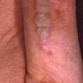
Table 13.1 Common patterns of bacterial infection in the skin.
| Infection | Clinical photograph | Clinical presentation | Organisms | Management |
| Infected eczema | 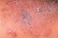 | Background inflammatory atopic dermatitis with excoriations and marked crusting and exudate | Staphylococcus aureus Streptococcus pyogenes | Antiseptic wash Topical antibiotic/steroid combination cream. Oral flucloxacillin or erythromycin |
| Impetigo | 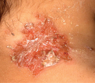 | Mainly children, especially face and limbs. Highly contagious. Yellow crusted lesions surrounded by normal skin | S. aureus S. pyogenes | Antiseptic wash Topical antibiotic Oral flucloxacillin or erythromycin |
| Bullous impetigo | 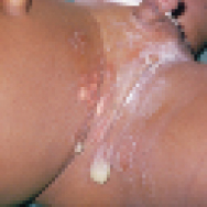 | Children and adults. Face, limbs and flexures affected. Erythema with bullae which rupture leaving superficial erosions and crusts | S. aureus with exfoliative toxins A/B (may become generalised—staphylococcal scalded skin syndrome) | Oral flucloxacillin or erythromycin |
| Boils (abscesses) | 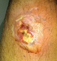 | Tender, inflamed indurated nodules with central pus may be single or multiple. If recurrent and recalcitrant consider toxin producing bacteria | S. aureus Consider Panton valentine leukocidin Toxin producing S. aureus | Antiseptic wash Oral flucloxacillin or erythromycin If PVL positive give nasal bactroban and consider giving clindamycin plus rifampicin for 4–6 weeks |
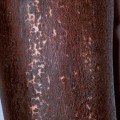
| Bacterial folliculitis | 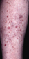 | Hair-bearing sites particularly legs, beard area and scalp. May result from shaving damage to skin. In recurrent infections look for S. aureus nasal carriage | S. aureus Pseudomonas aeruginosa (differential diagnosis Malassezia spp) | Topical antibiotics Acetic acid cream EarCalm® for P. aeruginosa Oral flucloxacillin or erythromycin Avoid shaving if possible |
| Ecthyma | 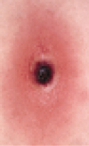 | Children, the elderly/debilitated. Mainly on the legs. Initially small bullae with necrotic dry adherent crust and underlying ulceration Heal slowly with scarring | S. pyogenes S. aureus | Antiseptic wash Oral penicillin V or erythromycin |
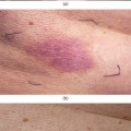
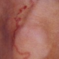
| Erysipelas | 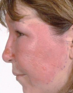 | Face or lower leg. Portal of entry is broken skin (trauma and tinea pedis) well-demarcated bright erythema | S. pyogenes (group A Strep. but also B, C, G) S. aureus (less common) | Intravenous benzyl penicillin or erythromycin |
Clinical presentation
Patients with a bacterial skin infection may recall an episode of trauma to the skin such as a graze, laceration, insect bite or implantation of foreign material, or they may have a history of ongoing skin disease. A more detailed history may reveal contact with potentially contaminated water via bathing, animal contact, travel abroad or other family members/close contacts similarly affected. However, many patients will not have any obvious source from the history alone.
Acute bacterial infections in the skin generally produce some or all of the classical characteristics of acute inflammation: erythema, swelling/oedema, heat/warmth and pain/discomfort. Patients may develop systemic symptoms such as fever and malaise. Many cutaneous infections start as an isolated lesion that then spreads to involve the surrounding previously uninvolved skin. Multiple lesions may be present in a follicular distribution.
Bacterial investigations
Taking bacterial swabs for microscopy and culture can be very useful in managing patients with probable cutaneous infections. Microbiological testing can identify the bacterial species, antibiotic resistance/sensitivity patterns and bacterial toxin production. This information allows medical practitioners to make informed decisions regarding patient management. Lesional skin and carrier sites can be swabbed. Nasal swabs may identify Staphylococcus aureus carriers who can suffer from recurrent infections because of bacterial shedding from the nose. When taking swabs they should be moistened in the transport media before contact with the skin and each surface of the swab should be rotated on the infected skin surface. Methicillin-resistant S. aureus (MRSA) may be community or hospital acquired. Panton Valentine Leukocidin (PVL) is a toxin produced by some strains of S. aureus which cause the bacteria to be highly virulent and highly transmissible. Patients with PVL-positive S. aureus often present with multiple/recurrent boils not settling with short courses of flucloxacillin (Figure 13.1). Often other family members are similarly affected. Request PVL testing when sending swabs to microbiology. In severe skin infections or when you suspect mycobacterial infections, take a skin biopsy for culture and polymerase chain reaction (PCR).
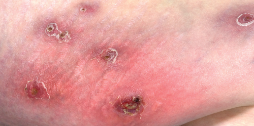
Figure 13.1 Mulitple abscesses due to PVL Staphylococcus aureus infection.
General approach to management
The treatment approach depends on the extent and the severity of the cutaneous infection.
Antiseptic
Related posts:

Stay updated, free articles. Join our Telegram channel

Full access? Get Clinical Tree


