Disorders of Hair, Adnexae, Nails, and Cartilage
Ifeoma U. Perkins
Jarish Cohen
Paradi Mirmirani
TRICHORRHEXIS NODOSA
Definition and Epidemiology
Trichorrhexis nodosa (TN) is an inherited or acquired fragile hair shaft disorder characterized by hair breakage that occurs as a result of damage to hair cuticles.1 The disease can have a more proximal or distal distribution along the hair shaft. There is a wide age range at presentation. Acquired TN is common in patients of African descent (proximal type) and Caucasian and Asian descent (distal type).
Etiology
Acquired TN can be the result of physical trauma including excessive brushing, heat, pulling, or twisting.2 It can also be the result of chemical trauma including permanent straightening or waving. Inherited TN can be caused by arginosuccinic aciduria, a rare metabolic disorder of the urea cycle causing hyperammonemia.3 If undiagnosed in infancy, neonates can present with failure to feed leading to lethargy and coma in some instances. Congenital TN can also be an incidental finding in patients with other hair shaft disorders including pili torti, monilethrix, and trichothiodystrophy.
Clinical Presentation
TN may present with small, white, or gray flecks on the hair shafts. Hair damage is usually noticed as a change in the quality of the hair or as “dry ends” or “flyaway hair” and may have a “shaggy” or “skimpy” appearance (Figure 14-1).
Hair breakage can result in focal or diffuse alopecia depending on the extent of scalp involvement. A tug test, in which a swatch of hair is held in one hand and the proximal end of the hair is tugged with the other hand, will lead to fragments of shortened hairs that are easily broken off.
Histologic Findings
Each node is formed by cortical fiber separation and fraying of the hair shaft in a manner analogous to the bristles of two paint brushes being pushed together (Figure 14-2).
CAPSULE SUMMARY
TRICHORRHEXIS NODOSA
TN occurs as a result of physical or chemical trauma. The clinical presentation is that of small nodes or white flecks on the affected hair shafts that can result in alopecia of various severity. Light microscopy shows fragile nodes with frayed cortical fibers with a “pushed together” appearance.
MONILETHRIX
Definition and Epidemiology
Monilethrix is a hair shaft disorder characterized by brittle, beaded hairs that emanate from keratotic, follicular papules.4 The condition usually manifests in the neonatal period or early childhood. In rare instances, babies are born
bald with beaded hairs appearing later in life. The course can be seasonal and is variable with some affected patients improving in adulthood, whereas in others it can worsen.
bald with beaded hairs appearing later in life. The course can be seasonal and is variable with some affected patients improving in adulthood, whereas in others it can worsen.
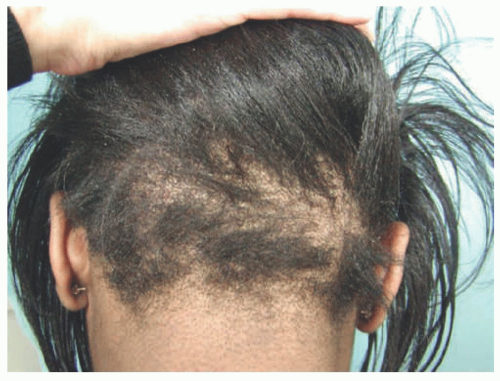 FIGURE 14-1. Trichorrhexis Nodosa. Localized area of short hair-blunt ends with positive tug test. From Mirmirani P, Huang KP, Price VH. A practical, algorithmic approach to diagnosing hair shaft disorders. Int J Dermatol. 2011;50(1):1-12. |
Etiology
Monilethrix is usually inherited in an autosomal dominant fashion with high penetrance and variable expressivity. In the autosomal dominant variant, mutations in 3 type II basic hair keratin genes, hHb1, hHb3, and hHb6 (chromosome 12q13) have been described, with the latter being the most commonly altered.5,6,7,8,9 An autosomal recessive variant has been reported and is caused by a mutation in desmoglein 4.10,11 Desmoglein 4 is a transmembrane cell adhesion molecule expressed in the hair cortex and upper cuticle, which is also associated with localized autosomal recessive hypotrichosis.8
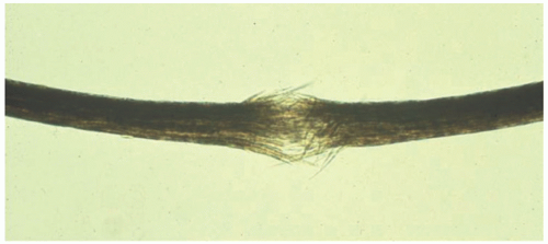 FIGURE 14-2. Trichorrhexis nodosa. Hair mount reveals brush-like ends in opposition. From Mirmirani P, Huang KP, Price VH. A practical, algorithmic approach to diagnosing hair shaft disorders. Int J Dermatol. 2011;50(1):1-12. |
Clinical Presentation
Hair shafts rarely grow more than 2 to 3 cm. The hairs break easily, resulting in severe alopecia (Figure 14-3). In severe variants, the affected hair can involve the entire scalp, secondary sexual hair, eyebrows, and eyelashes.12 Neonates who develop monilethrix initially present with normal lanugo hair and later have characteristic findings when they grow terminal hair. Follicular hyperkeratosis may also be found on the body. Other features may include koilonychia, mental retardation (rare), syndactyly, cataracts, as well as tooth and other nail abnormalities.13
Histologic Findings
Hair shafts show regularly spaced, elliptical nodes that are roughly 0.7 to 1.1 mm apart. The nodes are of normal caliber and have a typical air-filled medulla, whereas the areas of constriction show an absence of medulla (Figure 14-4).
CAPSULE SUMMARY
MONILETHRIX
Monilethrix is a hair shaft fragility disorder that typically presents in early childhood and can progress to severe alopecia. The autosomal dominant form is caused by mutations in type II basic keratin genes, and the autosomal recessive variant is caused by mutations in desmoglein 4. Affected hair shafts show elliptical nodes at regular intervals, and internodal regions that are of smaller caliber and lack the hair shaft medulla.
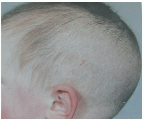 FIGURE 14-3. Monilethrix. Short broken hairs noted predominantly in the occipital scalp. Courtesy of Paradi Mirmirani, MD, Kaiser Permanente, Vallejo CA. |
TRICHORRHEXIS INVAGINATA
Definition and Epidemiology
Trichorrhexis invaginata is a rare congenital hair shaft abnormality characterized by short, brittle hairs that manifests in patients with Netherton syndrome (NS). NS, and thus trichorrhexis invaginata, affects children. Progressive alopecia and atopic eczema are present at a young age.
Etiology
NS is an autosomal recessive disorder caused by mutations in the SPINK5 gene, which encodes the serine protease inhibitor LEKTI.14,15 LEKTI is necessary for proper skin barrier function and desquamation.16 Although its function in the follicle is not well characterized, LEKTI appears to be highly expressed in sites of keratinization such as the inner root sheath and upper infundibulum.17
Clinical Presentation
NS is characterized by the triad of trichorrhexis invaginata, ichthyosis linearis circumflexa, and atopic diathesis.18 Patients are born with congenital ichthyosiform erythroderma,19 neonatal dehydration, failure to thrive, and recurrent infections.20 Ichthyosis linearis circumflexa consists of migratory polycyclic erythematous patches with surrounding serpiginous double-edged scale.21 An atopic diathesis may include eczema-like eruptions, atopic dermatitis, asthma, pruritus, allergic rhinitis, angioedema, urticaria, elevated serum IgE, and/or hypereosinophilia.20 The hair findings in NS, including TN, are not present in every hair. Multiple hairs should be tested, and the most high-yield location to pluck and examine is reported to be the lateral eyebrows.22
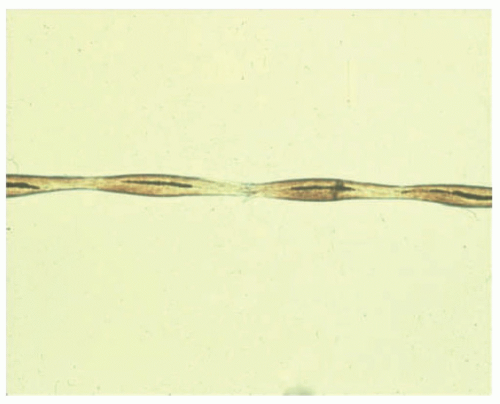 FIGURE 14-4. Monilethrix. Hair mount reveals beaded nodes at regular intervals. From Mirmirani P, Huang KP, Price VH. A practical, algorithmic approach to diagnosing hair shaft disorders. Int J Dermatol. 2011;50(1):1-12. |
Histologic Findings
In trichorrhexis invaginata, nodes appear at irregular intervals and are composed of a distal portion of the hair shaft that has sunken into the proximal portion creating a “ball-and-socket” appearance22 (Figure 14-5). Nascent nodes form under torsion, and the distal aspect of the hair shaft undergoes intussusception into the proximal aspect.23
CAPSULE SUMMARY
TRICHORRHEXIS INVAGINATA
Trichorrhexis invaginata is one of the triad of signs that define NS, the others being ichthyosis linearis circumflexa, and atopic diathesis. Patients with NS have defects in the SPINK5 gene, which encodes LEKTI, a serine protease inhibitor integral to normal barrier function. The characteristic light microscopic feature is that of nodes showing the distal portion of the hair shaft herniating into the proximal portion, forming a ball-andsocket joint appearance.
PILI TORTI
Definition and Epidemiology
Pili torti is a hair shaft defect characterized by short, brittle, and spangled hairs and is associated with various syndromes, including Menkes disease, Björnstad syndrome, and p63 mutation-associated ectodermal dysplasia syndromes (referred to in the literature by multiple monikers such as ectrodactyly, ectodermal dysplasia, and cleft lip;
Rapp-Hodgkin syndrome; and ankyloblepharon ectodermal dysplasia cleft lip/palate syndrome/Hay Wells Syndrome). Pili torti usually presents in early childhood, but can sometimes occur at birth or arise after puberty.
Rapp-Hodgkin syndrome; and ankyloblepharon ectodermal dysplasia cleft lip/palate syndrome/Hay Wells Syndrome). Pili torti usually presents in early childhood, but can sometimes occur at birth or arise after puberty.
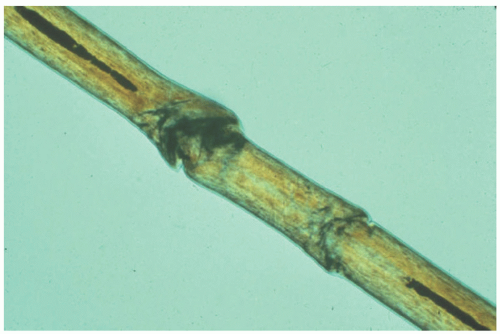 FIGURE 14-5. Trichorrhexis invaginata. Ball and socket appearance of hair shaft as it collapses on itself. Courtesy of Paradi Mirmirani, MD, Kaiser Permanente, Valejo CA. |
Etiology
The twisted appearance may be caused by mitochondrial dysfunction and the buildup of reactive oxygen species particularly in the inner root sheath.24 Menkes disease is caused by a mutation in the X-linked ATP7A gene, which encodes a copper-transporting ATPase.25,26,27,28,29,30 Defects in this protein may account for hair twisting by defective incorporation of copper into keratin disulfide bonds and/or by the aforementioned mitochondrial dysfunction mechanism. Mutations in the BCS1L gene underlie the phenotype of Björnstad syndrome.24,31 Hay-Wells and/Rapp Hodgkins syndromes are caused by mutations in the TP63 gene, which is expressed by many ectodermally derived structures such as the hair follicle.32
Clinical Presentation
Variable hair fragility manifests as patchy alopecia, with coarse stubble predominantly seen in the temporal and/or occipital areas presumably due to friction, with areas of hairs of up to 5 cm of length (Figure 14-6). The hairs of the eyelashes and eyebrows can also be involved. In addition to pili torti, patients with Menkes disease have severe developmental and neurologic impairment, connectivetissue abnormalities, and tortuous blood vessels.25 After lanugo hair is shed, infants develop stubbly, sparse, hypopigmented hair that has been described as “steel wool” in appearance and texture because it is difficult to comb and fractures with friction.33 In Björnstad syndrome, pili torti and bilateral sensorineural loss usually manifest before the age of two.34 The degree of hair shaft fragility correlates to the severity of hearing loss.34 TP63 gene mutation-related
ectodermal dysplasias have variable expressivity, but features can include irregular strands of tissue connecting the upper and lower eyelids, termed ankyloblepharon filiforme adnatum, hypohidrosis, erythroderma, palmoplantar keratoderma, and cleft lip, cleft palate, or both. There is often also a severe and recalcitrant scalp dermatitis in these patients.35
ectodermal dysplasias have variable expressivity, but features can include irregular strands of tissue connecting the upper and lower eyelids, termed ankyloblepharon filiforme adnatum, hypohidrosis, erythroderma, palmoplantar keratoderma, and cleft lip, cleft palate, or both. There is often also a severe and recalcitrant scalp dermatitis in these patients.35
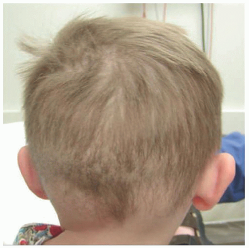 FIGURE 14-6. Björnstad syndrome; Pili torti. Short hair in the occipital scalp with spangled appearance. From Mirmirani P, Huang KP, Price VH. A practical, algorithmic approach to diagnosing hair shaft disorders. Int J Dermatol. 2011;50(1):1-12. |
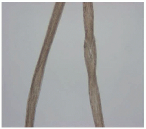 FIGURE 14-7. Björnstad syndrome; Pili torti. Hair mount reveals flattened and twisted hair on the long axis. From Mirmirani P, Huang KP, Price VH. A practical, algorithmic approach to diagnosing hair shaft disorders. Int J Dermatol. 2011;50(1):1-12. |
Histologic Findings
Affected hairs are flattened and twisted on the horizontal axis typically by 180β turns. Usually runs of 3 to 10 twists are present at irregular intervals along the hair shaft (Figure 14-7).
CAPSULE SUMMARY
PILI TORTI
Pili torti is a hair fragility condition that can be seen in various syndromes, including Menkes disease, Björnstad syndrome, and p63 mutation-related ectodermal dysplasia syndrome. Pili torti manifests clinically as short, brittle hair that is prone to fracturing, which can eventuate in patchy alopecia accentuated in the temporal and occipital regions. Light microscopy shows abnormal twisting of the hair at irregular intervals.
PILI BIFURCATI
Definition and Epidemiology
Pili bifurcati is a rare hair shaft disorder characterized by thin, short fragile hairs eventuating in diffuse alopecia. Pili bifurcati affects patients in childhood.
Etiology
The underlying pathogenic defect is unknown.
Clinical Presentation
Histologic Findings
The affected hairs show bifurcated hair shafts. On scanning electron microscopy, a single hair shaft splits into two smaller shafts, which then fuse downstream to again form a single hair shaft.38 The two bifurcated hair shafts are completely covered by their own cuticles.
CAPSULE SUMMARY
PILI BIFURCATI
Pili bifurcati is a hair shaft fragility disorder characterized by short coarse hair and diffuse alopecia. The hair shafts show nodes of bifurcation with downstream fusion into a single hair shaft.
TRICHOTHIODYSTROPHY
Definition and Epidemiology
Trichothiodystrophy is a hair shaft abnormality characterized by brittle hairs and is associated with neuroectodermal disorders. Trichothiodystrophy presents at birth.
Etiology
Trichothiodystrophy is caused by mutations in xeroderma pigmentosum-B, xeroderma pigmentosum-D, and p8/TTDA helicase/ATPase subunits of the DNA repair/basal transcription factor IIH, which are three genes involved in transcription initiation and nucleotide excision repair.39 The gene defects result in hair with a significant reduction in sulfur-rich cysteine matrix proteins. Alterations in these genes also give rise to xeroderma pigmentosum and Cockayne syndrome.
Clinical Presentation
Depending on the specific gene mutation, patients with trichothiodystrophy can present with a complex, sometimes referred by the acronym PIBIDS: photosensitivity, ichthyosis, brittle hair, intellectual impairment, decreased fertility, short stature, or IBIDS, when photosensitivity is not present. The hair findings include short, brittle hairs of varying length on the scalp, eyebrows, and eyelashes.39,40
Histologic Findings
Affected hairs show a clean transverse break through the hair shaft and an absence of cuticle cells at the fracture site, a defect termed trichoschisis (Figure 14-8). Hairs may be focally flattened or ribbon-like41 (Figure 14-9). When viewed under polarized light in the dark or “extinguished” position, an alternating light and dark banding pattern (tiger tail banding) can be seen42,43 (Figure 14-10).
CAPSULE SUMMARY
TRICHOTHIODYSTROPHY
Trichothiodystrophy is a brittle hair disorder caused by a deficiency in sulfur incorporation into the hair shaft due to mutation in genes involved in transcription initiation and DNA repair. Patients typically present in early childhood with ichthyosis and varying degrees of neurologic impairment, as well as short brittle hair.
MARIE-UNNA HEREDITARY HYPOTRICHOSIS
Definition and Epidemiology
Marie-Unna hereditary hypotrichosis (MUHH) is a hair shaft abnormality characterized by coarse, unruly hair, and progressive hair loss. The disorder usually presents in childhood.
Etiology
A subset of patients with MUHH have mutations in the U2HR locus, an upstream regulator of the Hr gene, but the exact mechanism of the disease is not known.44 Mutations in the epidermal growth factor receptor kinase substrate 8-like protein 3 have also been associated with MUHH.45
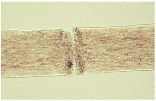 FIGURE 14-8. Trichothiodystrophy: light microscopy reveals trichoschisis. Courtesy of Paradi Mirmirani, MD, Kaiser Permanente, Vallejo CA. |
Clinical Presentation
MUHH can present as an isolated phenomenon or as part of an autosomal dominant inherited syndrome. Typically, patients are born with normal-appearing to slightly coarse hair, although there are cases with sparse to no hair.46,47 Over time, hair is progressively lost and becomes coarse and wiry. In addition to the scalp, hair can be lost from the eyebrows, eyelashes, and the body. By puberty, a nonscarring alopecia is present, with the vertex and scalp margins being the most affected sites.48
Histologic Findings
Affected hairs are deeply pigmented, are of varying caliber, and may be twisted or bent.49
CAPSULE SUMMARY
MARIE-UNNA HEREDITARY HYPOTRICHOSIS
MUHH is a hair shaft anomaly that manifests as coarse wiry hair that is gradually lost, resulting in nonscarring alopecia. The hairs in MUHH are twisted and bent and exhibit dark pigmentation.
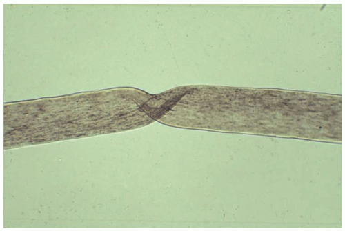 FIGURE 14-9. Trichothiodystrophy: light microscopy reveals ribboning. From Mirmirani P, Huang KP, Price VH. A practical, algorithmic approach to diagnosing hair shaft disorders. Int J Dermatol. 2011;50(1):1-12. |
PILI ANNULATI
Definition and Epidemiology
Also called “ringed hair,” pili annulati is a rare hair shaft disorder characterized by light and dark bands within the hair shaft, leading to a “shiny,” “spangled,” or “sandy” appearance.
Epidemiology
The disorder is present at birth or arises during infancy.
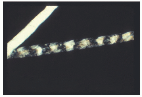 FIGURE 14-10. Trichothiodystrophy: polarizing microscopy reveals tigertail banding. From Mirmirani P, Huang KP, Price VH. A practical, algorithmic approach to diagnosing hair shaft disorders. Int J Dermatol. 2011;50(1):1-12. |
Etiology
Pili annulati is inherited in an autosomal dominant fashion. The disease-causing locus has been mapped to the telomeric region on chromosome 12q.50
Clinical Presentation
The hairs in pili annulati show alternating light and dark banding along the hair shaft with a variable number of hairs affected, often imparting a shiny appearance to the hair (Figure 14-11). It can affect the entire scalp hair or be patchy and can also involve the axillae, beard, and pubic region.51,52 Hair growth is normal and hairs are typically pliable and strong, but there may be some degree of hair fragility. TN-like fractures may be induced in the dark bands.
Histologic Findings
The clinically apparent light bands appear as dark bands on hair mount because of the inverse effects of reflected and transmitted light (Figures 14-12 and 14-13). The bands correspond to air-filled cavities within the hair shaft that can be appreciated on electron microscopy.53,54,55
CAPSULE SUMMARY
PILI ANNULATI
Pili annulati is characterized by a characteristic banding of hair, resulting in a shiny or sandy appearance. Affected hairs have air-filled cavities in the hair shaft and are typically of normal length and strength, but can have some degree of susceptibility to fracture.
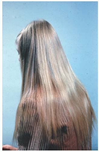 FIGURE 14-11. Pili annulati. Hair has a shiny appearance. From Mirmirani P, Huang KP, Price VH. A practical, algorithmic approach to diagnosing hair shaft disorders. Int J Dermatol. 2011;50(1):1-12. |
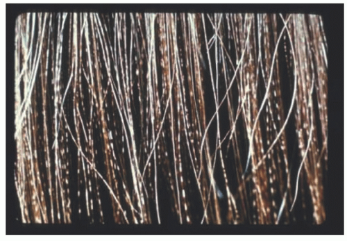 FIGURE 14-12. Pili annulati. Closeup of the hair shafts reveal spangled appearance. From Mirmirani P, Huang KP, Price VH. A practical, algorithmic approach to diagnosing hair shaft disorders. Int J Dermatol. 2011;50(1):1-12. |
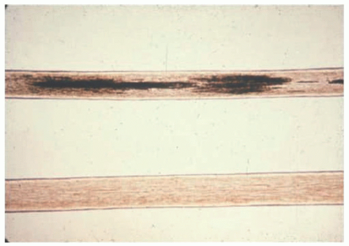 FIGURE 14-13. Pili annulati. Hair mount reveals dark and light bands in comparison with normal hair shaft. From Mirmirani P, Huang KP, Price VH. A practical, algorithmic approach to diagnosing hair shaft disorders. Int J Dermatol. 2011;50(1):1-12. |
WOOLLY HAIR
Definition and Epidemiology
Woolly hair is a hair shaft disorder characterized by fine, tightly wound hairs that may be associated with other disorders or syndromes. The disorder usually presents in childhood.
Etiology
Mutations that underlie the autosomal recessive presentations of wooly hair affect P2RY5 gene56,57 and lipase H gene.58,59 The P2RY5 gene encodes a G protein-coupled receptor that is expressed in Henle and Huxley layers of the inner root sheath of the hair follicle. Lipase H gene mutations are also present in isolated hypotrichosis and likely plays a role in hair follicle growth58,59 Somatic mosaic HRAS mutations can cause the association of woolly hair and epidermal nevi.60
Clinical Presentation
Woolly hair can present in isolation on the scalp in a partial or focal manner, such as in the wooly hair nevus, or in a syndromic manner61,62 (Figure 14-14). Associated findings and syndromes include palmoplantar keratoderma,63 keratosis pilaris atrophicans fasciei,64 Noonan syndrome,65 Carvajal syndrome,66 cardiofaciocutaneous syndrome,67 Naxos disease,68 keratosis follicularis spinulosa decalvans,69 and epidermal nevi.60
Naxos disease is an autosomal recessive disorder characterized by wooly hair from birth, diffuse nonepidermolytic palmoplantar keratoderma, and arrhythmogenic right ventricular dysplasia/cardiomyopathy.70,71 The latter tends to present in late puberty. In contrast, patients with Carvajal syndrome exhibit epidermolytic palmoplantar keratoderma in addition to wooly hair and cardiac defects.66,72
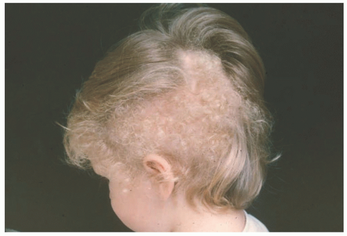 FIGURE 14-14. Woolly hair nevus. Discrete patch of tightly curled hair in an otherwise normal field of scalp. From Mirmirani P, Huang KP, Price VH. A practical, algorithmic approach to diagnosing hair shaft disorders. Int J Dermatol. 2011;50(1):1-12. |
Histologic Findings
There are no specific pathologic findings on hair mount.
CAPSULE SUMMARY
WOOLLY HAIR
Patients with wooly hair syndrome display fine tightly coiled hairs. The disorder may present in isolation or may be associated with other cutaneous disorders or syndromes.
ACQUIRED PROGRESSIVE KINKING OF THE HAIR
Definition and Epidemiology
Acquired progressive kinking of the hair (APKH) is a rare disorder characterized by a rapid onset of curly, unruly hair. The disorder presents in children and young adults with a peak in adolescence.
Etiology
The underlying pathogenic defect is unknown.
Clinical Presentation
Histologic Findings
Hairs are apparently normal on light microscopy, but partial twisting along the long axis of the hair shaft and shallow canalicular grooves can be seen on scanning electron microscopy.76,77
CAPSULE SUMMARY
ACQUIRED PROGRESSIVE KINKING OF THE HAIR
APKH is a rare acquired hair shaft disorder that manifests with patches of unruly, frizzy hair typically in adolescence. Electron microscopy shows irregular twisting of the hair and grooves along the long axis.
UNCOMBABLE HAIR SYNDROME
Definition and Epidemiology
Uncombable hair syndrome is a hair shaft disorder characterized by unruly hair that is difficult to style. The condition is also known as pili trianguli et canaliculi, and spun glass hair. The disorder usually manifests in childhood, but acquired cases that present later in life have been reported.
Etiology
Uncombable hair syndrome is caused by mutations in peptidylarginine deiminase 3 (PADI3), transglutaminase 3 (TGM3), and trichohyalin (TCHH). PADI3 and TGM3 mediate posttranslational modification of TCHH, and all three
genes are involved in the formation of the hair shaft.78 An autosomal recessive inheritance pattern is implicated in the majority of cases.
genes are involved in the formation of the hair shaft.78 An autosomal recessive inheritance pattern is implicated in the majority of cases.
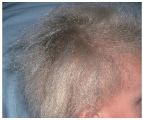 FIGURE 14-15. Uncombable hair syndrome: hair has an unruly, spangled appearance and is difficult to comb. Courtesy of Ken Greer, MD, University of Virginia Health Sciences Center, Charlottesville, VA. |
Clinical Presentation
The hair is usually blonde to light brown with a frizzy, spangled texture, and projects outward from the scalp79,80 (Figure 14-15).
Related posts:
Stay updated, free articles. Join our Telegram channel

Full access? Get Clinical Tree


