Introduction
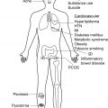
Over time, repeated episodes of hidradenitis suppurativa (HS) disease flares can lead to a number of debilitating cutaneous, systemic, as well as psychological and social complications. Given the progressive course of this disease, early diagnosis and initiation of appropriate treatment is critical to limiting the development of serious complications.
Cutaneous Complications
The typical skin lesions associated with HS include tender subcutaneous nodules and sterile abscesses; however, long term, poorly controlled disease can lead to cutaneous complications including sinus tracts, fistulas, lymphedema, scarring/contractures, and squamous cell carcinomas (SCC).
Sinus Tracts
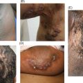
Sinus tracts are advanced HS lesions that manifest as subcutaneous tunnels that often drain malodorous discharge and can coalesce to form large areas of honeycombing under the skin ( Fig. 9.1 ). The development of sinus tracts may result from the connection of multiple ruptured follicles over time. The presence or absence of, as well as the extent of, sinus tract formation is used to help determine the severity of disease. Patients with at least one sinus tract are automatically staged Hurley Stage II disease, whereas the presence of multiple interconnected sinus tracts are staged Hurley Stage III disease. However, not all sinus tracts can be visualized or detected by manual palpation, suggesting that a subset of patients may be incorrectly classified as having lower stage disease and undertreated as a result. Ultrasound imaging can help detect subclinical lesions and be used as a tool to more accurately stage patients and aid in management.
Although biologics have been reported to help aid in the closure of some sinus tracts, most patients require surgical intervention for definitive treatment. If extensive surgery of the perineal/perianal area is being considered, preoperative magnetic resonance imaging (MRI) is recommended to rule out the presence of anal fistulas. Wide surgical excision provides a low rate of recurrence, however, extensive surgery often requires general anesthesia and can be associated with postoperative pain and contractures. Deroofing is a tissue-sparing technique that can be utilized in cases where wide excision may result in significant complications and/or burden.
Fistulas
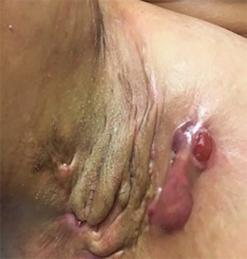
A fistula is defined as an abnormal connection between two epithelial-lined surfaces and can occur as a complication after surgery, infection, or from chronic inflammatory conditions such as inflammatory bowel disease and HS. Anal and urethrocutaneous fistulas can occur in patients with chronic, poorly controlled anogenital HS. An anal fistula is an abnormal tract between the perianal skin and the anal canal, whereas a urethrocutaneous fistula is an abnormal tract between the skin and the urethra.
Anal fistulas should be suspected in any patient with HS that has persistent perianal pain and draining of purulent or bloody discharge from a sinus opening near the anal orifice. If an anal fistula is suspected, an MRI of the anogenital region should be obtained to confirm the diagnosis. For example, if Crohn’s disease is on the differential, further imaging may be needed to characterize the type of fistula as this could determine treatment.
Anal fistulas are often resistant to medical therapy and require surgical intervention. A multidisciplinary approach involving a colorectal surgeon is recommended. Various surgical techniques have been described depending on the complexity and extent of the fistula. See Chapter 23 for a discussion on operating room-based surgical procedures. Simple inter-sphincteric or low trans-sphincteric anal fistulas can be managed with a fistulotomy or fistulectomy. A fistulotomy (similar to de-roofing of a sinus tract) involves “laying open” the fistula tract. A probe is inserted into the tract and an incision is made over the entire length of the fistula. The tract is then curetted to prevent recurrence and allowed to heal by secondary intent. During a fistulectomy, the entire fistulous tract is cut out. More complex anal fistulas, including trans-sphincteric or supra-sphincteric fistulas, have increased risk of fecal incontinence with surgical intervention. A modified seton procedure, which involves dissecting out the fistula tract while preserving the anal sphincter muscle, has been used to manage patients with HS with complex anal fistulas. This procedure, in conjunction with excision of remaining HS affected tissue, has been used to successfully manage complex anogenital HS disease with anal fistulas.
Urethrocutaneous fistulas are much rarer complications of HS, with fewer than ten cases reported in the literature. A urethrocutaneous fistula should be suspected if a patient has difficulty passing urine or experiences leaking of urine during micturition. An ascending urethrogram should be obtained if a urethrocutaneous fistula is suspected. Once diagnosis is confirmed, a multidisciplinary treatment approach involving a urologist is recommended.
Lymphedema
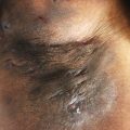
Lymphedema is characterized by soft tissue swelling that occurs when lymph fluid is unable to drain due to blockade or destruction of local lymphatics. It commonly occurs on the arms and legs, but can affect every part of the body, including the genitals ( Fig. 9.2 ). Genital lymphedema is a rare and debilitating complication of long-standing HS and can lead to recurrent infections, ulceration, increased risk of malignancy, as well as significant psychosocial morbidity.
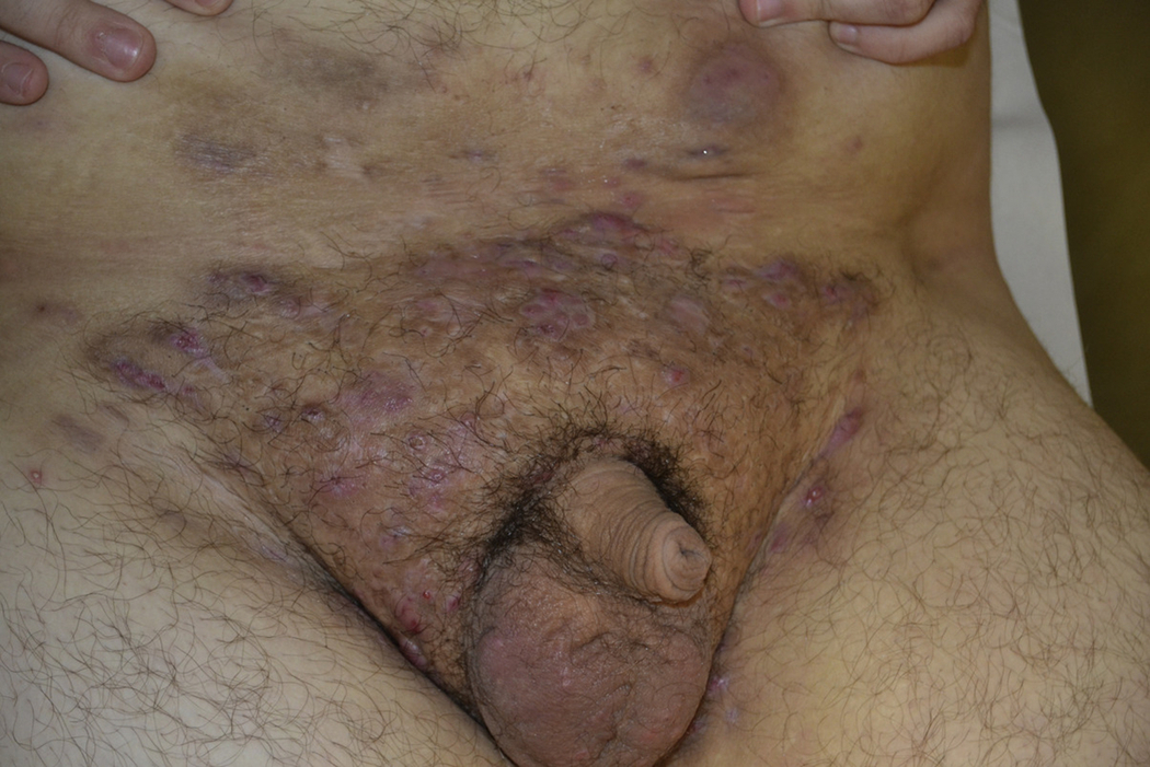
A systematic review of 27 patients with HS found that men have a higher incidence of lymphedema than women, with the scrotum (59%) and penis (44%) most frequently affected, followed by the labia majora (15%), perineum (11%), groin (11%), buttocks (7%), and rarely the abdomen (4%). Interestingly, all cases only involved the lower part of the body. Over 20% of patients had more than 2 locations affected. Typically, lymphedema is thought to occur in patients with long-standing HS; however, rapid progression of lymphedema occurring after 3 to 4 years from disease onset has been reported, suggesting that lymphedema may not only be a complication of chronic HS, but also of aggressive HS.
Clinically, lymphedema presents with swelling of the skin, frequently with overlying induration characterized by a “woody appearance.” A severe variant of scrotal lymphedema, scrotal elephantiasis, has been described in patients with filariasis, following inguinal node irradiation or surgical lymphatic destruction, and rarely in patients with HS. Scrotal elephantiasis is characterized by massive scrotal swelling with gross genital deformation, oftentimes presenting with obscuration of the penis.
Lymphatic obstruction can also cause lymphangiectasias (dilated superficial lymphatic channels) which manifests as clear vesicles on an indurated plaque and is common on scrotal skin or the labia majora in HS patients with genital lymphedema. Verrucous papules, plaques, and nodules can also be present. Providers should have a low threshold to biopsy these polypoid lesions as they can mimic verrucous carcinoma and SCC.
Lymphedema is often managed with physical compression; however, this is not practical for managing lymphedema of the anogenital area, given the technical limitations associated with adequately compressing this area. Patients also often have insufficient response to medical therapy including oral antibiotics, oral corticosteroids, acitretin, and biologic therapy. Surgical intervention is often required with removal of the affected tissue and superficial lymphatics, followed by various methods to cover the surgical defects. Split-thickness skin grafts are most often used, followed by skin flaps, and lastly healing by secondary intention. Overall, patients have favorable responses to surgical intervention with adverse events (including penile edema, wound breakdown, and cellulitis) reported in 14% of patients. Carbon dioxide (CO 2 ) laser excision with secondary-intention healing has also been reported in the literature to successfully treat genital HS lesions with associated lymphedema.
Scarring, Contractures, and Mobility Limitations
Scarring is a common complication seen in patients with HS. Recurrent episodes of inflammation followed by wound healing lead to scar formation, which can cause significant physical symptoms and limitations as well as psychological symptoms in patients with HS.
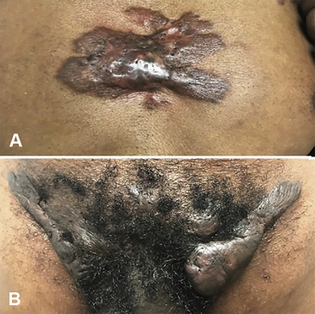
Inflammatory HS lesions typically heal with different types of scars, including atrophic and hypertrophic scars. Atrophic scars present as shallow, indented, dyspigmented papules, and plaques, while hypertrophic scars present as firm plaques or rope-like bands that can involve entire anatomic locations in severe cases. Keloids can also occur in HS patients prone to keloid formation (see Fig. 9.3 ).
Scar formation is not only disfiguring and painful but can also lead to limb contractures and reduced mobility, especially in the groin and axillae. Scarring in the anogenital area can predispose to stricture formation of the urethra, anus, and rectum. A stricture is an abnormal narrowing of a body passage which can occur secondary to scar tissue. Patients with strictures can present with difficulty or pain with defecation and/or urination. A thorough history and physical exam is necessary to assess for physical deformity and functional limitations related to scarring, contractures, and strictures, especially in patients with chronic, severe disease.
Scarring can have a profound emotional impact on patients with HS. Patients may feel embarrassed about the appearance of visible scars and feel compelled to hide them by wearing long-sleeved clothing and may limit certain activities such as swimming. The pursuit of new intimate relationships is also seen as a great burden in patients with scarring secondary to HS.
In order to prevent the physical and psychological complications associated with scarring in HS patients, early and aggressive treatment is needed. Once scarring and contractures form, medical management is often insufficient. Although intralesional triamcinolone injections may help flatten some hypertrophic scars, definitive treatment often requires surgical intervention. Fractional CO 2 lasers have also shown efficacy in the treatment of many different types of scars, including HS-related scarring. Patients who are prone to developing keloids may require additional adjunct treatments, including a trial of biologic therapy and or/intralesional triamcinolone injections.
Cutaneous Squamous Cell Carcinoma
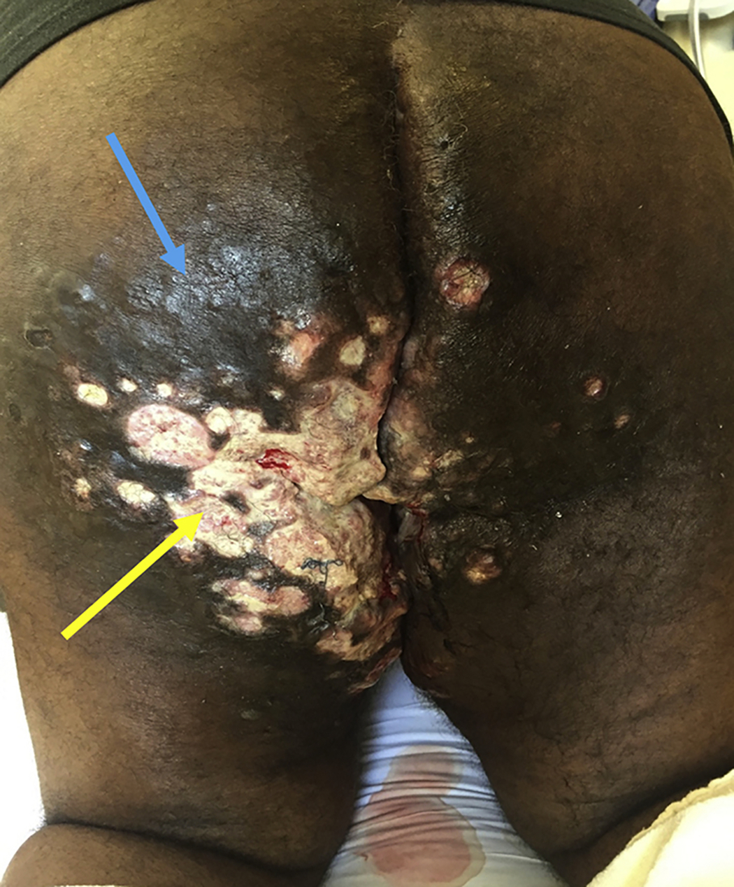
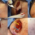
SCC is a rare, yet serious complication of chronic HS, with studies showing a prevalence rate ranging from < 1% to 4.6%. Although HS is three times more common in women, SCC much more commonly occurs in men, which may be attributed to the higher burden of disease men experience in the perianal/perineal area. HS-associated SCC typically presents in the gluteal and perianal region ( Fig. 9.4 ), though cases involving the perineal, vulvar, groin, thighs, and sacrum have been described. Interestingly, SCC of the axillary area is rarely seen, which suggests regional factors may play a role in the formation of SCC.
HS-associated SCC can be referred to as a Marjolin ulcer, a cutaneous malignancy that arises in chronically inflamed wounds or scarred tissue. A review of 85 cases of SCC arising in patients with HS found the average age at time of diagnosis of SCC was 52 years. The latency period between onset of HS and SCC development was 26 years. However, a case developing after only 3 years of active HS has been reported in the literature.
Chronic inflammation and scarring in HS lesions predispose to SCC formation. Other possible contributing risk factors include tobacco exposure and human papillomavirus (HPV) infection. In a retrospective review of 80 cases of SCC complicating HS, 80% of patients reported tobacco use. Similarly, a study by Lavogiez et al. found that a significant number (10 out of 13) of patients with HS that were heavy smokers (> 30 pack-year history) developed SCC. HPV is a known risk factor for the development of anogenital SCC, independent of HS. It has been hypothesized that a similar association may exist for HS-related SCC. Lavogiez et al. found HPV in all eight cases of SCC-related HS, with seven of eight cases identified as high-risk HPV. In contrast, no cases of HPV were detected by Kohorst et al. looking at 12 patients with HS-related SCC. Importantly, many of the biologic agents used to treat HS, such as tumor necrosis factor (TNF) alpha inhibitors, may also increase patient risk for reactivating latent infections, such as HPV. Further studies are needed to better elucidate a possible relationship between HPV and HS-related SCC.
Clinically, any nonhealing, ulcerative, or indurated lesion forming within a chronic wound or scar should raise suspicion for an SCC. SCC in chronic HS can also present with localized pain, which can make it challenging to distinguish from an inflammatory lesion of HS. Therefore, a low threshold for biopsy of any suspicious lesion is recommended. Multiple biopsies are often needed to diagnose SCC; therefore, if clinical suspicion is high, a repeat biopsy is recommended. A case reported in 1964 described a case of SCC that was missed with the initial 7 biopsies.
Although histologically the majority of SCCs arising in HS patients are characterized as well- or moderately-differentiated, these SCCs tend to be more aggressive and have an increased risk of metastasis and mortality. Patients can present with nodal metastasis at time of SCC diagnosis, and mortality rates up to 50% to 60% have been reported, suggesting early detection and aggressive intervention are required.
Once a diagnosis of a SCC is made, a multidisciplinary treatment approach including medical and surgical oncology is recommended. Prior to surgery, imaging with MRI or positron emission tomography (PET) scans may be helpful in establishing the extent of disease. A large and deep surgical excision with a minimum margin of 2 cm is recommended. Sentinel lymph node evaluation should be considered given the high prevalence of lymph node metastasis at time of diagnosis. Furthermore, adjuvant radiotherapy under the care of a radiation oncologist should be considered in select cases. Despite aggressive surgical intervention, recurrence rates of these SCCs are high, necessitating close surveillance.
Systemic Complications
The chronic, inflammatory nature of HS can predispose to multiple systemic complications including anemia, systemic amyloidosis/nephrotic syndrome, serious infection/sepsis, and chronic pain. As with cutaneous complications, early recognition and management of HS is needed to prevent these complications ( Table 9.1 ).
| Cutaneous Complications | Description | Management |
|---|---|---|
| Sinus tracts |
|
|
| Anal and urethrocutaneous fistulas |
|
|
| Lymphedema |
|
|
| Scarring, limb contractures and mobility limitations |
|
|
| Cutaneous SCC |
|
|
Related posts:


Stay updated, free articles. Join our Telegram channel

Full access? Get Clinical Tree


