Fig. 24.1
(a) A nodular cyst-like proliferation with papillary projections infiltrating the dermis. (b) Papillary projections with tubular structures lined by cuboidal or columnar epithelium. (c) The glandular differentiation
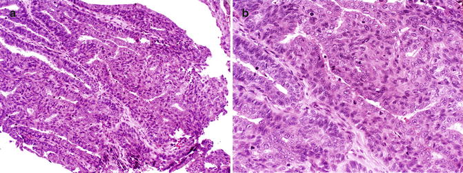
Fig. 24.2
(a) Cytologic atypia within solid areas. (b) Cytologic atypia with numerous mitoses
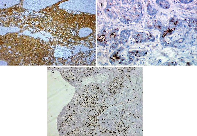
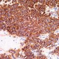
Fig. 24.3
(a) The tumor cells are diffusely positive for MNF116. (b) CEA highlights the luminal border of tubules. (c) The intensity of Ki67 expression is a marker of aggressive behavior
Differential Diagnosis
ADPC should be differentiated from benign adnexal tumors such as hidradenoma, papillary eccrine adenoma, tubular apocrine adenoma, apocrine cystadenoma, and metastatic adenocarcinoma. In particular, hidradenomas whose digital involvement is extremely rare lack papillary projections and consist of two cell types: polygonal cells with eosinophilic cytoplasm and clear cells. p63 has been reported as a useful marker for distinguishing primary ADPC from metastatic adenocarcinomas. A predominantly solid pattern and focal or absent papillary structures may be a diagnostic pitfall in ADPC, particularly on small biopsy specimens.
Prognosis
Histopathologic features were not found to be predictive of outcome. The neoplasm tends to locally recur in up to 50 % of cases with an incomplete excision. The rate of metastasis is reported to range from 14 to 41 %, with both widespread metastasis and frequent metastasis to the lungs.
Related posts:


Stay updated, free articles. Join our Telegram channel

Full access? Get Clinical Tree


