The diagnosis and management of the following non-infectious vulvar ulcers are reviewed: vulvar aphthae in adult and pediatric patients, aphthae associated with Behçet’s disease, vulvar ulcers resulting from Crohn’s disease, and vulvar ulcers associated with human immunodeficiency virus infection. There are many resources providing excellent reviews of infectious ulcers; therefore this topic will not be covered here.
Ulcers versus erosions
Ulcers should be differentiated from erosions both clinically and histologically. This is accomplished on the basis of depth. An erosion is a defect in the epidermis only, resulting in a red, smooth, moist, superficial, atrophic plaque. Ulcers are deeper, extending into the underlying dermis, and appear necrotic at the base with either yellow, fibrinous material or an eschar adherent to it. Large, deep, or long-standing ulcers heal with scarring, while erosions and superficial ulcers typically heal without scarring. Erosive diseases of the vulva are reviewed elsewhere.
Types of ulcers
Causes of vulvar ulcers are diverse and can be divided into infectious and noninfectious categories. Infectious causes include primary syphilis, lymphogranuloma venereum (LGV), chancroid, granuloma inguinale (GI), and herpes simplex virus (HSV). In the United States, HSV is the most common of these. Noninfectious ulcers include those caused by drug reactions or adverse effects, autoimmune or inflammatory disease, trauma, and aphthous ulcers.
Aphthous Ulcers
Oral aphthae (aphthosis, aphthous stomatitis, recurrent aphthous ulcers) have similar characteristics to vulvar aphthae. Commonly known as cancer sores, they are painful, recurring lesions of the oral mucous membranes. This is the most common lesion affecting oral mucosa, occurring in up to 20% of the general population and 60% of select groups. They are typically very tender and can become painful enough to interfere with speech, mastication, or swallowing.
Both oral and vulvar aphthae have been classified into three groups: minor, major, and herpetiform. Minor aphthae are smaller than 1 cm and heal within 7 to 10 days without scarring. Major aphthae are greater than 1 cm, deep, extremely painful, and heal with scarring in 10 to 30 days. Herpetiform aphthae are multiple, grouped, small ulcers that heal without scarring, and despite the name, are not associated with HSV.
Aphthae are classified further based on clinical course, as either simple or complex. Most patients have simple aphthosis, or several episodes per year of either minor, major, or herpetiform aphthae separated by disease-free periods. Complex aphthosis is defined as either almost constant presence of three or more ulcers, or recurrent oral and genital aphthae and exclusion of Behçet’s disease (BD).
Vulvar aphthae are thought to be relatively rare, and they are reported infrequently. Clinicians who specialize in caring for women and girls with vulvar disease, however, believe that vulvar aphthae are, in fact, not rare. Although studies looking at incidence and characteristics of vulvar aphthae are few, authors agree that most cases meet criteria for major aphthae (>1 cm). Some patients can further be classified as having complex aphthosis, because they have recurrent vulvar ulcers and concurrent oral ulcers in the absence of other features of BD.
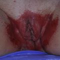
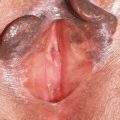
In the case of vulvar aphthae, keratinized, hair-bearing, as well as mucosal skin can be involved. Reported sites include the vagina, introitus, forchette, labia minora, labia majora, and perineal body. The most common location is the medial aspect of the labia minora, often presenting bilaterally on opposing labial surfaces as “kissing aphthae” ( Figs. 1 and 2 ). Ulcers are typically 1 to 2 mm deep with well-demarcated, ragged borders. Borders can become overhanging and heaped up, sometimes simulating an ulcerated malignant tumor. The base of the ulcer ranges from black, necrotic tissue, to gray or yellow adherent exudates. The surrounding skin is usually erythematous, edematous, warm, and tender. Associated regional lymphadenopathy and cellulitis can occur.
As with the oral variant, the cause of vulvar aphthae is not known and even less well studied. They can occur in adult women, but are more common in adolescent or premenarchal girls who are neither sexually active/abused, nor immunocompromised. Their discovery can be alarming and often precipitates an anxiety-provoking, costly, and unsuccessful search for sexual abuse or sexually transmitted infections. Patients and even many clinicians do not recognize noninfectious causes of vulvar ulcers; education about this entity is essential.
Lipschutz first reported vulvar aphthae in 1913 in adolescent girls in whom no specific cause was identified. Many authors have questioned the association with a viral infection, based on frequently associated antecedent fever, myalgias, and malaise. Epstein-Barr virus (EBV) has been most commonly implicated. Halvorsen and colleagues identified and reviewed 26 individual case reports of EBV-associated aphthae, a diagnosis which was supported by initial and convalescent antibody titers and Monospot testing. Few reports describe isolation of EBV directly from the base of the ulcer either by culture or by polymerase chain reaction (PCR).
Various theories have been postulated to explain the development of vulvar ulcers associated with EBV. EBV is a ubiquitous virus, and both the oropharynx and genital tract have been shown to harbor EBV even in the absence of symptoms. Therefore, direct transmission from sexual partners, either by genital–genital or orogenital contact, has been suggested as the cause. Many of the reported cases, however, occurred in patients before the onset of any kind of sexual activity. Of 26 reported cases, only 6 reported prior sexual activity. It has been postulated, based on these cases, that either autoinoculation or hematogenous spread from the patient’s own oropharyngeal infection is the source for these vulvar lesions.
Efforts to prove an association with specific viruses or other infections in larger cohorts have failed to confirm this theory. In a retrospective review of nine patients with nonsexually transmitted vulvar ulcers, eight of whom were premenarchal, none had evidence for EBV infection. In a larger review 2 years later, Huppert and colleagues studied 20 patients with vulvar aphthae, 5 of whom were premenarchal. Although 7 out of 20 patients appeared ill on initial examination, and 19 out of 20 had systemic complaints compatible with a viral illness (the most common being fever, malaise, and headache); only 2 had acute EBV and 2 had possible (equivocal) cytomegalovirus (CMV) infections. A thorough search for bacterial and fungal infections was negative in all study participants.
Other viruses have been implicated in vulvar aphthae. One author reported the case of a 13-year-old girl with influenza A and concomitant major vulvar aphthae. However, the patient had a recurrence of the lesions 2 months after the initial infection without a systemic viral illness. There is one report of a large ulcer of the labium minus associated with typhoid fever ( Salmonella paratyphi ) in a 25-year-old woman with a recent travel history. Most likely, many different viral infections, either respiratory or gastrointestinal, can precipitate vulvar aphthae, making a specific search extremely difficult.
Noninfectious causes have also been sought to explain vulvar aphthae in young girls. Friction and occlusion from tight-fitting clothing has been proposed as a physical cause in this group. Cebesoy and colleagues noted that 10 out of 10 premenarchal girls between the ages of 9 and 13 with vulvar aphthae associated with no known cause or underlying systemic illness reported frequently wearing tight-fitting pants or polyester underwear. Seven out of 10 had kissing ulcers, and authors proposed that chronic irritation was possibly playing a role.
Evaluation of Vulvar Aphthae
The most important aspect of the evaluation of a patient with vulvar aphthae is a complete history and physical examination, with particular attention to signs or symptoms of an underlying associated systemic condition: BD human immunodeficiency virus (HIV), malabsorption, ulcerative colitis, Crohn’s disease (CD), celiac disease, cyclic neutropenia, periodic fever syndromes, and leukemia. Regardless of the debate over a true association with a recent viral infection, signs and symptoms such as fever, malaise, myalgias, upper respiratory disease, and gastrointestinal symptoms should be elicited. Questions pertaining to triggering factors or exposures such as trauma, abuse, and sexually transmitted infections should be posed carefully. Excluding BD becomes important in patients who have recurrent oral and vulvar ulcers, and may not be possible on the first episode. This is discussed in greater detail. Obtaining a detailed history may not reveal an underlying etiology, and this should not discourage the patient or physician. Patients and parents should be reassured that vulvar aphthae are similar to oral aphthae in that a cause is often not identified.
An extensive laboratory work-up is often undertaken, creating great anxiety on the part of the patient and parents, and generating enormous, unnecessary medical expense. These searches are usually futile and should be tailored to the history and examination. Excluding HSV with viral culture or PCR is prudent. In sexually active or high-risk patients, screening for syphilis and HIV is appropriate. LGV, GI and chancroid are rare in the United States and should only be pursued in cases with a high index of suspicion. Based on current literature, search for EBV and CMV is not recommended. Routine bacterial and fungal cultures reveal skin flora or nonpathogenic bacteria and do not add benefit.
Obtaining a biopsy may be difficult, particularly in a young patient. Results are typically nonspecific: chronic active inflammation and necrosis without vasculitis. If a biopsy is performed, care should be taken to obtain a full-thickness punch or incisional specimen that includes skin from the periphery of the ulcer, rather than from the center of the lesion. In a high-risk patient in whom infection is suspected, biopsy with special staining for organisms can be a helpful way to make the diagnosis.
Chronic or recurrent vulvar aphthae in the absence of HIV, cyclic neutropenia, inflammatory bowel disease, or BD (primary complex aphthosis) is rare. Therefore, search for an underlying cause (secondary complex aphthosis) should be thorough. Continuous management to assess for the evolution for BD is essential, since patients may initially present with vulvar aphthae, and later meet criteria for BD. Similar to the evaluation for primary, isolated episode of vulvar aphthae, history and physical examination are the most important components of a work-up for chronic or recurrent aphthae.
Laboratory evaluation of chronic vulvar aphthae should include HSV PCR or culture and HIV testing. Neutropenia and hematologic deficiencies should be ruled out with a complete blood count with differential, serum iron, folate, zinc, and vitamin B12. Consider searching for celiac with antibody testing and HLA-B27 for BD.
Treatment of Vulvar Aphthae
Many treatments for oral aphthae have not been rigorously studied and for vulvar aphthae have not been studied at all. Therefore, treatment of vulvar aphthae is based on that of oral aphthae, case reports, and mostly anecdotal experience. National registries and randomized trials are needed to assess the benefit of various treatment modalities.
The main components of treatment are supportive care, including adequate pain management, and the reduction of ulcer duration. Treatment of recurrent or chronic ulcers is directed at resolution and prevention.
Supportive care begins with patient and parent reassurance that aphthae are not infectious, communicable, or sexually transmitted. Making the comparison with more commonly known “canker sores” can be helpful. Rest and proper fluid and nutrition are recommended, especially in the setting of systemic symptoms. Twice daily sitz baths in plain, warm water are the best method for gentle cleansing. Cleansing with a hand-held shower head is a good alternative. Soaps and other topical over-the-counter agents are not tolerated and should be discouraged because of their irritant or allergic potential.
Pain can be severe and must be addressed adequately. Barrier agents are helpful in managing painful ulcers. Orabase (carmellose sodium) is a denture adhesive paste that provides a barrier. It can be used either alone or compounded with a topical corticosteroid.
Developed in the 1980s, sucralfate is a basic aluminum salt of sucrose octasulfate, which binds preferentially to proteins on ulcerated tissue and promotes healing by multiple different mechanisms. It has antibacterial properties, stimulates fibroblast and keratinocyte proliferation, and promotes angiogenesis. Sucralfate has been extensively studied for use in disease of the gastrointestinal tract, mostly peptic ulcer disease. Given its success in this area, clinicians have adopted it for treating a myriad of other erosive and ulcerative conditions including burns, chronic venous ulcers, and severe adult erosive diaper dermatitis. There is one published report of its successful use in three patients with vaginal ulcerations caused by pessary use, laser ablation, and an ulcer of unknown etiology. These patients were treated with sucralfate 10% suspension douches twice daily and experienced complete resolution of long-standing ulcers that had been refractory to previous treatments. Similar to Orabase, sucralfate also can be compounded with a topical steroid.
Polyvinylpyrrolidone–sodium hyaluronate gel (Gelclair) is a viscous oral gel with two barrier-forming ingredients that adhere to mucosal surface. It is a class 1 medical device approved in 2001. Since that time, multiple studies have shown its efficacy in relieving pain secondary to oral mucositis from chemotherapy. One open trial of 30 patients, including 4 with severe diffuse aphthae of unknown cause and 10 with severe acquired immunodeficiency syndrome (AIDS)-associated oral lesions noted significant reduction in pain scores at 5 to 7 hours and at 1 week after use. This is a novel and interesting agent that could also be evaluated for pain control in vulvar ulcers.
Topical anesthesia is the second important component of pain management, and can be delivered in a number of formulations. Various compounded topical “caine” anesthetics are effective. Lidocaine can be dispensed to the patient for use at home up to four times daily in various preparations and vehicles: 2% gel, spray, or viscous solution; 4% cream; or 5% ointment. Gel or ointment is preferred. Huppert describes success with benzocaine 20% oral gel.
External dysuria can be severe. Voiding under water in a bathtub and use of topical anesthetics and barrier agents before micturition are helpful solutions.
Pain can be managed with nonsteroidal anti-inflammatory drugs or oral narcotic agents. Some patients, particularly young girls, may require short hospitalization for effective pain management, placement of a Foley catheter, rest, and wound care. Patients who appear to have secondary cellulitis of surrounding tissue warrant antibiotics.
While the immunopathogenesis of aphthae is poorly understood, medical treatment is directed at diminishing inflammation. Of the topical agents available, a potent inhibitor of inflammatory mediators, amlexanox 5% oral paste (Aphthasol), is the most extensively studied. For oral aphthae, it is superior to both placebo paste and to no treatment at all for reduction of pain, ulcer size, and ulcer duration. Additional benefit can be obtained by starting the paste at the onset of prodromal symptoms rather than waiting for active ulceration. Using this method, Murray and colleagues found that only 35% of patients who treated mucosa with prodromal symptoms went on to develop an ulcer, and healing time was 4.1 days faster in those who did develop an ulcer. Efficacy of amlexanox paste is equal to clobetasol ointment for the treatment of oral aphthae. Amlexanox is another agent that should be evaluated for use in vulvar aphthae.
The mainstay of topical therapy for vulvar aphthae is corticosteroids. Double-blind, placebo-controlled trials showed that topical corticosteroids significantly reduce oral ulcer duration and pain when compared with placebo. Class 1 topical steroids should be used in their ointment form to reduce exposure to irritant or allergic components of creams, gels, or foams. Physicians should not hesitate to use ultrapotent steroids on the vulva in this circumstance, presuming the patient or parent understands appropriate focal application. Although lacking supportive data both for oral and vulvar aphthae, intralesional and oral steroids also may have benefit, and studies are needed to assess their use over current topical agents.
Tetracycline antibiotics have been used in various formulations for treating oral aphthae. In trials, the mouthwashes are dosed four times daily for 1 to 2 minutes. They significantly reduce pain and duration of recurrent ulcers compared with placebo. Formulas for compounding mouthwashes have been described. Use of topical antibiotics for vulvar ulcers has not been studied; however, many case reports describe using oral antibiotics for presumed infection during management of acute vulvar aphthae. The failure of antiseptic mouthwashes to improve pain or ulcer duration for oral aphthae suggests that the antimicrobial effect is not as important as perhaps the anti-inflammatory effect of these antibiotics. Trials exploring the use of either oral or topically applied antibiotics for vulvar ulcers are warranted.
Treatment of Chronic/Recurrent Aphthae
Chronic vulvar aphthae unassociated with an underlying illness (primary complex aphthosis) is rare, and studies to support therapeutic decisions do not exist. Physicians who manage these patients must extrapolate from experience in the treatment of chronic oral aphthae. Because the mechanism of ulcer formation is likely similar in these two anatomic sites and can occur simultaneously in the same patient, the data on treatment of chronic oral aphthae are reviewed here.
The approach to managing complex aphthosis is different from managing a solitary episode or simple aphthae. Complex aphthosis, by definition, is chronic and recurrent, thus requiring systemic agents to heal long-standing lesions, reduce recurrences, and induce remission. The main systemic agents used include thalidomide, dapsone, colchicine, pentoxifylline, and tumor necrosis factor (TNF) inhibitors. Treatment choice is based on patient profile, medical history, and previous treatment trials. As with any chronic dermatologic condition requiring ongoing systemic therapy, rotating agents to reduce adverse effects is helpful.
Comparisons of systemic therapeutic modalities for chronic aphthae are rare. Letsinger and colleagues published one of the first reports of a large series of this kind. This paper reviewed treatment of 42 patients with primary complex oral aphthosis. The authors proposed a therapeutic strategy starting with colchicine 0.6 mg two to three times daily as tolerated, progressing to dapsone dosed from 25 to 100 mg daily, and then to a combination of these two agents at full tolerated doses. The authors found that the two drugs combined were much more effective than either one alone, and nearly as effective as thalidomide, the final step in their suggested therapeutic ladder. Fifty-seven percent and 59% of patients achieved at least 50% improvement in these last two treatment groups, respectively.
Since the publication of this study, a second review of systemic treatments for 21 patients with severe recurrent oral aphthae has been published. Patients initially were treated with prednisone for 2 weeks starting at 0.5 mg/kg/d, which was reduced to half that dose for a second week. Simultaneously, one of four systemic agents was started and maintained for 6 months: thalidomide, dapsone, colchicine, or pentoxifylline. The authors found that thalidomide was the most efficient and best-tolerated drug, with seven out of eight (87.5%) patients experiencing complete remission. Eight of nine patients treated with dapsone (89%) and eight of nine patients treated with colchicine (89%) experienced excellent-to-moderate improvement in their disease. Three out of five (60%) patients treated with Pentoxifylline had excellent-to-moderate improvement. Dapsone was best for inducing remission, with three patients remaining ulcer free for up to 9 months after discontinuation of the drug. Otherwise, most patients relapsed within weeks to several months of cessation of therapy.
Introduced in the 1950s as a sedative, thalidomide was subsequently banned worldwide in 1962 after the discovery of its devastating teratogenic effects. Thalidomide saw a renewed acceptance for its use in erythema nodosum leprosum (ENL) and was approved and classified as an orphan drug for this indication by the US Food and Drug Administration in 1998. ENL remains the only approved indication for this drug; however, it has re-emerged as an alternative off-label therapy for many other dermatologic conditions. Careful monitoring using the System for Thalidomide Education and Prescribing Safety (STEPS) program is mandatory to ensure appropriate use and prevent pregnancy. The mechanism of action of thalidomide is multifactorial and poorly understood, but in the case of aphthae, is probably mediated in part by its anti-TNF effect.
Its effectiveness in complex aphthosis initially was shown in three open-label trials using thalidomide doses ranging from 100–400 mg/d. Most patients had significant improvement or complete resolution of lesions and pain within several weeks of starting therapy. Larger randomized trials definitively established the efficacy of this drug. On 100 mg/d, 48% and 44% of patients experienced complete remission compared with only 9% and 8% of patients receiving placebo. These studies reported relapse times of 20 to 30 days after stopping therapy.
Successful use of TNF inhibitors in BD has led to their off-label use in complex aphthosis. Scheinberg reported success using etanercept for two patients with chronic recurrent aphthous stomatitis, one meeting criteria for BD. Both patients discontinued thalidomide, despite its effectiveness, because of adverse effects, and were subsequently treated with etanercept. Complete remission occured in both patients within 3 and 5 weeks of therapy. Recurrence was noted after discontinuation within 5 and 7 weeks. Each patient quickly responded after a second reintroduction of etanercept. Robinson and colleagues subsequently reported success using etanercept for another case of severe, recalcitrant oral aphthae. Treatment with adalimumab led to complete resolution of a 7-year history of recalcitrant, severe major aphthae in an 18-year-old. Such encouraging reports are increasing in number, and larger trials using these agents are desperately needed for this disabling condition.
Behçet’s Disease
BD is a multisystem disease of unknown etiology, with the highest prevalence in the Mediterranean, particularly Turkey, with a rate of 1 case per 250 people in the population over 12 years old. A pathognomonic test for BD does not exist; therefore the diagnosis is made based on the satisfaction of established criteria. International study group criteria for BD were established in 1990. By this consensus, patients must have recurrent oral aphthae at least three times in 1 year, plus two of the following:
Recurrent genital aphthae
Uveitis or retinal vasculitis
Erythema nodosum-like lesions or papulopustular skin lesions
Positive pathergy test.
Ulcers involving oral and genital skin and mucosa are a hallmark of the disease. Second to oral ulcers, genital ulcers are the next most common feature of BD, occurring in 57% to 93% of patients. Recognizing these lesions is essential, because they may precede other features of the disease. Overlooking this association may result in delay in diagnosis and an increase in mortality. Aphthae associated with BD typically occur more frequently and more often in crops compared with recurrent aphthous stomatitis (RAS). Genital ulcers of BD typically start as a tender nodule, become deep and painful, and eventually heal with scarring. Searching for scars on genital skin, even in the absence of active, clinical disease, is an important part of the examination. Ulcers typically are found on the labia majora, but can occur anywhere on the vulva, perineum, or perianal skin. They can also occur intravaginally, potentially leading to fistula formation with the urethra or bladder. Exceptionally deep external genital ulcers can lead to labial destruction.
Treatment of aphthae of BD is based on experience in treating RAS and has been reviewed in detail elsewhere. In the opinion of Alpsoy, who has written extensively about this disease, colchicine alone or plus benzathine penicillin is a good starting point for women with genital ulcers. Thalidomide and dapsone are second choices with immunosuppressive agents (cyclosporine, azathioprine, and TNF inhibitors) to follow for unresponsive patients. Topical barriers, antibiotics, anesthetics, and corticosteroids can be used as described for treating primary aphthosis.
TNF-alpha, derived from gamma–delta Th-1 lymphocytes, is postulated to play a role in the pathogenesis of BD. Accordingly, off-label use of TNF inhibitors is increasingly being used for BD. Although primarily used for ophthalmic, gastrointestinal, and other more severe systemic manifestations, there are multiple reports of successful use of TNF inhibitors, particularly infliximab, for treating severe oral and genital aphthae of BD. Complete remission is observed with maintenance therapy. A representative case is illustrated by Robertson and colleagues, who successfully used infliximab to treat severe orogenital aphthae associated with BD. The patient was a 65-year-old woman who had failed treatment with dapsone, thalidomide, azathioprine, cyclosporine, and colchicine. There was marked improvement after the first infusion with infliximab (scheduled at 0, 2, and 6 weeks at 5 mg/kg). By the third infusion, she was completely clear of ulcerations for the first time in 10 years.
Likewise, etanercept has shown to be useful for treating aphthae of BD. Two female patients enjoyed complete remission of oral and vulvar ulcers when etanercept, 25 mg twice weekly, was added to their regimens of methotrexate in one case and methotrexate and prednisolone in the second. Other data indicate that etanercept may be best reserved for clearing oral aphthae rather than genital aphthae. A recent randomized controlled trial comparing 25 mg twice weekly etanercept with placebo for mucocutaneous BD in 40 men showed that the drug had little effect on genital ulcers, while oral ulcers began clearing within 1 week of treatment.
Infliximab was effective in a case resistant to etanercept. This is most likely due to the effect of infliximab on both soluble and membrane-bound TNF, and therefore, more powerful TNF blockade. With this in mind, a reasonable approach is to treat patients with etanercept as a first-line agent, due to its excellent safety profile, and move to infliximab if response is suboptimal.
HIV-Associated Aphthous Ulcers
Most studies examining HIV-associated ulcers are conducted in Africa and involve the risk of transmission and coinfection with herpes simplex, syphilis, or chancroid. The literature investigating vulvar aphthae associated with HIV is scant, consisting primarily of case reports. HIV-associated aphthae are similar to ulcers that affect the oral mucosa, esophagus, and rectum in these patients, and pathogenesis may be the same.
They are typically large, painful, and recurrent ( Fig. 3 ). Patients are generally severely immunocompromised with a low CD4 count and AIDS-defining infections. Thirty-seven percent have coexistent oral ulcers. HIV-associated aphthae can persist for weeks or months, becoming necrotic and causing disabling pain. Local destruction by nonhealing ulcers can be severe, with formation of a recto-vaginal fistula reported in one woman and a labial–vaginal fistula extending to the ischiorectal fossa in another. Five fistulas resulting from HIV-associated aphthae were reported in a national survey involving 29 severely immunocompromised women: four recto-vaginal fistulas requiring diverting colostomies and one vaginal–perineal fistula.