Epidermolysis bullosa acquisita (EBA) is a rare autoimmune subepidermal blistering disease characterized by immune deposits on anchoring fibrils of cutaneous and mucosal basement membrane zones. It is due to circulating antibodies directed to type VII collagen. Clinical manifestations include a classical form with skin fragility, blisters and scars on trauma-prone surfaces, an inflammatory form, and a cicatricial pemphigoid–like form. Specialized tests available in only certain laboratories are necessary to confirm a diagnosis of EBA, such as immunoelectron microscopy, immunoblotting, or ELISA using recombinant proteins. A frequent association between EBA and Crohn disease has been observed.
Epidermolysis bullosa acquisita (EBA) is a rare autoimmune subepidermal bullous disease involving the skin and the mucous membranes. It is characterized by the deposition of autoantibodies directed to the anchoring fibrils of the basement membrane zone of stratified squamous epithelia. These autoantibodies recognize type VII collagen, which is the major component of anchoring fibrils. The incidence of EBA is estimated to be between 0.2 and 0.5 new cases per million inhabitants per year. Several clinical presentations have been reported; classical EBA includes skin fragility, blisters over the trauma-prone surfaces, and milium cysts. Making a definitive diagnosis of EBA may be difficult because specialized tests only available in certain laboratories are necessary to confirm the clinical suspicion. This orphan disease is often misdiagnosed, explaining an important delay in its diagnosis and the difficulty of performing therapeutic trials.
Pathogenesis
Type VII Collagen
Type VII collagen is a homotrimer composed of 3 identical α chains. Each chain includes a 145-kDa central helical region characterized by the repeated Gly-X-Y amino acid sequence; this region is flanked by a large 145-kDa amino-terminal noncollagenous (NC1) domain and a small 34-kDa carboxyl-terminal noncollagenous (NC2) domain. Type VII collagen molecules form dimers through their NC2 domains and are stabilized by disulfide bridges. Then these antiparallel dimers aggregate laterally to form the anchoring fibrils. The NC1 globular domains are located at the end of these fibrils and interact with other extracellular matrix proteins, such as laminin 332, type IV collagen, type I collagen, and fibronectin. These domains probably stabilize the basement membrane zone to the underlying dermis. Autoantibodies of patients with EBA preferentially recognize epitopes located in the NC1 domain but epitopes in the NC2 domain and the helical region also are reported. Binding of autoantibodies to type VII collagen induces a reduction in the number of anchoring fibrils and consequently a skin fragility and a cleavage between epidermis and dermis.
Pathogenicity of Autoantibodies
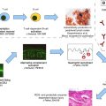
Because EBA is often associated with low titers of autoantibodies directed to the basement membrane zone, the pathogenicity of these autoantibodies has been difficult to demonstrate for a long time. Cases of spontaneous EBA have been described in dogs, such as great Danes, but this animal model did not permit a better understanding of the pathogenesis of this disease. Pathogenicity of autoantibodies that target type VII collagen was proved when two research teams demonstrated that (1) iterative passive transfer of antibodies directed to murine type VII collagen induces subepidermal blisters in mice, (2) repeated injections of autoantibodies from EBA patients in hairless mice produce a phenotype reminiscent of EBA, and (3) immunization of mice with recombinant murine type VII collagen results in a subepidermal phenotype closely resembling human EBA. The development of an active experimental model permitted dissection of the molecular events after the binding of autoantibodies to the basement membrane zone and the demonstration that dermoepidermal cleavage is dependant on the capacity of antibodies to activate the alternative pathway of complement.
Related posts:
 A Globally Available Internet-Based Patient Survey of Pemphigus Vulgaris: Epidemiology and Disease Characteristics
 Diagnosis and Clinical Features of Pemphigus Foliaceus
Linear IgA Disease: Clinical Presentation, Diagnosis, and Pathogenesis
Pemphigoid Gestationis: Pathogenesis and Clinical Features
Diagnosis and Clinical Features of Pemphigus Foliaceus
Linear IgA Disease: Clinical Presentation, Diagnosis, and Pathogenesis
Pemphigoid Gestationis: Pathogenesis and Clinical Features
 Pathogenesis of Epidermolysis Bullosa Acquisita
Nail Involvement in Autoimmune Bullous Disorders
Pathogenesis of Epidermolysis Bullosa Acquisita
Nail Involvement in Autoimmune Bullous Disorders
Stay updated, free articles. Join our Telegram channel

Full access? Get Clinical Tree


