Fig. 19.1
Approaches and aesthetic incisions in conservative surgery. The incisions are indicated according to the place of resection and the Langer lines of the breast. It is interesting to point out the approach of the upper and medial quadrant through the periareolar region to avoid the scars in the region described by Grisotti as “no man’s land” (in blue)
Know the techniques of gland shaping to avoid defects secondary to the loss of part of the gland after resection.
Know the fundamentals and effects of radiotherapy in conservative treatment: several publications have analyzed the changes in the irradiated mammary gland according to its volume and the homogeneity of the dose delivered. In a prospective and randomized trial, Moody et al. [8] compared the adverse effects of radiotherapy in small, medium-sized, and large mammary glands, and found moderate and severe negative aesthetic results in only 6 % of small breasts and in up to 39 % of large ones. Gray et al. [9] evaluated 267 irradiated patients after conservative surgery. They observed a significant reduction in aesthetic results in patients with macromastia and inadequate treatment, with areas of overirradiation or underirradiation, about 10–15 % as a consequence of the lack of homogeneity of the dose owing to the size of the breast. Following these parameters, we can obtain approximately 70 % good results, leaving 30 % of patients with remaining deformities that would require a secondary surgical correction [10]. Oncoplastic surgery of the breast had its origin in the intent to prevent these unsatisfactory results of breast conservation observed in these 30 % of patients.
The crucial factor to develop and implement these techniques and the sequence related to other treatments (chemotherapy, radiotherapy) motivated further interdisciplinary analysis to evaluate its safety and results. It is in the limitations of conservative surgery related to the breast and tumor volume or to the site of the lesion (e.g., central tumors), which are classic relative contraindications for conservative treatment, where oncoplastic surgery of the breast achieves breast conservation and immediate reconstruction with oncological safety in adverse anatomical conditions.
On the other hand, oncoplastic surgery of the breast is also indicated in the following cases: superficial tumors that need a skin resection, secondary resections in breasts with multiple scars, widening of resection because of positive margins, and in patients with previous breast augmentation surgery and breast cancer who need oncologically safe margins and breast conservation.
In summary, and to respond to the difficult question of how do we decide who needs immediate reconstruction with breast conservative treatment, we can list three basic situations in which the oncoplastic surgery finds can apply it:
1.
Problems related to the site of the tumor (central, in the midline, upper medial quadrants, etc.) or to tumor volume/breast volume relation [4].
2.
In the treatment of locally advanced cancer treated with induction chemotherapy and salvage surgery, preserving the breast with wide resection margins and good local control.
3.
Special situations such as skin resections in superficial tumors, patients with multiple previous scars, resections with wide margins in patients with ductal carcinoma in situ or secondary to tumorectomy with positive margins, or breast cancer in patients with previous breast augmentation surgery.
Following the previous exposition, we recommend that when the patient has risk factors that increase the possibility of sequelae after breast-conserving surgery, immediate breast reconstruction with oncoplastic techniques is preferable.
19.2 Etiology and Classification of the Sequelae
There are many factors that can be determinant in producing a deformity in the breast tat has been operated on. The most important is probably the gland resection itself that produces a reduction in the breast volume. In a planned resection, it is important to calculate the approximate tumor volume and the healthy tissue margin around it, for example: if we resect a tumor of 2-cm diameter with a margin of 1 cm, this is equivalent to 30 g of gland volume, but if we enlarge the margin to 2 cm, the defect enhances up to more than 100 g with a different impact on the final result. The tumor site is the second determinant factor: there are sites of the breast where the defect can be repaired favorably, such as the upper and lateral quadrants, and others such as the medial region or lower quadrants where the structural alteration is maximal and its correction difficult. The size of the breast is also important: many results are conditioned by this factor, the damage being less when the relationship between the breast and the tumor volume is larger. A body mass index greater than 30 is also related to a higher number of sequelae [11].
Breast retraction and fibrosis are the usual changes after radiotherapy, but there are some factors that can increase the sequelae secondary to this treatment. A total dose of 66 versus 50 Gy worsens the cosmetic results [12] and, as mentioned before, the gland and fat tissue volume also have a negative influence on this last issue [8, 9]. Chemotherapy can worsen the results, administered either simultaneously or sequentially with radiotherapy [13].
When patients seek consultations because of sequelae of a conservative treatment, there are some parameters related to the patient’s anatomy that have to be evaluated, as well as the characteristics of the breast tat has been operated on and the symmetry of both breasts and the nipple–areola complex. Deferred breast reconstruction of these deformities is limited by five determinant factors: the lack of skin or gland tissue, scar retraction, radiodermatitis, and fibrosis.
Evaluation of sequelae is highly subjective, and the concordance between surgeons and patients or between different surgeon is generally low [14]. In recent years some informatic models have been designed (3dMD, BAT Software) to systemize this evaluation and improve the planning of reconstruction [15, 16]. A number of classifications have been proposed with the intention to evaluate the defects and plan corrections as shown in Table 19.1. In all of them there is generally coincidence in the evaluation of minor sequelae (type I or II), involving only asymmetries without or with minimal changes in the shape of the treated breast, except for Berrino’s classification [17], which added the displacement of the nipple–areola complex (Fig. 19.2). Most of the “problematic” patients present with major sequelae that range from moderate deformities to severe sequelae with sclerosis of the whole breast that even sometimes needs mastectomy. For these sequelae the classifications are confusing and the indications for corrections range between simple treatments such as lipofilling and mastectomies with immediate reconstruction with microsurgical or pedicled flaps associated or not associated with prosthetic material [17–19].
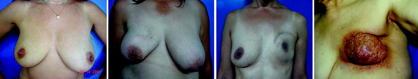
Table 19.1
Cosmetic sequelae after conservative treatment of breast cancer: classification
Berrino [17] | Clough et al. [18] | Fitoussi et al. [19] | |
|---|---|---|---|
Type I | Malposition and distortion of the NAC mainly due to postoperative fibrosis and scar contracture | Asymmetrical breasts with no deformity of the treated breast | Low ipsilateral deformity does not affect the shape or volume of the breast |
Type II | IIa: localized tissue insufficiency is observed, which may be due to skin deficiency IIb: subcutaneous tissue deficiency IIab: both | Deformity of the treated breast, compatible with partial reconstruction and breast conservation | Good shape and sufficient volume, but with obvious asymmetry in relation to the contralateral healthy breast |
Type III | Deformity is characterized by breast retraction and shrinkage and is mainly due to the effects of radiotherapy on residual breast parenchyma | Major deformity of the breast, requires mastectomy | Asymmetry does not maintain the shape and volume, frequent dislocation of the NAC |
Type IV | Severe radiation-induced damage to the skin, NAC, and subcutaneous and glandular tissues is present | – | Greater deformity, lack of native tissue, scarring and radiation effects |
Type V | – | – | Severe deformity from both surgery and prior radiotherapy, where the breast is too small and/or completely sclerosed |
Fig. 19.2
Cosmetic sequelae after conservative treatment for breast cancer: classification. Left asymmetry without deformity (Clough et al. type I, Fitoussi et al. types I and II). Left-Center asymmetry with moderate deformity and mild dislocation of the nipple–areola complex (Berrino types I and II, Clough et al. type II, Fitoussi et al. type III). Right-Center breast deformity and asymmetry as well as of the nipple–areola complex (Berrino type III, Fitoussi et al. type IV). Right fibrosis and severe actinic sclerosis with severe disappearance of the nipple–areola complex (Berrino type IV, Clough et al. type III, Fitoussi et al. type V)
19.3 Timing of Reconstruction of the Partial Mastectomy Defect: Our Experience
In our institutional experience after using the classifications mentioned in the previous for some years, we tried to simplify the evaluation of the sequelae and systemize the reconstruction techniques by employing a more functional concept related to each particular patient.
We analyzed the following parameters: age, biotype, time between the first medical consultation and the surgery and primary radiotherapy, grade of complexity of the sequelae, previous reconstruction intents, and presence of a prosthesis in the previously irradiated breast. Generally, in relation to all these parameters, we waited for a least 1 year after radiotherapy had finished before recommending reconstruction, with the condition that the breast was stable and did not show signs of edema or radiodermatitis, and that physical examination and imaging (mammography, ultrasonography, MRI) confirmed the absence of local recurrences.
We divided the patients in two large groups based on the type of sequelae and also the complexity of the reconstruction technique needed for each particular patient: group A had minor defects and group B had major defects.
In group A we included the sequelae that did not compromise or only produced a mild change in the shape of the breast, with or without asymmetry of the nipple–areola complex or the breast. We divided this group into three subgroups:
1.
Breast asymmetry without alteration of the shape of the breast that had been operated on.
2.
Minor sequelae in the breast that had been operated on without asymmetry of the nipple–areola complex, with or without associated breast asymmetry.
3.
Minor sequelae in the breast that had been operated on with asymmetry of the nipple–areola complex, associated or not associated with breast asymmetry.
In group B we included the sequelae that compromised moderately or severely the breast’s shape with asymmetry of the nipple–areola complex. In this group we also added the damage produced by severe actinic sclerosis and fibrosis, and a special subgroup that corresponds to patients with prior reconstruction attempts with unsatisfactory results, who generally have implants and ask for a second procedure. We can divide this group into three subgroups:
1.
Moderate or severe sequelae in the shape and volume of the treated breast without or with moderate actinic damage.
2.
Moderate or severe sequelae in the shape and volume of the breast that has been operated on without or with moderate actinic damage and a previous reconstruction attempt with or without implants.
3.
Severe actinic damage with loss of the shape and alteration of the volume of the treated breast. Marked sclerosis and fibrosis.
It is important to explain that in both groups the cause of breast asymmetry can be due to several factors related not only to the primary treatment but also to the biotype of the patient, changes in body weight, adjuvant oncological treatments, age, etc. (see Table 19.2).
Table 19.2
Cosmetic sequelae after conservative treatment for breast cancer: IAR functional classification
Minor defects | Major defects | Examples | |
|---|---|---|---|
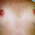
a-I | Breast asymmetry without altering the shape of the breast operated on | – | 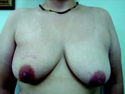 |
a-II | Mild sequelae in the shape of the breast operated on without asymmetry of the NAC. They can be associated or not associated with breast asymmetry | – |  |
a-III | Mild sequelae in the shape of the breast operated on without asymmetry of the NAC, They can be associated or not associated with breast asymmetry | – | 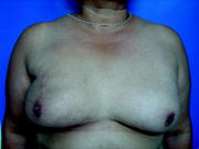 |
b-I | – | Moderate or severe sequelae in the shape and volume of the treated breast without or with moderate actinic sequelae | 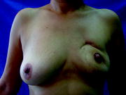 |
b-II | – | Moderate or severe sequelae in the shape and volume of the treated breast without or with moderate actinic damage with a previous attempt at reconstruction with or without implant insertion |  |
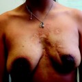
b-III | – | Severe actinic sequelae with loss of the shape and marked alteration in the volume of the treated breast. Marked sclerosis and fibrosis |  |
Analyzing the patients according to this classification, we used a treatment algorithm to choose the most suitable surgical technique (see Figs. 19.3 and 19.4).
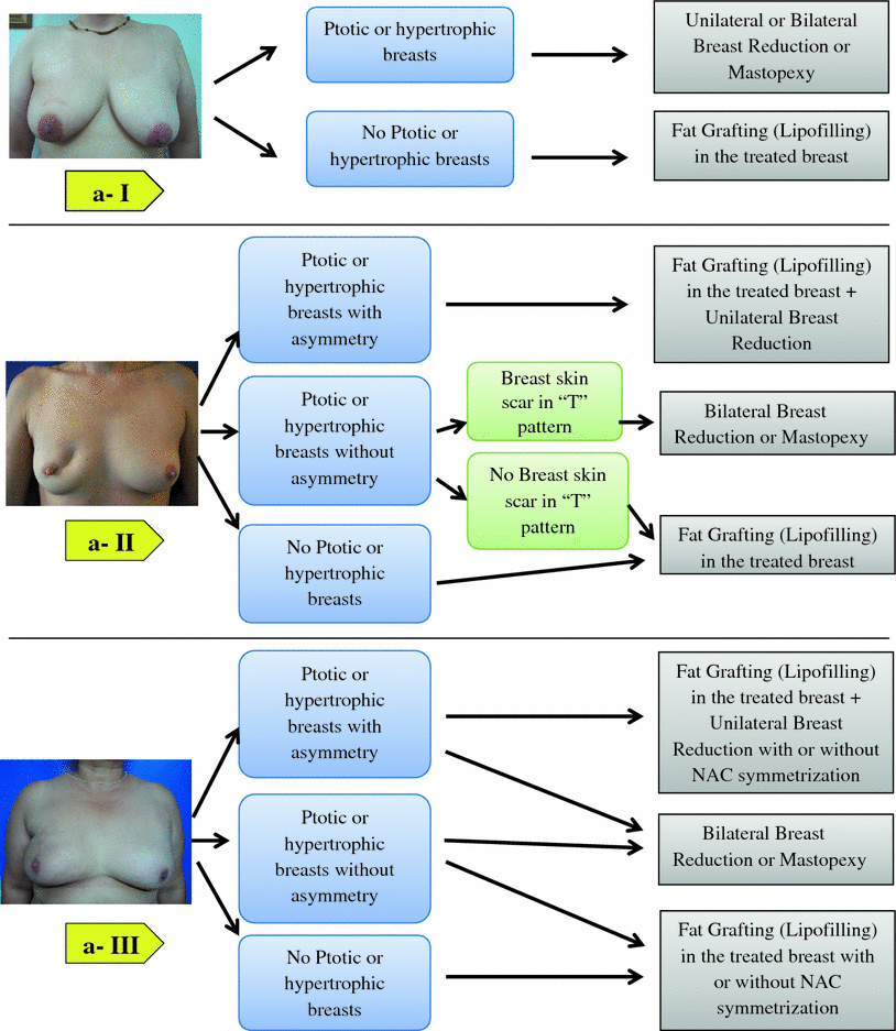
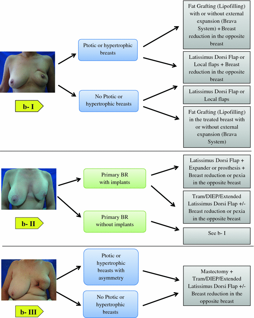
Fig. 19.3
Cosmetic sequelae after conservative treatment for breast cancer. Management algorithm for repair of partial mastectomy defects. Minor defects. NAC nipple–areola complex
Fig. 19.4
Cosmetic sequelae after conservative treatment for breast cancer. Management algorithm for repair of partial mastectomy defects. Major defects. BR breast reconstruction, DIEP deep inferior epigastric perforator, Tram transverse rectus abdominis myocutaneous
Related posts:

Stay updated, free articles. Join our Telegram channel

Full access? Get Clinical Tree


