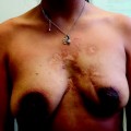
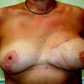
Fig. 30.1
Long-term result of breast reconstruction 13 years after free deep inferior epigastric perforator (DIEP) flap
Free flap reconstruction can be associated with significant complications and morbidity. There is an increased risk of general complications such as deep vein thrombosis, pulmonary embolism, pneumonia and acute respiratory distress syndrome. Specific complications of free flaps include microsurgical problems requiring expedient re-exploration of the anastomosis, haematoma, fat necrosis and partial or total flap loss. Significant complications may add to logistical problems and increase healthcare costs. Donor site complications such as wound-healing problems and mesh infection may prolong the recovery process and increase the length of inpatient admission. Other problems include asymmetry, abdominal weakness, bulging and incisional hernia. The main disadvantage of autologous breast reconstruction is donor site morbidity, particularly with myocutaneous flaps, where despite a degree of expendability, harvest still leads to loss of function of the donor muscle. A high level of microsurgical expertise is imperative, and even with this, there always remains a small and quantifiable failure rate in free tissue transfer.
30.2 Indications
Autologous tissue can be used in both immediate and delayed breast reconstruction. Other indications include:
Risk reducing mastectomy
Large contralateral breast
Substantial soft tissue defect
Previous complications of implant-based reconstruction
Latissimus dorsi muscle cannot be used because of atrophy or previous pedicle damage
Salvage surgery for locally advanced or recurrent breast cancer.
Absolute contraindications for free tissue breast reconstruction include:
Physiologically old
Cardiorespiratory disorders
Vasospastic disorders
Significant thrombophilia
Inadequate recipient vessels.
Relative contraindications include obesity, smoking, diabetes, autoimmune disease, psychosocial problems, distortion of vascular anatomy by previous scars, an inexperienced surgeon and unresectable locally advanced breast cancer [2, 3]. The potentially detrimental effects of adjuvant radiotherapy on autologous tissue breast reconstruction remain a subject of ongoing research and debate [4, 5]. The indications for postmastectomy radiotherapy are gradually expanding, and its effect on the reconstructed breast mound may increasingly become a matter of significant consideration particularly with respect to the decision of immediate versus delayed free flap reconstruction, as well as the appropriate timing of delayed reconstruction [6].
30.3 Recipient Vessels
Recipient vessels for microanastomosis of the free breast flaps include:
1.
Subscapular axis
Circumflex scapular vessels
Thoracodorsal vessels
Serratus branch
2.
Internal thoracic/mammary vessels
3.
Perforator to perforator anastomosis.
30.4 Perforator Flaps
Free perforator flaps are perfused by fascial or myocutaneous perforators and evolved as a refinement of conventional myocutaneous free flaps. Flap harvest requires accurate dissection of perforator vessels, which are often small, through muscle or fascia to yield adipocutaneous flaps from a multitude of donor sites. Although an exacting technique, this enables tissue harvest without the need to sacrifice muscle or fascia [7].
Use of the patient’s own tissues gives the most natural, durable and often finest cosmetic result in breast reconstruction. Donor site morbidity remains the most significant cost, and the ideal of minimising this has led to the evolution of myocutaneous to perforator flap design. Even with perforator flaps, a degree of donor site morbidity remains and due consideration should be given to this in preoperative selection. Traditional donor sites used for breast reconstruction such as the lower abdomen, upper lateral back, buttocks, peri-iliac and lateral thigh areas are ideally suited for the application of the perforator flap concept as tissue may be raised without the need to sacrifice underlying muscle or fascia. Only what is needed is harvested and the donor site anatomy and function are respected. Currently the most widely used perforator flaps for breast reconstruction are the free deep inferior epigastric perforator (DIEP) and superior gluteal artery perforator (S-GAP) or inferior gluteal artery perforator (I-GAP) flaps.
30.5 Breast Reconstruction with Lower Abdominal Tissue
The lower abdomen is typically the most abundant source of tissue for autologous breast reconstruction. In particular, with delayed breast reconstruction where a large amount of skin may be required to restore the original breast envelope, the lower abdomen is often the only donor area which can provide sufficient tissue in a single-stage operation. A large and natural-feeling breast mound can be created without the need for an implant by harvest of an area of tissue that is normally discarded in an aesthetic abdominoplasty procedure. The donor area is usually well accepted, and not uncommonly, it may even be a cosmetic improvement. Although this technique can provide excellent long-term results, donor site morbidity should not be underestimated [8, 9].
30.5.1 Anatomy of the Lower Abdomen
The lower abdominal soft tissue is supplied by myocutaneous perforators through the rectus abdominis muscle and direct cutaneous vessels from three main vascular axes (Fig. 30.2). The deep inferior epigastric artery (DIEA), a branch of the external iliac vessel, is the primary source of vascular supply to the rectus abdominis muscle. The deep superior epigastric artery (DSEA), which is the terminal branch of the internal mammary artery, is a lesser vessel of supply and anastomoses with the DIEA within the substance of the muscle by means of microvascular communications (choke vessels) in the muscle segment cranial to the umbilicus. Additionally, direct cutaneous supply of the abdominal apron exists through the superficial inferior epigastric artery (SIEA). The triple blood supply to the lower abdominal tissue allows it to be used in a variety of techniques [10–16]:
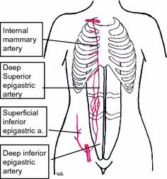
Fig. 30.2
Vascular supply of the lower abdominal apron
Pedicled transverse rectus abdominis myocutaneous (TRAM) flap
Free TRAM flap
Free DIEP flap
Free SIEA flap.
The evolution from pedicled TRAM flaps to free perforator flaps has been driven by the aim to reduce donor site morbidity and to preserve the integrity and function of the anterior abdominal wall. The complications encountered with techniques using lower abdominal tissue for breast reconstruction are related to:
Extent of muscle resection
Extent of fascia resection
Use of synthetic mesh to repair the abdominal wall.
These factors should be borne in mind when selecting the appropriate procedure for an individual patient (Table 30.1). Whichever variant of lower abdominal flap is used, sensory cutaneous innervation to the anterior abdominal wall is disrupted in all cases.
Table 30.1
Flap survival and donor site morbidity after abdominal tissue breast reconstruction
Pedicled TRAM flap (%) | Free TRAM flap (%) | Free DIEP flap (%) | |
|---|---|---|---|
Total flap loss | <1 | 5–7 | 1–5 |
Partial loss | 28–60 | 6–8 | 6 |
Fat necrosis | 27–40 | 7–13 | 6–10 |
Abdominal bulge | 8–28 | 5–8 | 0.3–5 |
Abdominal hernia | >6 | 4–6 | 0–1.4 |
30.5.2 Pedicled TRAM Flap
The pedicled TRAM flap relies on blood supply through the deep superior epigastric vessels (DSEA) within the substance of the rectus abdominis muscle to supply a horizontal ellipse of lower abdominal skin and fat (Fig. 30.3). The flap is transferred to the chest wall through an appropriately sized subcutaneous tunnel [10–12]. This procedure is relatively short (3–4 h) and does not require microvascular transfer. However, perfusion of the flap relies on microscopic intramuscular connections between the DSEA and the DIEA, which results in reduced vascularity (particularly to the suprapubic area) and thereby limits the amount of tissue which can be transferred safely. The reported incidence of fat necrosis is up to 42 %.

Fig. 30.3
Pedicled transverse rectus abdominis myocutaneous (TRAM) flap
With bipedicled TRAM flaps or in bilateral reconstructions, both rectus abdominis muscles are sacrificed, causing a permanent, severe reduction of abdominal wall function which has been found to interfere with activities of daily living, sports and housework. This can also interfere with the biomechanics of the paravertebral musculature, resulting in a high incidence of back pain. Other specific complications of this procedure include an extended recovery time, intercostal nerve compression and complications related to prosthetic mesh where required to repair the abdominal wall following pedicled TRAM flap harvest [9, 17–19]. The development of reliable free tissue transfer techniques has provided an alternative to the pedicled TRAM flap in an attempt to reduce abdominal wall damage and lower the risk of partial flap necrosis. The bipedicled TRAM flap carries substantial and significant donor morbidity and its use should be avoided as far as possible [20].
30.5.3 Free TRAM Flap
The deep inferior epigastric vessels are the dominant blood supply for the free TRAM flap. The lower abdominal skin is transferred with a segment of rectus abdominis muscle (Fig. 30.4). The deep inferior epigastric vessels are of good length and calibre for anastomosis to branches of the subscapular axis or the internal mammary vessels (Fig. 30.5) [13]. Depending on the extent of fascial harvest, the rectus sheath may require insertion of prosthetic mesh for closure.
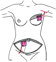

Fig. 30.4
Free TRAM flap
Fig. 30.5
Anastomosis to internal mammary vessels
This technique provides better tissue perfusion compared with the pedicled TRAM flap, and a larger portion of the abdominal apron can be transferred safely, with reduction in the risks of partial flap or fat necrosis. This consideration is particularly important for the reconstruction of a large breast. The amount of rectus abdominis muscle that is harvested can be reduced or minimised, thus causing less interference with abdominal wall function [21–26]. This technique requires a higher level of surgical expertise and microsurgical skills. The free TRAM flap has complications which include mesh infection, abdominal bulging and hernia formation.
30.5.4 Free DIEP Flap
The free DIEP flap spares the entirety of the rectus abdominis muscle. The lower abdominal skin ellipse is transferred by means of perforators of the deep inferior epigastric vessels dissected meticulously through a split in the rectus abdominis fascial sheath and muscle [14, 15, 27–29]. This technique is particularly indicated for young, athletic patients and in bilateral breast reconstruction (Fig. 30.6).
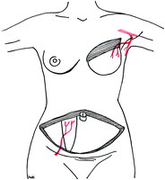
Fig. 30.6
Free DIEP flap
Although the DIEP flap still carries all the potential complications inherent in any free tissue transfer, donor site complications and morbidity are reduced. No muscle or fascia is harvested and synthetic mesh is not required for donor site closure. Preservation of the rectus sheath and muscle maintains abdominal and paravertebral muscle strength and reduces donor site morbidity, postoperative pain and in-hospital stay. The vascularity of the transferred tissue is not compromised, and the incidence of fat necrosis is no different from that with the conventional free TRAM flap [1, 7, 27–29].
Perfusion studies have been performed measuring the circulation in the DIEP flap using laser Doppler velocimetry as well as oxygen tension using implantable microcatheter probes. The perfusion of the DIEP flap has been found to be comparable to that of TRAM flaps. The muscle-sparing harvest does not jeopardise flap perfusion and survival even when the entire lower abdominal soft tissue ellipse is sustained on only one or a few perforators (Fig. 30.7) [30, 31].
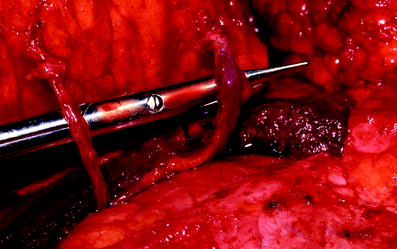
Fig. 30.7
Myocutaneous perforators of DIEP flap
Disadvantages include requirements for increased operating time, and an even higher level of surgical expertise owing to the technically exacting nature of perforator vessel dissection.
30.5.5 Free SIEA Flap
The lower abdominal skin ellipse can be transferred without muscle based on the SIEA and superficial inferior epigastric vein, a branch of the femoral vessels (Fig. 30.8) [32]. Since the free SIEA flap is raised without breach of the muscular or aponeurotic part of the abdominal wall, there is no risk of postoperative functional or mechanical weakness. Donor site morbidity is minimal and comparable to that for an abdominoplasty procedure. The operation is also substantially quicker, with a relatively simple dissection of the vascular pedicle. Unfortunately the SIEA has a high degree of anatomical variability in terms of its presence, course and calibre. It is absent in a third of patients, and the vascular pedicle is short, with a very small diameter of 1.5–2 mm. Because of its location, it is easily damaged by previous surgery in the inguinal region. The smaller vessel calibre may result in decreased flap perfusion with a higher risk of partial or total flap necrosis [32–34].
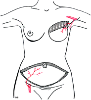
Fig. 30.8
Free superficial inferior epigastric artery flap
30.6 Breast Reconstruction with Buttock Tissue
When abdominal flaps are unavailable or inappropriate, a good second choice for free flap breast reconstruction is the skin and fat from the buttock area. The myocutaneous gluteal flaps have rarely been favoured in the past owing to their very short pedicle, awkward dissection, sizeable discrepancy in vessel diameter for anastomosis, potential for sciatic nerve damage and the need to sacrifice a significant portion of the gluteus maximus muscle. The gluteal perforator flaps are technical refinements which overcome the many disadvantages of the myocutaneous variant. Elimination of the muscle component of the traditional gluteal myocutaneous flap lengthens the vascular pedicle to approximately 8 cm. This improves intraoperative exposure and facilitates microvascular anastomosis to the internal mammary vessels or its perforators without the need for vein grafts [35–37]. Even thin patients with small breasts will usually have an adequate amount of fat in the gluteal region which enables this flap to be used. Donor site morbidity is relatively low, the scar is well hidden and postoperative recovery is quicker compared with abdominal flap harvest.
30.6.1 S-GAP Flap
The S-GAP flap comprises skin and fat harvested from the upper-third buttock area and leaves an acceptable donor site scar (Fig. 30.9). The maximum flap dimensions are 10 cm × 32 cm, with a weight of up to 800 g.
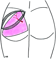
Fig. 30.9
Free superior gluteal artery flap
Related posts:

Stay updated, free articles. Join our Telegram channel

Full access? Get Clinical Tree


