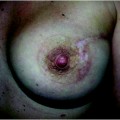
Fig. 36.1
Preoperative view of the patient before inferior right breast quadrantectomy, intraoperative radiotherapy, and bilateral reshaping
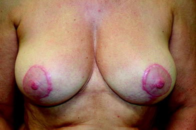
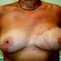
Fig. 36.2
Result at 7 months after the surgery with bilateral hypertrophic scars
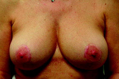
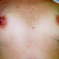
Fig. 36.3
Result at 2 years after three sessions of intralesional corticosteroid injections
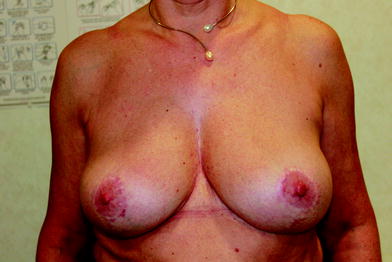
Fig. 36.4
Final result at 4 years
36.3.5.2 Bleomycin
This can cause necrosis of keratinocytes with a mixed inflammatory infiltrate in skin of healthy subjects. In keloid and hypertrophic scars, the effect of bleomycin may be due to a reduction of collagenase synthesis and/or increased destruction owing to inhibition of lysyl oxidase or TGF-β1. Despite the mechanism being unclear and lack of evidence, a few clinical trials showed a high regression rate, minimum complications, and recurrence in scar treatment with the multipuncture method. Bleomycin is often combined with triamcinolone for intralesional injection.
36.3.5.3 5-Fluorouracil
The effects on scar are due to inhibition of fibroblast proliferation. Combined injection with triamcinolone acetonide is often performed to reduce the dose of 5-fluorouracil. Injection of 5-fluorouracil is also combined with surgical excision. However, it is contraindicated in young women with the possibility of pregnancy and age under 18 years. Subcutaneous injection should be avoided. The side effects include a burning sensation and purpura formation.
36.3.5.4 Verapamil
This is a calcium channel antagonist, which can decrease collagen synthesis in extracellular matrix. It stimulates synthesis of the collagen-degradation enzyme procollagenase and increases TGF-β activity. It can be used as a monotherapy or combined with surgery or pressure therapy for keloid treatment. The data on the concentration and complications are limited and should be verified.
36.3.6 Laser
There are several laser applications for scar treatments, as a monotherapy or in combination with other modalities [24–26]. The effects of the laser on scar are mainly limited to the depth and the superficial layer action. Many types of laser used, including ablative lasers, dermal remodeling nonablative lasers, vascular lasers, ultraviolet-B lasers, and intense pulsed light lasers. In general, the selection of laser therapy depends essentially on the patient and the characteristics of the scars. The skin photo type is very important because melanin has a wide absorption spectrum and can be targeted by visible, ultraviolet and infrared light. Isotretinoin affects collagen metabolism and wound repair and its use must be avoided for the 6 months prior to an ablative laser procedure. Anticoagulant and antiplatelet therapies should be discontinued to prevent postlaser purpura. Fractional nonablative laser therapy has been reported with significant improvement in clinical and histopathological appearance [27] in a broad range of posttraumatic scars and surgical scars.
36.3.7 Surgery
A surgeon can improve the scar in both form and more importantly function. Wound management should be considered as a systematic role from preoperative planning, the intraoperative procedure, and immediate postoperative care until late follow-up [28, 29].
In scar contracture, the principal role of the surgeon is to restore the functions of the patient. Scar contracture release should be performed together with an intense rehabilitation program and if possible other scar therapeutic modalities to prevent contracture recurrence. Surgical management of scar release includes scar revisions, split-thickness skin grafts, full-thickness skin grafts, local flaps, pedicle flaps, and distant microsurgical flaps. The selection of this reconstructive ladder depends on the patient and the scar characteristics. A free flap or perforator flap can give a favorable result in a massive area, deep scarring tissue, and poor surrounding tissue. Dermabrasion, minor scar revision, or simple serial excision with or without tissue expansion can be an effective option in scar management.
36.3.8 Lipofilling
The lipofilling technique has been used for many years and has rapidly become popular especially in aesthetic surgery [30]. In the era of tissue engineering, progenitor and stem cells are being studied and are rapidly gaining interest. The fat is removed by liposuction from the subcutaneous tissue, usually from the abdomen or from the thighs according to the morphology of the patient. The specimen obtained is subjected to soft centrifugation to remove blood cell contaminants and obtain an adipocyte-enriched preparation. Recently, a number of new techniques have been described, mostly based on enzymatic treatments, with the ultimate goal to improve adipocyte purification. After harvesting and processing, the purified fat is injected into the scar area. The lipofilling procedure claims not only to improve the volume deficit but also to improve the color and surface of the scar area.
36.3.9 Radiotherapy
The employment of radiotherapy in the treatment of benign skin disorders, including keloids, is presently allowed only under certain conditions and is subject to compliance with strict protection rules [31, 32]. A series of studies have demonstrated the efficacy and preliminary safety of ionizing radiation beams in their protocols. The combination of surgical and radiotherapeutic treatment causes a synergistic effect relating to scar treatment. Radiotherapy can be delivered at a time when connective tissue is more radiosensitive, by decreasing fibroblast proliferation and causing a rapid mast cell degranulation which reduces histamine levels and is capable of accelerating collagen formation. The total doses of ionizing radiation differ among the different protocols reported in the literature. However, an increase of the total dose of ionizing radiation administered could theoretically enhance the risk of radiogenic skin cancer for the patients treated. There is a report debating the risk of radiotherapy in the treatment of keloids with regard to the carcinogenicity of radiation. However, no clinical trial has demonstrated this finding in an analytic study despite the potential theoretical risk outlined. Regular X-ray irradiation, electron beam irradiation, and brachytherapy after excision of keloid are performed with favorable outcomes.
36.4 European Institute of Oncology Experience
Hypertrophic scar has been treated surgically followed by brachytherapy according to two different techniques. A total of 51 patients with breast scar are included in the database, and in the first period low-dose radiation (with isolation of the patient) was used in 31 patients, whereas recently a group of 20 patients were treated with high-dose radiation (without isolation). The recurrence rate was 15.7 % (eight patients), with the global aesthetic result considered as good in 58 % of cases (Figs. 36.5, 36.6


Related posts:

Stay updated, free articles. Join our Telegram channel
Full access? Get Clinical Tree
