Although the metastatic rate of cutaneous squamous cell carcinoma (SCC) is low, detailed examination for the presence of micro- and macrometastasis of lymph nodes is crucial in avoiding the devastating outcomes and in planning appropriate treatment. Cutaneous SCC of the head and neck can spread to parotid lymph nodes, cervical lymph nodes, or both, depending on the location of the primary tumor. Therefore, clinical and radiologic evaluation of the parotid and neck should be performed in patients with cutaneous SCC. Optimal treatment of metastatic cutaneous SCC of the head and neck should consist of complete surgical resection with adjuvant radiotherapy.
- •
There are certain characteristics to a primary lesion of cutaneous squamous cell carcinoma (SCC) that imply a higher risk of lymphatic metastasis, such as size, thickness, and histopathologic features (differentiation, perineural involvement, desmoplasia, and so forth).
- •
The presence of lymph node disease is a poor prognostic factor in cutaneous SCC.
- •
Cutaneous SCC of the head and neck can spread to parotid lymph nodes, cervical lymph nodes, or both, depending on the location of the primary tumor. Therefore, clinical and radiologic evaluation of the parotid and neck should be performed in patients with cutaneous SCC.
- •
The optimal treatment for metastatic cutaneous SCC of the head and neck should be complete surgical resection with adjuvant radiotherapy.
Introduction
Skin cancer is the most common malignancy in the white population, and is related to chronic solar ultraviolet radiation. Although the head and neck comprises only 9% of the total body surface area, most nonmelanoma skin cancers are encountered in this region as a result of sun exposure. The incidence of cutaneous squamous cell carcinoma (SCC) of the head and neck accounts for approximately 20% to 25% of all nonmelanoma skin cancers. Cutaneous SCC of the head and neck rarely metastasizes to regional lymph nodes (approximately 5% of patients), but when it does the parotid lymph node bed is most frequently involved. Patients with metastatic cutaneous SCC have a poor prognosis with an overall 5-year survival rate of 34.4%. Therefore, the detection of macrometastasis or micrometastasis of parotid and/or cervical lymph nodes is crucial. Because the involvement of lymph nodes in cutaneous SCC of the head and neck is relatively uncommon, and as it often takes a long time from the initial tumor identification and treatment to the ultimate development of metastases, it is worthwhile highlighting the disease process to bring it to the forefront of the clinician’s mind. The aim of this article is to review the lymphatic drainage of skin, the principles of lymphatic metastasis, risk factors and patterns of tumor spread, and the staging and therapeutic implications of metastatic cutaneous SCC of the head and neck.
Cutaneous lymphatic system of the head and neck
The lymphatic system is an important component of the immune system, playing a vital role in infection, inflammation, and cancer metastasis. It provides a pathway for the drainage of lymph, a white, “milky” fluid that contains proteins, lipids, cells, cellular debris, and foreign products. The lymphatic system includes a terminal capillary network, precollector and collector lymph vessels, lymphatic ducts, and lymph nodes. The superficial lymphatic plexus is located in the upper dermis (near the arterial plexus) and collects lymph from the interstitium. Lymphatic capillaries have an endothelial leaflet layer, and an incomplete basement membrane and muscle layer; however, they do not have intraluminal valves. These capillaries are generally tortuous, and several lymphatic capillaries join in the dermis to form lymphatic vessels called precollectors. Precollector lymphatic vessels have a complete endothelial layer, basement membrane, and muscle layer with intraluminal valves, which are separated by approximately 2-mm intervals. Thereafter, lymphatic fluid enters collector lymphatic vessels at the cutis-subcutis boundary. The collector lymphatic vessels have a continuous basal membrane, valves, and smooth muscle cells, which play a vital role in lymph transport by propelling lymph toward afferent vessels that ultimately drain into lymph nodes. Thus, unidirectional lymph flow in lymphatic vessels is maintained by an intrinsic pump mechanism, and retrograde lymph flow is prevented by internal valves. The velocity of lymph flow is variable throughout the body and is relatively slow in the head and neck (approximately 1.5 cm/min).
In the head and neck region there are more than 300 lymph nodes, subclassified into lymph node groups. Lymph node groups are generally named according to their locations: suboccipital, retroauricular, parotid, facial (buccal), retropharyngeal, and cervical (submental, submandibular, upper jugular, middle jugular, lower jugular, anterior jugular, spinal accessory chain, and supraclavicular). The patterns of lymphatic drainage in the head and neck are categorized as follows :
- 1.
Main lymphatic pathway: drains anterior face, oral cavity, and nasal mucosa
- 2.
Posterior accessory lymphatic pathway: drains the postauricular region, posterior scalp, suboccipital region, nasopharynx, and oropharynx
- 3.
Anterior lymphatic pathway: drains the oral cavity, median part of the lower lip, and the skin overlying the chin
- 4.
Superficial lateral pathway: drains the posterior scalp and initially enters the suboccipital and retroauricular nodes
The lymphatic drainage pathways for the skin of the head and neck region are usually predictable, and are presented in Table 1 .
| Anatomic Region | Lymph Node Group |
|---|---|
| Eyelid and eyebrows | Parotid, submandibular |
| Anterior ear and preauricular region | Parotid |
| Helix and lateral aspect of the auricle | Retroauricular, suboccipital |
| Cheek, nose, upper lip | Parotid, submandibular |
| Lateral part of lower lip | Submandibular |
| Median lower lip, chin | Submental |
| Lateral aspect of the head and forehead | Parotid, upper jugulodigastric |
| Frontal and parietal parts of scalp | Parotid |
| Posterior part of scalp and head | Suboccipital, retroauricular, upper, middle and lower jugulodigastric, supraclavicular |
| Neck | Upper, middle, and lower jugulodigastric |
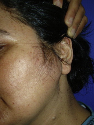
The parotid gland has a high-density lymphatic network. It has been demonstrated that intraparotid lymph nodes are “trafficking” the lymphatic metastasis of cutaneous SCC, in particular for tumors located on the lateral part of the face ( Fig. 1 ). Marks dissected 1 to 11 lymph nodes from the parotid gland in a series of 17 radical parotidectomies. He found that on average, one node remains in the deep lobe of the parotid following superficial parotidectomy. Similarly, McKean and colleagues found that almost all lymph nodes of the parotid gland are located lateral to the facial nerve, in the so-called superficial lobe of the gland.
Cutaneous lymphatic system of the head and neck
The lymphatic system is an important component of the immune system, playing a vital role in infection, inflammation, and cancer metastasis. It provides a pathway for the drainage of lymph, a white, “milky” fluid that contains proteins, lipids, cells, cellular debris, and foreign products. The lymphatic system includes a terminal capillary network, precollector and collector lymph vessels, lymphatic ducts, and lymph nodes. The superficial lymphatic plexus is located in the upper dermis (near the arterial plexus) and collects lymph from the interstitium. Lymphatic capillaries have an endothelial leaflet layer, and an incomplete basement membrane and muscle layer; however, they do not have intraluminal valves. These capillaries are generally tortuous, and several lymphatic capillaries join in the dermis to form lymphatic vessels called precollectors. Precollector lymphatic vessels have a complete endothelial layer, basement membrane, and muscle layer with intraluminal valves, which are separated by approximately 2-mm intervals. Thereafter, lymphatic fluid enters collector lymphatic vessels at the cutis-subcutis boundary. The collector lymphatic vessels have a continuous basal membrane, valves, and smooth muscle cells, which play a vital role in lymph transport by propelling lymph toward afferent vessels that ultimately drain into lymph nodes. Thus, unidirectional lymph flow in lymphatic vessels is maintained by an intrinsic pump mechanism, and retrograde lymph flow is prevented by internal valves. The velocity of lymph flow is variable throughout the body and is relatively slow in the head and neck (approximately 1.5 cm/min).
In the head and neck region there are more than 300 lymph nodes, subclassified into lymph node groups. Lymph node groups are generally named according to their locations: suboccipital, retroauricular, parotid, facial (buccal), retropharyngeal, and cervical (submental, submandibular, upper jugular, middle jugular, lower jugular, anterior jugular, spinal accessory chain, and supraclavicular). The patterns of lymphatic drainage in the head and neck are categorized as follows :
- 1.
Main lymphatic pathway: drains anterior face, oral cavity, and nasal mucosa
- 2.
Posterior accessory lymphatic pathway: drains the postauricular region, posterior scalp, suboccipital region, nasopharynx, and oropharynx
- 3.
Anterior lymphatic pathway: drains the oral cavity, median part of the lower lip, and the skin overlying the chin
- 4.
Superficial lateral pathway: drains the posterior scalp and initially enters the suboccipital and retroauricular nodes
The lymphatic drainage pathways for the skin of the head and neck region are usually predictable, and are presented in Table 1 .
| Anatomic Region | Lymph Node Group |
|---|---|
| Eyelid and eyebrows | Parotid, submandibular |
| Anterior ear and preauricular region | Parotid |
| Helix and lateral aspect of the auricle | Retroauricular, suboccipital |
| Cheek, nose, upper lip | Parotid, submandibular |
| Lateral part of lower lip | Submandibular |
| Median lower lip, chin | Submental |
| Lateral aspect of the head and forehead | Parotid, upper jugulodigastric |
| Frontal and parietal parts of scalp | Parotid |
| Posterior part of scalp and head | Suboccipital, retroauricular, upper, middle and lower jugulodigastric, supraclavicular |
| Neck | Upper, middle, and lower jugulodigastric |
The parotid gland has a high-density lymphatic network. It has been demonstrated that intraparotid lymph nodes are “trafficking” the lymphatic metastasis of cutaneous SCC, in particular for tumors located on the lateral part of the face ( Fig. 1 ). Marks dissected 1 to 11 lymph nodes from the parotid gland in a series of 17 radical parotidectomies. He found that on average, one node remains in the deep lobe of the parotid following superficial parotidectomy. Similarly, McKean and colleagues found that almost all lymph nodes of the parotid gland are located lateral to the facial nerve, in the so-called superficial lobe of the gland.
Lymphatic metastasis of cutaneous SCC of the head and neck
Lymphatic metastasis is one of the major determinants in the staging, treatment, and prognosis of head and neck malignancies. Lymphatic metastasis is generally initiated when the tumor directly penetrates the basement membrane of the epithelium and invades the extracellular matrix. This process is facilitated by the impairment of adhesive properties of tumor cells, secretion of proteolytic enzymes, and increase in intratumoral interstitial fluid pressure. Tumor cells enter the lymph through peritumoral and intratumoral lymphatic capillaries, which are dilated and hyperplastic, by means of hydrostatic pressure gradients and active movement. The development and proliferation of peritumoral and intratumoral lymphatic capillaries are triggered by lymphangiogenesis, which is mediated by growth factors, cytokines, and chemokines mainly secreted from tumor cells and/or host cells. Recent studies have demonstrated that the vascular endothelial growth factor (VEGF)-C/VEGF-D/VEGF receptor-3 signaling axis plays a vital role in lymphangiogenesis, and promotes formation of tumor lymphatics and metastatic spread of tumor cells to lymph nodes. Vascular endothelial growth factor-C and/or VEGF-D expression in tumor cells stimulates VEGF receptor-3, a cell-surface receptor tyrosine kinase located on lymphatic endothelial cells, and activates growth of lymphatic vessels. In addition, VEGF-A, platelet-derived growth factor BB, and hepatocyte growth factor have been linked to lymphangiogenesis and lymphatic metastasis. Finally, tumor cells are transported to the subscapular sinus of lymph node via lymphatic vessels. Unfortunately, lymph nodes are poor barriers for tumor spread. Therefore, only small numbers of tumor cells are destroyed while most tumor cells can easily settle and proliferate in the subscapular sinus of the first-echelon lymph node. Further metastasis to other lymph nodes is almost inevitable once the cortex of a sentinel lymph node (SLN) is invaded.
Patient with high-risk cutaneous SCC of the head and neck
Most patients with cutaneous SCC of the head and neck are at low risk of developing local recurrence, lymph node metastasis, and distant metastasis. However, there is a subset of patients who are at high risk for local recurrence and nodal metastasis, and in whom long-term survival is very low. These patients should be carefully evaluated for the presence of macrometastasis or micrometastasis in regional lymph nodes. In such patients, SLN biopsy or elective parotidectomy/neck dissection should be considered, even when lymph node metastasis is not detected on clinical or radiologic evaluation. This high-risk cutaneous SCC patient group is defined by patient-related, tumor-related, and previous treatment–related risk factors ( Table 2 ).
Related posts:

Stay updated, free articles. Join our Telegram channel

Full access? Get Clinical Tree


