Cutaneous Manifestations of Systemic Disease
Herbert P. Goodheart
Peter G. Burk
Overview
The cutaneous surface, nails, hair, and oral cavity often afford clues to many underlying disorders. The skin is sometimes referred to as a “window” to disease. For example, the presence of jaundice, palmar erythema, pruritus, and spider telangiectasias points to liver disease. The appearance of pyoderma gangrenosum, erythema nodosum, or severe aphthous stomatitis may indicate inflammatory bowel disease.
By appreciating skin signs of systemic diseases, the health care provider can often lead his or her patient to an early diagnosis and expedient and appropriate treatment.
This chapter reviews some of the cutaneous manifestations of systemic diseases and it highlights recent developments in their management.
 ENDOCRINE DISEASES
Diabetes-associated lesions
Necrobiosis lipoidica diabeticorum
Diabetic bullous disease
Diabetic neuropathic ulcers
Acanthosis nigricans
Eruptive xanthomas
Diabetic dermopathy
Perforating folliculitis (Kyrle’s disease)
Disseminated granuloma annulare
Scleredema of Buschke-Löwenstein
Thyroid Disease
Pretibial myxedema
Warm, moist, and velvety skin
Alopecia with diffuse hair loss
Nails
Hair
 LIPID ABNORMALITIES
Eruptive xanthomas
Planar xanthomas
Xanthelasma
Tuberous xanthomas
Tendinous xanthomas
 CONNECTIVE TISSUE DISEASES
Systemic lupus erythematosus
Drug-induced lupus erythematosus
Subacute cutaneous lupus erythematosus
Chronic cutaneous lupus erythematosus
Neonatal lupus erythematosus
Dermatomyositis
Scleroderma and morphea
CREST syndrome and progressive systemic sclerosis
 ERYTHEMA NODOSUM
 CUTANEOUS SARCOIDOSIS
 INFLAMMATORY SKIN DISORDERS
Reiter’s syndrome
Pyoderma gangrenosum
Exfoliative dermatitis
 NEUROCUTANEOUS SYNDROMES
Neurofibromatosis
Tuberous sclerosis
Endocrine Diseases
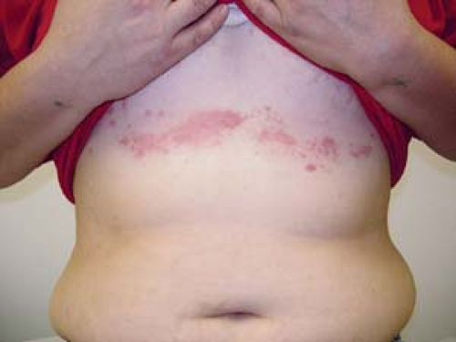
 25.1 Necrobiosis lipoidica diabeticorum. This diabetic patient has early lesions that consist of yellow-red plaques. Epidermal atrophy and telangiectasias tend to occur later. |
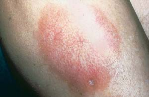 25.2 Necrobiosis lipoidica diabeticorum. This diabetic patient has more advanced lesions than those seen in Figure 25.1. Epidermal atrophy and telangiectasias are seen here. |
Basics
Many different skin lesions are seen in conjunction with endocrine diseases. Some cutaneous lesions are directly related to the degree of endocrine dysfunction and may be caused by an excess or lack of a hormone acting on a specific tissue, such as warm and moist skin associated with hyperthyroidism or dry and cool skin associated with hypothyroidism.
In a disease such as diabetes, it may be difficult to link these skin findings to a specific degree of endocrine dysfunction (e.g., necrobiosis lipoidica diabeticorum with the level of hyperglycemia).
Diabetes Mellitus
Diabetes mellitus is a disease characterized by a disturbance in the production of insulin or resistance to insulin activity, which results in abnormal glucose metabolism.
Diabetes causes cellular changes, such as microangiopathy of small blood vessels. This causes organ damage, including retinal disease, renal dysfunction, and possibly cutaneous lesions.
Diabetes can also affect immune function and can lead to an increase of bacterial, fungal, and yeast infections.
Diabetes-Associated Lesions: Necrobiosis Lipoidica Diabeticorum
Necrobiosis lipoidica diabeticorum (NLD) is characterized by yellow-red to brown, translucent plaques with epidermal atrophy and telangiectasia (Fig. 25.1). As lesions progress, the center becomes depressed and yellow (Fig. 25.2). Ulceration is not uncommon.
NLD appears most commonly on the pretibial areas, but it may arise on other sites.
NLD is more common in women than in men.
Treatment for NLD is not very satisfactory. The condition is typically chronic with variable progression and scarring.
NLD is seen more frequently in type 1 than in type 2 diabetes and may occur before the onset of clinical diabetes. Lesions may ulcerate. A minority of patients have no clinical evidence or family history of diabetes; in these patients, the term necrobiosis lipoidica is used. Currently, the term necrobiosis lipoidica is used to encompass all patients with the same clinical lesions regardless of whether diabetes is present or not.
The lesions of NLD can be similar to those of morphea (see later discussion) and other localized sclerosing lesions.
High-potency topical steroids or intralesional steroid injections are used and can lessen the inflammation of early active lesions and the active borders of enlarging lesions, but these have little beneficial effect on atrophic lesions that are burned out. In fact, with atrophic lesions, steroid use may cause further atrophy.
Because localized trauma can cause NLD to ulcerate, protection of the legs with support stockings may be helpful.
Antiplatelet aggregation therapy with aspirin and dipyridamole has produced varied results but overall has shown some beneficial effects.
Pentoxifylline (Trental) is a drug used for the treatment of intermittent claudication. Pentoxifylline is believed to decrease blood viscosity by increasing fibrinolysis and red blood cell deformity. It also inhibits platelet aggregation.
Topical application of bovine collagen is believed to improve granulation tissue by supporting fibroblast activity and promote wound debridement by increasing the number of macrophages and neutrophils at the wound site.
Ticlopidine, nicotinamide, clofazimine, and perilesional heparin injections have been used in uncontrolled studies and appeared to benefit some patients with NLD.
Diabetes-Associated Lesions: Diabetic Bullous Disease
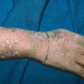
Diabetic bullous disease (bullosis diabeticorum) manifests as large, tense, subepidermal, noninflammatory blisters (Fig. 25.3).
Lesions most often arise spontaneously on the lower extremities, especially the ankles and feet. The development of multiple lesions at several locations may occur.
Lesions tend to arise in patients with long-standing diabetes mellitus who have multiple complications of the disease.
Bullous diabetic lesions can be differentiated from bullous pemphigoid by the characteristic location of lesions and negative direct immunofluorescence of skin biopsies.
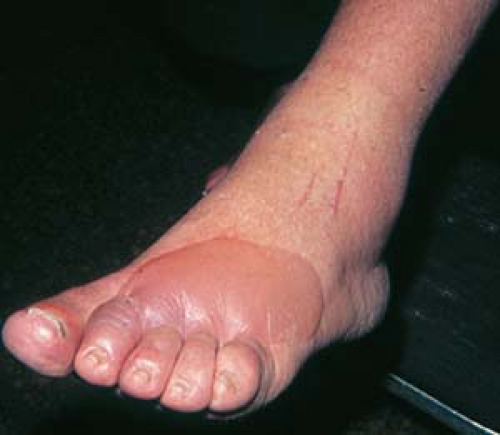 25.3 Diabetic bullous disease. This large, tense blister arose spontaneously in a characteristic location. |
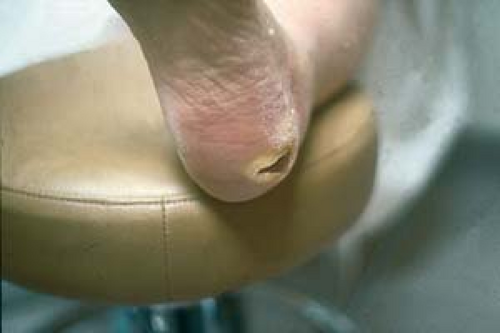 25.4 Diabetic ulcer of the heel (mal perforans). These lesions are noted at sites of pressure, such as the heel in this patient. |
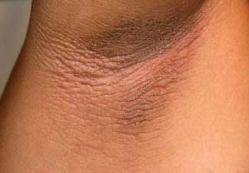 25.5 Acanthosis nigricans. Note the characteristic hyperpigmentation in a typical location. This patient has insulin-resistant diabetes. (Image courtesy of Bernard Cohen, M.D.) |
 25.6 Cutaneous candidiasis. These satellite pustules were positive for Candida albicans. |
Blisters generally heal spontaneously within 2 to 6 weeks of onset.
Topical antibiotics are recommended until the lesions heal.
Diabetes-Associated Lesions: Diabetic Neuropathic Ulcers
Diabetic neuropathic ulcers (mal perforans) (Fig. 25.4) most often occur at sites of pressure (e.g., the heel), particularly in areas of poor sensory function and poor circulation. They are usually painless as a result of peripheral neuropathy.
Diabetic foot ulcers occur as a result of the loss of protective sensation.
Other factors, such as mechanical changes in conformation of the bony architecture of the foot and atherosclerotic peripheral arterial disease, may be contributing causes.
Diagnosis
Diabetic neuropathic ulcers should be distinguished from infections and other ulcerations and cutaneous neoplasms that may present as ulcerations.
It is beyond the scope of this discussion to describe all therapies for diabetic foot ulcers. However, management can include glycemic control, special footwear, topical wound management, daily saline soaks, surgical debridement, and skin grafting when necessary.
Becaplermin (Regranex*), a recombinant human platelet-derived growth factor, is available in gel form for topical therapy (in conjunction with good ulcer care) and is reported to promote healing of diabetic neuropathic foot ulcers.
Hyperbaric oxygen therapy may be useful.
Other Findings in Diabetic Patients
Acanthosis nigricans (Fig. 25.5) sometimes occurs in insulin-resistant diabetes (also see Figs. 14.19 and 14.20).
Cutaneous candidiasis (Fig. 25.6) may also occur (see also Figs. 7.31, 7.32 and 7.33) (see Chapter 7, “Superficial Fungal Infections”).
Eruptive xanthomas are seen as skin markers for various primary genetic disorders such as certain types of hyperlipidemias or secondary to diabetes (see later discussion).
Diabetic dermopathy is characterized by small brownish, atrophic, scarred, hyperpigmented plaques (Fig. 25.7)
Lesions occur primarily on the anterior lower legs in patients with type 1 and type 2 diabetes.
It is typically a late manifestation of diabetes and is usually asymptomatic.
Diabetic dermopathy must be differentiated from lesions caused by trauma.
Perforating folliculitis (Kyrle’s disease) consists of firm, rough, hyperkeratotic papules, which are often hyperpigmented in dark-skinned people.
There is a high incidence of perforating folliculitis in patients with long-standing diabetes who are undergoing long-term hemodialysis.
Lesions are found most commonly on the extensor surfaces of the extremities. Itching can be intense.
Disseminated granuloma annulare consists of annular dermal papules (see Chapter 4, “Inflammatory Eruptions of Unknown Cause”). It occurs both in patients with clinical diabetes and sometimes in individuals with only abnormal glucose levels.
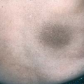
Scleredema of Buschke-Löwenstein is a rare manifestation of diabetes mellitus. The lesion is a sclerotic, thickened plaque characteristically seen on the upper back.
Diagnosis
The diagnoses of many of these entities are generally made on clinical grounds.
A skin biopsy may be necessary to confirm the diagnosis.
Laboratory Evaluation
Serum glucose levels and glycosylated hemoglobin A1 are determined to confirm the diagnosis of diabetes mellitus.
Skin biopsies of lesions of necrobiosis lipoidica and granuloma annulare demonstrate palisading granulomas with degeneration of collagen.
Skin biopsy of perforating folliculitis demonstrates basophilic material in the dermis, with transepidermal elimination.
Skin biopsies of diabetic dermopathy show thickening of blood vessels and mild perivascular infiltrate.
Diabetic bullous lesions have subepidermal blistering on hematoxylin and eosin staining of skin biopsy tissue; direct immunofluorescence of skin biopsies in these lesions is negative for immunoglobulins.
Diabetic neuropathic ulcers are rarely examined by biopsy.
Perforating folliculitis can be differentiated from other keratotic papules by the size and distribution of lesions and by a skin biopsy.
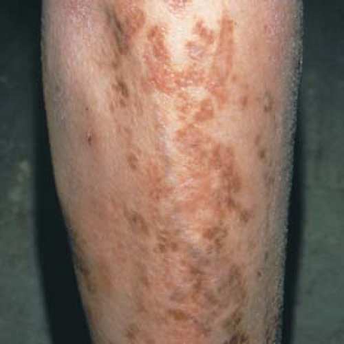 25.7 Diabetic dermopathy. Small, asymptomatic, brownish, atrophic, scarred, hyperpigmented plaques are seen on the shins of this diabetic patient. |
Careful consideration should be given before performing a skin biopsy on a patient with diabetes, particularly on areas such as the lower extremities.
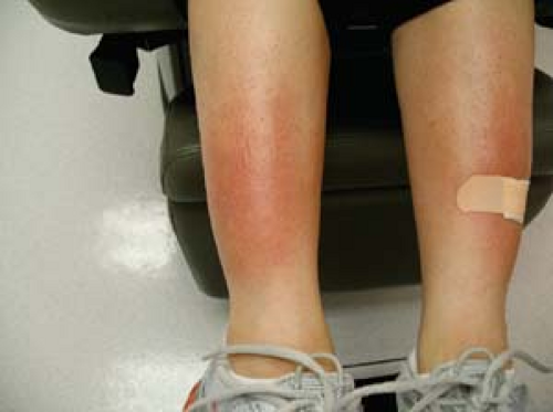 25.8 Pretibial myxedema, Graves’ disease. Erythematous plaques with early involvement. (Image courtesy of Ashit Marah, M.D.) |
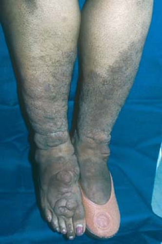 25.9 Pretibial myxedema, Graves’ disease. Note the progressive exuberant hypertrophy with folding of the skin on the shins and tops of the feet. |
Thyroid Disease
Basics
Thyroid hormones profoundly influence the growth and differentiation of epidermal and dermal tissues.
Abnormal levels of thyroid hormone produce striking changes in the texture of the skin, hair, and nails.
Some of the associated skin alterations in thyroid disease are the result of a deficiency or a high toxic level of tissue thyroid hormone. Other skin disorders seen with thyroid disease, such as vitiligo and alopecia areata, are associated clinical findings that are not directly related to thyroid hormone function but are seen on occasion in patients with thyroid disease.
Findings such as pretibial myxedema are caused by circulating autoimmune γ-globulin, which acts as a thyroid-stimulating hormone.
Hyperthyroidism may be caused by Graves’ disease, subacute thyroiditis, toxic goiter, and thyroid carcinoma.
Hypothyroidism may be caused by iodine deficiency (cretinism), Hashimoto’s thyroiditis, pituitary dysfunction with thyroid-stimulating hormone deficiency, and surgical or radiation ablation of the thyroid.
Patients with thyroid disease may be hyperthyroid at one point in their clinical course and hypothyroid at another time.
Description of Lesions
Hyperthyroid skin lesions may include the following:
Warm, moist, and velvety skin
Alopecia with diffuse hair loss
Nail changes with onycholysis (Plummer’s nails)
Hyperpigmentation
Pretibial myxedema lesions—flesh-colored, waxy infiltrated translucent plaques (Figs. 25.8 and 25.9)
Hyperthyroid findings may include the following:
Nervousness and tremor
Weight loss
Tachycardia with atrial fibrillation
Proximal muscle weakness
Graves’ disease
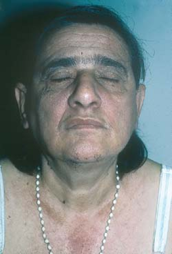
Exophthalmos (Fig. 25.10)
Hypothyroid skin lesions may include the following:
Myxedema of the skin with generalized thickening and a dry, coarse feel; yellow skin secondary to carotenemia
Hair changes—coarse, sparse hair; lateral third of eyebrows lost
The following conditions may be associated with thyroid disease:
Alopecia areata
Vitiligo
Connective tissue diseases
Multiple endocrinopathy syndrome
Urticaria
Distribution of Lesions
Pretibial myxedema lesions are found most frequently on the lower legs.
Clinical Manifestations
Hyperthyroid skin changes that result from the hypermetabolic state (e.g., warm, moist, flushed skin) occur during the active thyrotoxic stage of thyroiditis, during active Graves’ disease, and in patients with toxic goiters. These skin changes may gradually resolve when the patient returns to a euthyroid state.
Graves’ disease lesions (pretibial myxedema) occur in up to 4% of patients with this disease. The skin lesions and eye lesions usually do not resolve, even after treatment of the thyroid disease brings a return to a euthyroid state.
Hypothyroid skin changes (e.g., cool, dry skin) are related to the length and severity of the clinical hypothyroid state. These skin lesions gradually improve some months after the patient returns to a euthyroid state.
Diagnosis
Diagnosis of both hyperthyroid and hypothyroid disease is made by specific thyroid function tests.
Graves’ disease is diagnosed clinically and with confirmatory thyroid function tests.
Laboratory Evaluation
Elevated thyroid-stimulating hormone levels are the most sensitive screening test for hypothyroidism.
Serum thyroid hormone levels can be most accurately measured by obtaining free thyroxine and free triiodothyronine levels.
Antithyroglobulin antibodies and antithyroid microsomal antibodies are often positive in Graves’ disease and Hashimoto’s thyroiditis.
Long-acting thyroid stimulator is elevated in 50% of patients with Graves’ disease.
Skin biopsies in Graves’ disease show increased staining of hyaluronic acid with mucin stains in the reticular and papillary dermis.
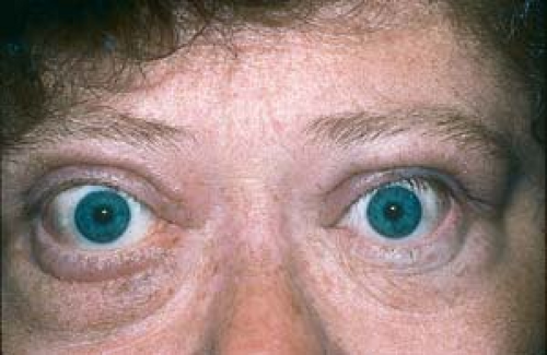 25.10 Exophthalmos. This patient has Graves’ disease. Note the lid retraction and proptosis. |
Pretibial myxedema must be differentiated from other skin diseases with increased mucin production, such as papillary mucinosis and scleredema.
Functional symptoms (e.g., increase or decrease of sweating, dry skin, hair and nail changes) of hyperthyroidism and hypothyroidism may improve after appropriate treatment of thyroid disease and return to a euthyroid state.
Treatment of pretibial myxedema lesions can be attempted with high-potency topical steroids and intralesional steroids, although the response is generally poor.
Cutaneous Manifestations of Lipid Abnormalities: Xanthomas
Basics
Abnormalities of lipid metabolism, with high circulating levels of various lipoproteins, can result in deposition of cholesterol and other lipids in the skin, tendons, and other organs.
Xanthomas result from the deposition of cholesterol and other lipids found in tissue macrophages in the skin and tendons. There is also a high correlation between abnormal lipoproteinemia and the development of atherosclerosis.
Lipoprotein abnormalities have been classified into primary (genetic) lipoproteinemia and secondary lipoproteinemia resulting from underlying diseases.
Primary lipoproteinemias are phenotypic expressions of various genetic disorders of lipid metabolism with the following characteristics:
Type I, familial lipoprotein lipase deficiency: elevated chylomicrons
Type IIA, familial hypercholesterolemia: elevated low-density lipoproteins
Type IIB, familial hyperlipidemia: elevated low-density lipoproteins and very-low-density lipoproteins
Type III, familial dysbetalipoproteinemia: elevated intermediate-density lipoproteins
Type IV, endogenous familial hypertriglyceridemia: elevated triglycerides
Type V, familial combined hyperlipidemia: elevated chylomicrons and elevated very-low-density lipoproteins
Secondary hyperlipoproteinemias result from disturbances in cholesterol and triglyceride metabolism caused by cholestatic liver disease, diabetes mellitus, pancreatitis, multiple myeloma, and nephrotic syndrome. These disorders may mimic any of the genetic lipoprotein abnormalities and may produce similar xanthomatous deposits in tissues.
Description of Lesions
Eruptive xanthomas are smooth, yellow, papular lesions (2 to 5 mm) (Figs. 25.11 A and B). There is sometimes a red halo around the lesions.
Planar xanthomas are flat to slightly palpable yellow lesions.
Xanthelasma (xanthoma palpebrarum) is a form of planar xanthoma (Fig. 25.12).
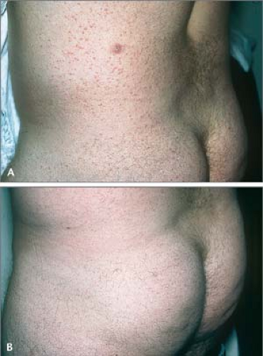
25.11 A and B Eruptive xanthomas. A: This 28-year-old male patient has a triglyceride level of 31,000 and a cholesterol level of 580 mg/dL. B: This is the same patient after 2 months of a low-fat diet and a cholesterol-lowering drug.
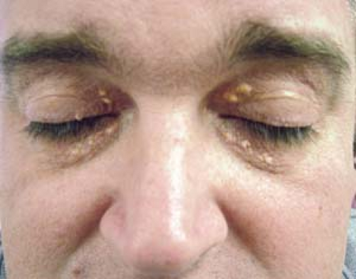
25.12 Xanthelasma. Periorbital yellow-orange plaques are present on the upper inner eyelids. This patient was normolipemic when this photograph was taken.
Tuberous xanthomas are small (0.5 cm) to large (3 to 5 cm), firm, yellow papules and nodules (Fig. 25.13).
Tendinous xanthomas are subcutaneous thickenings around tendons and ligaments.
Distribution of Lesions
Eruptive xanthomas appear most frequently over the knees, elbows, and buttocks.
Planar xanthomas are found in the palmar creases but may also be generalized.
Xanthelasma lesions are usually found on the eyelids and medial canthus.
Tuberous xanthomas are found on the elbows, knees, and buttocks.
Tendinous xanthomas affect the Achilles tendon, extensor tendons of the wrists, elbows, and knees.
Clinical Manifestations
Eruptive xanthomas tend to appear suddenly over the extensor surfaces and pressure points. These lesions are usually seen in association with very high levels of triglycerides (2,000 to 4,000 mg/dL). Uncontrolled diabetes mellitus and acute pancreatitis are both common underlying causes of their surfacing on the skin.
Planar xanthomas are usually asymptomatic. Palmar xanthomas are seen with type III lipoproteinemia. Diffuse planar xanthomas are found in patients with multiple myeloma.
Xanthelasma lesions grow slowly over years. More than 50% of patients with xanthelasma have normal lipoprotein levels.
Tuberous xanthomas also are slow growing. They are associated with familial hypercholesterolemia but can also occur in patients with high triglyceride levels.
Tendinous xanthomas occur in patients with hypercholesterolemias.
Diagnosis
The diagnosis is made by clinical evaluation of skin and subcutaneous lesions.
Skin biopsy is confirmatory for xanthomas.
Laboratory Evaluation
Fasting blood levels of triglycerides and cholesterol should be determined.
Lipoprotein electrophoresis demonstrates specific lipoprotein abnormalities.
Skin biopsy of xanthomas demonstrates collections of lipids in foamy macrophages in the dermis.
Serum glucose levels and glycosylated hemoglobin A1 are determined to rule out diabetes mellitus.
Serum amylase levels should be examined to rule out pancreatitis.
Serum protein electrophoresis should be performed to rule out multiple myeloma.
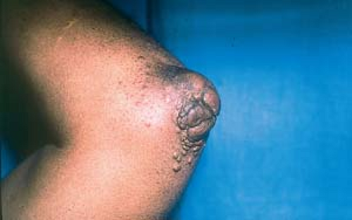 25.13 Tuberous xanthomas. These are firm papules and nodules in a patient with hyperlipidemia type II. |
Eruptive xanthomas must be differentiated from cutaneous sarcoid papules and cutaneous histiocytosis.
Tuberous xanthomas can be confused with rheumatoid nodules and subcutaneous granuloma annulare.
Patients with lipid disorders and xanthomas must be appropriately evaluated for primary and secondary lipoprotein abnormalities. Treatment of the underlying cause may reverse both eruptive and tuberous xanthomas over time.
Dietary restrictions and cholesterol-lowering drugs may reverse some changes associated with hypercholesterolemia.
Xanthelasmas of the eyelids can be removed by application of 25% to 50% trichloroacetic acid, by local electrodesiccation, laser therapy, and excision; however, lesions can recur.
Connective Tissue Diseases
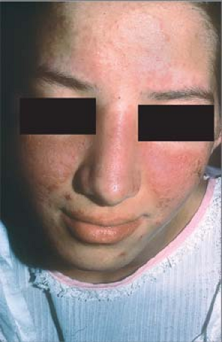 25.14 Systemic lupus erythematosus. A “butterfly rash” is evident. Note the sparing of the nasolabial areas that are often involved with seborrheic dermatitis. |
 25.15 Systemic lupus erythematosus. A photodistribution on the face and the “V” of the neck are evident in this patient. Note the violaceous color suggestive of connective tissue disease. Similar findings are noted in dermatomyositis. |
Systemic Lupus Erythematosus
Basics
Systemic lupus erythematosus (SLE) is a chronic, idiopathic, multisystemic, autoimmune disease associated with polyclonal B-cell activation. Fibrinoid degeneration of connective tissue and the walls of blood vessels associated with an inflammatory infiltrate involving various organs may result in arthralgia or arthritis, kidney disease, liver disease, central nervous system disease, gastrointestinal disease, pericarditis, pneumonitis, myopathy, and splenomegaly, as well as skin disease.
The cutaneous manifestations of SLE result from the production of multiple autoantibodies that deposit immune complexes at the dermal–epidermal junction.
Current investigators have reclassified lupus skin lesions into three distinct groups:
Acute cutaneous lupus erythematosus (ACLE) lesions are strongly associated with active SLE; however, ACLE lesions may occasionally be seen in subacute cutaneous lupus erythematosus (SCLE).
SCLE comprises the second category.
Chronic cutaneous lupus erythematosus (CCLE) traditionally had been referred to as discoid lupus erythematosus (DLE). DLE lesions may be seen in both SLE and CCLE.
SLE is seen in a 9:1 female-to-male ratio; it is more common in blacks and Hispanics.
Approximately 10% of patients with SLE have a first-degree relative with the disease. An association of lupus and human leukocyte antigens (HLA) -DR2 and -DR4 has been seen.
The following 4 of the 11 American Rheumatologic Association criteria for lupus are related to the skin and are considered lupus-specific lesions:
The classic malar or “butterfly” rash (Fig. 25.14) is a persistent erythema over the cheeks that tends to spare the nasolabial creases. Sometimes, this is the initial symptom of lupus, and it often occurs after sun exposure.
Photosensitivity (Fig. 25.15) occurs as an exaggerated or unusual reaction to sunlight. The reaction may resemble a drug eruption.
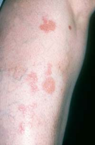
Discoid lesions (discoid lupus erythematosus) are erythematous lesions that evolve into scaly, atrophic scarring plaques (Fig. 25.16). Such discoid lesions affect 10% to 15% of patients with SLE.
Oral ulcerations may develop, often on the hard palate or nasopharynx.
Nonspecific lesions of lupus that may be seen in SLE and other connective tissue diseases include the following:
The “spider” type of telangiectasia is usually seen in SLE, scleroderma, and dermatomyositis. Macular (matlike) telangiectasias usually occur in scleroderma.
Periungual telangiectasias are seen in SLE, as well as in dermatomyositis and scleroderma.
Palmar telangiectasias are usually seen in SLE.
Vasculitis, palpable purpura, and vasculitic ulcers usually occur in SLE and scleroderma.
Raynaud’s phenomenon is associated with SLE and scleroderma.
Livedo reticulitis, panniculitis, thrombophlebitis, urticaria, urticarial vasculitis, frontal alopecia (“lupus hair”) and diffuse nonscarring alopecia, palmar erythema, and bullae are associated primarily with SLE.
Distribution of Lesions
The lesions of ACLE tend to occur in sun-exposed areas such as the face, dorsa of the forearms, the hands, and the “V” of the neck.
The lesions of DLE may occur on the face, scalp, ears, neck, or oral mucosa, or they may be widespread (see later discussion).
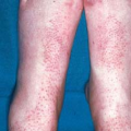
On the lower extremities, livedo reticularis (Fig. 25.17) and vasculitic ulcers (Fig. 25.18) may be seen.
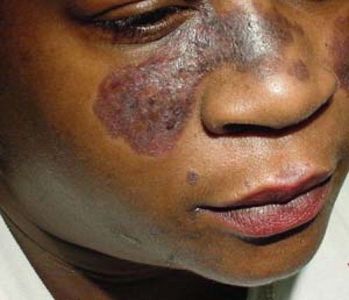
25.16 Systemic lupus erythematosus (discoid lupus erythematosus). Lesions of DLE, consisting of erythematous, scaly, disc-shaped scarring plaques, are present in this patient, who has SLE.
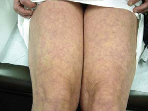
25.17 Systemic lupus erythematosus. Livedo reticularis is present.
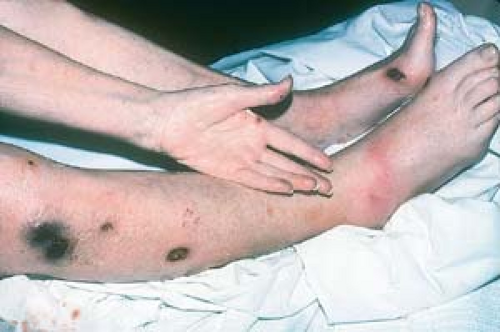
25.18 Systemic lupus erythematosus. Vasculitic ulcers are seen on this patient’s legs.
On the dorsal hands, violaceous plaques that spare the skin overlying the joints are characteristic of SLE. Conversely, in dermatomyositis, the joints are affected (Gottron’s papules). Ulcerated vasculitic lesions on the fingertips may also develop (Fig. 25.19).
Clinical Manifestations
Fatigue, fever, and malaise may be the presenting nonspecific symptoms.
Signs or symptoms are related to the specific organ or area involved (e.g., arthralgia).
Flare of lupus is common during pregnancy.
The following hematologic abnormalities may be associated with SLE: idiopathic thrombocytopenic purpura, hemolytic anemia, leukopenia, and clotting abnormalities, which may be related to the anticardiolipin syndrome. Other associated conditions and symptoms include rheumatoid arthritis, Sjögren’s syndrome, seizures, and the occurrence of multiple spontaneous abortions.
Diagnosis
According to the American Rheumatologic Association, a person has SLE if four or more of the following criteria are present:
“Butterfly” rash
Lesions of CCLE or DLE
Photosensitivity
Oral ulcers
Arthritis in two or more joints
Serositis
Renal disorder
Neurologic disorder
Hematologic disorder
Immunologic disorder: anti-DNA, anti-Smith (anti-Sm) antibody, or a false–biologic-positive syphilis serologic result
Antinuclear antibodies (ANAs)
Laboratory Evaluation
Antinuclear antibody (ANA) titers are positive in 95% of patients with SLE. The peripheral rim pattern is associated most strongly with lupus erythematosus, although other patterns commonly are present.
Anti-dsDNA (antibody to native double-stranded DNA) is present in 60% to 80% of patients and is more specific for SLE.
Anti-Sm antibody has a strong specificity for SLE. This is particularly relevant in patients in whom anti-dsDNA results are negative and may help exclude underlying systemic involvement.
Antiphospholipid antibodies are present in 25% of patients.
The erythrocyte sedimentation rate is usually elevated.

25.19 Systemic lupus erythematosus. Necrotic, painful, vasculitic ulcers are present on the fingertips.
Hypocomplementemia occurs in 70% of patients and is noted especially when there is renal involvement in active SLE.
The lupus band test involves the direct immunofluorescence of uninvolved, non–sun-exposed skin. When positive, it is suggestive of the presence of renal disease. This test has been largely supplanted by the aforementioned serologic tests.
Facial Lesions
Rosacea
See Chapter 1, “Acne and Related Disorders.”
Presence of acnelike papules and pustules in addition to malar erythema
Absence of systemic complaintsRelated posts:

Stay updated, free articles. Join our Telegram channel

Full access? Get Clinical Tree