Cutaneous Lymphoid Hyperplasia, Cutaneous T-Cell Lymphoma, Other Malignant Lymphomas, and Allied Diseases


The evaluation of lymphoid neoplasms typically requires clinicopathological correlation, and classification often re­­quires panels of immunostains. This section of the atlas will focus on the clinical appearance of cutaneous lymphomas that should prompt the physician to perform a biopsy. The distribution and morphology of the lesion are also helpful in classification of the neoplasm to ensure optimal therapy.


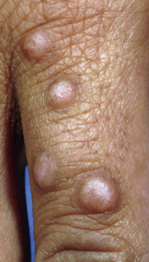
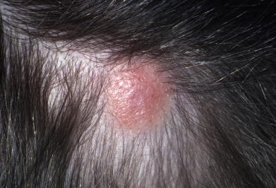
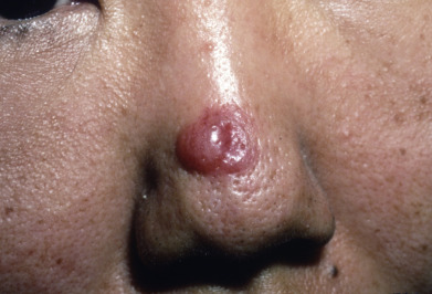
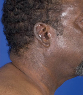
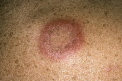
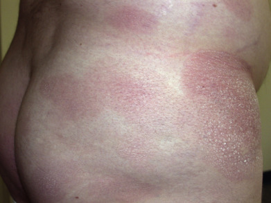
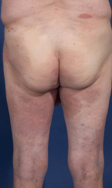
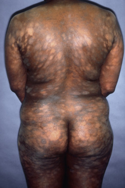
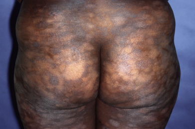
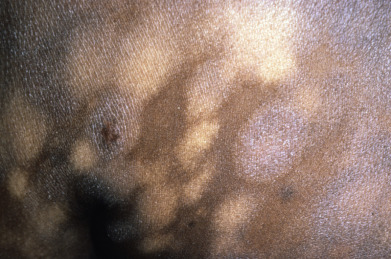
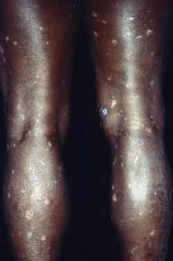
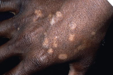
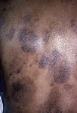
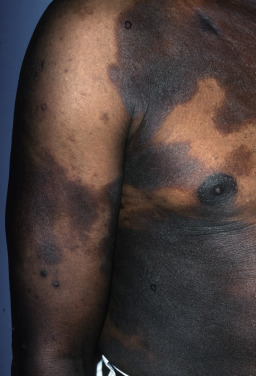
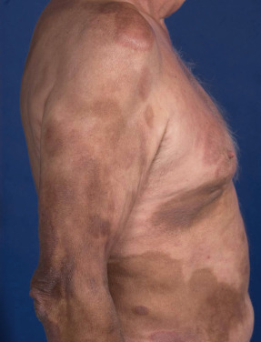
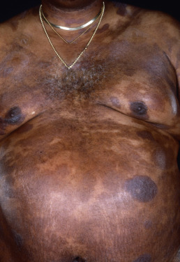
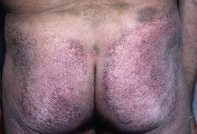
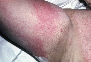
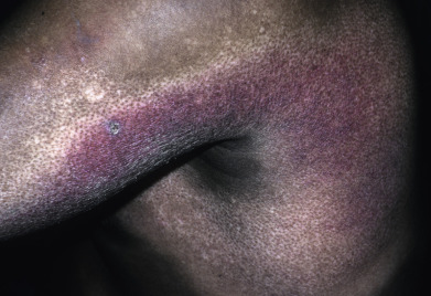
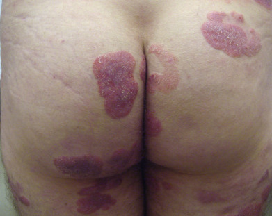
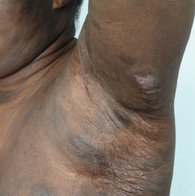
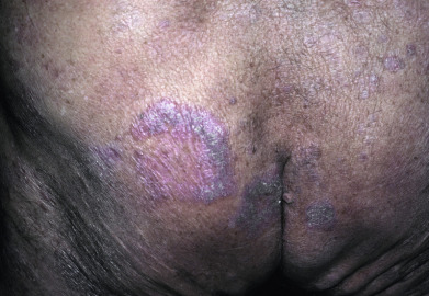
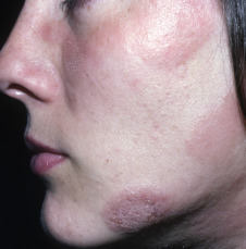
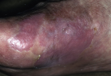
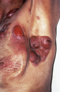
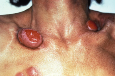
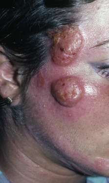
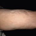
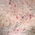
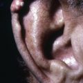
Mycosis fungoides commonly involve the trunk, buttocks, and proximal extremities. The lesions tend to be larger than 5 cm in diameter with a poikilodermatous appearance (mottled, hyperpigmentation and hypopigmentation, atrophy, and telangiectasia). B-cell lymphomas commonly present as plum-colored nodules with a smooth, shiny surface. Discrete nodules on the head and neck, trunk, or proximal extremities; arcuate lesions and nodules on the trunk; taught, shiny nodules on the lower extremities; and multinodular lesions on the legs are all seen in the wide variety of lymphoid neoplasms in this chapter. The lesions of lymphomatoid papulosis are often papulonecrotic, erupt in crops, and resolve spontaneously. Leukemias and myelomas often appear as skin-colored to purple papules and nodules, at times with accompanying hemorrhage into the lesions. This portion of the atlas will focus on the cutaneous manifestations of lymphoid proliferations.


Fig. 32.1


Cutaneous lymphoid hyperplasia, nodular B-cell pattern.


Fig. 32.2


Cutaneous lymphoid hyperplasia, nodular B-cell pattern.


Fig. 32.3


Cutaneous lymphoid hyperplasia, nodular B-cell pattern.


Fig. 32.4


Cutaneous lymphoid hyperplasia, bandlike T-cell pattern.


Fig. 32.5


Jessner lymphocytic infiltrate of the skin.


Fig. 32.6


Mycosis fungoides, patch stage.


Fig. 32.7


Mycosis fungoides, patch stage.


Fig. 32.8


Mycosis fungoides, patch stage.


Fig. 32.9


Mycosis fungoides, patch stage.


Fig. 32.10


Mycosis fungoides, patch stage.


Fig. 32.11


Mycosis fungoides, patch stage.


Fig. 32.12


Mycosis fungoides, patch stage.


Fig. 32.13


Mycosis fungoides, patch stage.


Fig. 32.14


Mycosis fungoides, patch stage.


Fig. 32.15


Mycosis fungoides, patch stage.


Fig. 32.16


Mycosis fungoides, patch stage.


Fig. 32.17


Mycosis fungoides, patch stage.

Courtesy Scott Norton, MD.


Fig. 32.18


Mycosis fungoides, patch stage with poikiloderma.


Fig. 32.19


Mycosis fungoides, patch stage with poikiloderma.

Courtesy Steven Binnick, MD.


Fig. 32.20


Mycosis fungoides, patch stage with poikiloderma.

Courtesy Steven Binnick, MD.


Fig. 32.21


Mycosis fungoides, plaque stage.


Fig. 32.22


Mycosis fungoides, plaque stage.


Fig. 32.23


Mycosis fungoides, plaque stage.

Courtesy Steven Binnick, MD.


Fig. 32.24


Mycosis fungoides, plaque stage.

Courtesy Steven Binnick, MD.


Fig. 32.25


Mycosis fungoides, plaque stage.

Courtesy Steven Binnick, MD.


Fig. 32.26


Mycosis fungoides, tumor stage.


Fig. 32.27


Mycosis fungoides, tumor stage.


Fig. 32.28


Mycosis fungoides, tumor stage.

Courtesy Steven Binnick, MD.


Sep 3, 2019 | Posted by in Dermatology | Comments Off on Cutaneous Lymphoid Hyperplasia, Cutaneous T-Cell Lymphoma, Other Malignant Lymphomas, and Allied Diseases

Full access? Get Clinical Tree

Get Clinical Tree app for offline access