Conservative Management of Postsurgical Lymphedema
Michelle Lee Hsia
Christopher Pham
Ketan M. Patel
History
Lymphedema is a distressing long-term health problem that many breast cancer survivors must manage. Cancer-related lymphedema is referred to as secondary lymphedema and usually involves traumatic injury to the native lymphatic circulation. Trauma can occur from surgical incisions, lymph node dissections (sentinel or axillary), radiation, or advanced cancer that compresses lymphatic structures. Approximately 5% to 20% of women develop upper extremity lymphedema after sentinel lymph node biopsy (SLNB) and axillary lymph node dissection (ALND), respectively, with the risk of those rates of increasing up to 2 years post surgery (1,2). Because of the higher risk of developing lymphedema after ALNDs compared to SLNBs, surgeons will try to perform SLNBs whenever appropriate. However, there are additional risk factors that can raise one’s risk, such as type of breast surgery (mastectomy vs. lumpectomy), number of lymph nodes removed, subsequent breast surgeries, radiation, and obesity (2,3). Because of these covariates, the true incidence of upper extremity lymphedema after surgery is ill defined.
Most patients who have lymphedema report onset of symptoms within the first 3 years of surgery (4). However, lymphedema has been reported to occur within days and up to 30 years after breast cancer treatment (5). Unfortunately, there is varying data on the incidence and prevalence of lymphedema after breast cancer, primarily due to differences in diagnosis, inability to compare different patient populations, and inadequate follow-up/delayed reporting by patients (4).
Due to the ambiguity of lymphedema incidence rates, patient education that focuses on risk factors and symptoms of lymphedema should be integrated in their care. A focus group exploring patients’ experiences with breast cancer–related lymphedema and its treatments found four substantive themes (6):
Patients felt there was a lack of information on lymphedema provided before, during, and after cancer treatment.
Patients reported common symptoms and triggers. Patients were able to recall specific triggers that marked the onset of their lymphedema and common activities that exacerbated their symptoms. These common symptoms were described as numbness, heaviness, tingling, aching, tightness, and burning (6).
Patients reported difficulty accessing care and resources. Finding therapists certified in lymphedema is difficult and insurance often limited the number of visits and compression garments covered.
Patients reported that lymphedema had a detrimental impact on quality of life. Patients commonly reported being unable to participate in day-to-day activities (either at home or at work) due to pain, limited range of motion, and/or strength. They also report an inability to enjoy leisure or social activities because of body image concerns, difficulty finding appropriate clothing, and disappointment in their performance of activities (6,7).
The optimal treatment of lymphedema likely lies in multidisciplinary care consisting of surgeons, oncologists, primary care physicians, occupational therapists, and physical therapists. Traditional and nontraditional therapies are outlined in this chapter to describe ways to treat and manage breast cancer-related upper extremity lymphedema. For most patients, a physician is required to make a referral to a lymphedema-certified occupational therapist or physical therapist. If a patient does seek services without a referral, it is still advisable for the rehabilitative team to consult and communicate with the patient’s oncologist or primary care physician.
Risk Reduction Education
Since there is a lack of understanding and research on why lymphedema occurs with some patients and not with others who are medically treated identically, there is also no consensus on how to prevent it from occurring. Preventative strategies that are given to patients lack scientific
evidence and follow a “common sense” approach (8). The National Lymphedema Network’s (NLN’s) position paper on Risk Reduction Practices suggest precautions patients should take after their surgeries to reduce the likelihood of developing lymphedema (Table 30-1).
evidence and follow a “common sense” approach (8). The National Lymphedema Network’s (NLN’s) position paper on Risk Reduction Practices suggest precautions patients should take after their surgeries to reduce the likelihood of developing lymphedema (Table 30-1).
TABLE 30-1 NLN Recommendations for Risk Reduction Practicesa | |||||||||||||||||||||||||
|---|---|---|---|---|---|---|---|---|---|---|---|---|---|---|---|---|---|---|---|---|---|---|---|---|---|
| |||||||||||||||||||||||||
Traditional Therapy
Complete Decongestive Therapy
Dynamic insufficiency, also known as high-volume insufficiency, is when the lymphatic load (amount of lymph fluid) exceeds the ability of an intact lymphatic system to transport (9). Mechanical insufficiency, known as low-volume insufficiency, is the reduction in transport capacity of the lymph fluid due to functional causes, such as surgery, radiation, trauma, or inflammation. In the case of combined dynamic and mechanical insufficiency, there is a simultaneous reduction in transport capacity and an increase in lymphatic load.
Once lymphedema is diagnosed, there is no cure. The primary goal of treatment is to return lymphedema to a latent and/or manageable stage through optimizing and utilizing intact lymphatic structures.
Complete decongestive therapy (CDT) (Fig. 30-1) is considered the standard treatment for lymphedema and consists of a combination of manual lymphatic drainage (MLD), skin care, compression, and decongestive exercise.
Manual Lymphatic Drainage
MLD is a manual treatment technique developed by Emil Vodder (1896,1897,1898,1899,1900,1901,1902,1903,1904,1905,1906,1907,1908,1909,1910,1911,1912,1913,1914,1915,1916,1917,1918,1919,1920,1921,1922,1923,1924,1925,1926,1927,1928,1929,1930,1931,1932,1933,1934,1935,1936,1937,1938,1939,1940,1941,1942,1943,1944,1945,1946,1947,1948,1949,1950,1951,1952,1953,1954,1955,1956,1957,1958,1959,1960,1961,1962,1963,1964,1965,1966,1967,1968,1969,1970,1971,1972,1973,1974,1975,1976,1977,1978,1979,1980,1981,1982,1983,1984,1985,1986). What initially started as manipulation of swollen lymph nodes for patients with chronic colds and sinus infections, transformed into an evidence-based practice of massaging the lymphatic system to drain lymph fluid. MLD consists of four basic “Vodder strokes” that work due to the working and resting phase of the strokes.
During the working phase of a stroke, a manual stretch is applied to the subcutaneous tissue, resulting in the manipulation of the anchoring filaments of lymph capillaries and the smooth musculature in the wall of the lymphangions (9). The stroke is applied directionally, with the purpose of redirecting lymph toward lymph nodes for drainage. The pressure is enough to stretch the subcutaneous tissues against the underlying fascia, however not to the extent of damaging the anchoring filaments or other lymphatic structures (Fig. 30-2) (9).
The resting phase of the stroke is the pressure being released. It is pressure free, with the elasticity of the skin moving the therapist’s hand passively back to the starting position. It is during this phase that initial lymph vessels absorb tissue fluid from the interstitial spaces.
Preparation Pathways and Manual Techniques
Using MLD, therapists are identifying healthy lymphatic areas to serve as facilitators to drain the protein-rich
lymph fluid in the interstitial spaces. These pathways and lymphatic structures are used to reroute the lymph fluid from blocked or damaged areas in the swollen arm and breast back to the venous system. In the example of unilateral, secondary upper extremity lymphedema after breast cancer, the therapist manipulates lymph nodes and collectors in healthy adjacent quadrants, including the ipsilateral supraclavicular (to stimulate return of lymph fluid into the venous system) and inguinal areas and the contralateral axillary area. The purpose is to increase lymphangiomotoricity—the working phase of stretching the subcutaneous tissue—to result in a “suction effect” of the lymph fluid in the affected area (9).
lymph fluid in the interstitial spaces. These pathways and lymphatic structures are used to reroute the lymph fluid from blocked or damaged areas in the swollen arm and breast back to the venous system. In the example of unilateral, secondary upper extremity lymphedema after breast cancer, the therapist manipulates lymph nodes and collectors in healthy adjacent quadrants, including the ipsilateral supraclavicular (to stimulate return of lymph fluid into the venous system) and inguinal areas and the contralateral axillary area. The purpose is to increase lymphangiomotoricity—the working phase of stretching the subcutaneous tissue—to result in a “suction effect” of the lymph fluid in the affected area (9).
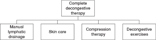 FIGURE 30-1 Complete decongestive therapy. |
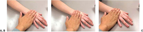 FIGURE 30-2 Manual stroke. A: Working phase—skin stretch surface area. B: Working phase—skin stretch proximal direction. C: Resting phase—release stretch, return to previous position. |
Despite lymphatic “watersheds” that separate quadrants in our bodies, some lymph fluid can cross watershed through lymph capillaries (Fig. 30-3). Areas with high lymph fluid accumulation cause the lymphatics to dilate in that area, with that pressure forcing lymph back into the lymph capillaries (dermal backflow) and across a watershed. After lymph nodes are manipulated or “activated,” manual strokes of redirecting fluid from a congested area to a healthy area are continued. Fluid travels from the affected axilla across the watershed to the contralateral axilla (interaxillary anastomoses) and ipsilateral inguinal area (axilloinguinal anastomoses). The initial preparation of lymph nodes and adjacent pathways can initiate a decrease in limb volume without the manipulation of the limb.
After the preparation of the lymphatic system, the working and resting strokes of MLD on the affected limb or breast/trunk are performed. Working manual strokes are completed against the lymph vessels natural flow patterns, redirecting fluid up to the lymph nodes (toward the axilla in our example). These manual strokes are applied in quadrants, starting at the shoulder and upper arm (Fig. 30-4). Each section is “drained” by applying traveling strokes distal to proximal, to clear and promote lymph movement in that area. The follow quadrants are the elbow joint, forearm, wrist, and hand/fingers. The purpose being each proximal section has been activated and cleared by the manual strokes, therefore as distal sections are being worked on, lymph fluid is encouraged to move within the pathways promoted up the arm.
When scar tissue obstructs lymphatic circulation, lymphostatic fibrosis can develop. Due to congestion within the structures, lymph fluid pools and attracts fat cells that bind in the surrounding tissues. The lymph fluid hardens and the tissues become denser. The inclusion of fibrosis-breaking techniques are added to MLD by providing a more intense “kneading” or “s-shape” maneuver to the skin that is intended to soften and break up areas of fibrosis (9). This should only be applied once decongestion of the lymphatic system has begun.
While generally safe, there are some considerations before performing MLD in patients with active oncologic disease. If a patient has active malignancies, working with the referring physician is advised. While there is no conclusive evidence that MLD would accelerate the spread of active malignancies to other parts of the body, there is reason to be cautious (9). MLD is contraindicated while patients are actively receiving radiation therapy because of issues regarding the sensitivity and integrity of the skin. MLD may be considered after radiotherapy once the skin has healed. Comorbidities (such as congestive heart failure, renal failure, deep vein thrombosis, etc.) that affect the lymphatic system should be considered before performing MLD and should be discussed with the referring physician before performing any maneuvers.
Self-Care
The lymphatics play a role in immunologic function and the removal of lymph nodes or damage to the native lymphatic structures increases the risk of soft tissue infection. Lymph nodes filter bacteria and toxins that invade our bodies and the immune system works to prevent or stop infection. If the remaining lymphatic structures cannot keep up with the lymphatic fluid that needs to filter to fight infection, the protein-rich environment can serve as an environment for bacteria to grow and spread (9).
As stated previously, the NLN advocates maintaining good skin care and addressing infections as urgent medical care. Based on expert opinion and physiologic principles, it is encouraged that patients avoid aggressive skin care, needle punctures, or blood draws on the affected arm, and monitor for cuts and scratches secondary to their impaired
lymphatic transport ability (10,11). Any damage to the skin, trauma or heat, can serve as an entrance to bacteria. During CDT, skin care is performed, and patients are educated on ways to integrate proper techniques into their daily routines. After proper cleaning, moisturizing is integral to maintain healthy and intact skin (to prevent dry and cracked skin). Patients should be comfortable inspecting their skin for any wounds or signs of infection (redness, tenderness, warmth). If a patient incurs a wound or blister in their upper extremity, they should follow basic hygiene protocols of cleaning with soap and water, applying an over-the-counter antimicrobial ointment, and covering it with a clean, dry bandage (9). While patients are at an increased risk of infection, not every injury will result in infection. Educating patients and making them active participants in their care is crucial.
lymphatic transport ability (10,11). Any damage to the skin, trauma or heat, can serve as an entrance to bacteria. During CDT, skin care is performed, and patients are educated on ways to integrate proper techniques into their daily routines. After proper cleaning, moisturizing is integral to maintain healthy and intact skin (to prevent dry and cracked skin). Patients should be comfortable inspecting their skin for any wounds or signs of infection (redness, tenderness, warmth). If a patient incurs a wound or blister in their upper extremity, they should follow basic hygiene protocols of cleaning with soap and water, applying an over-the-counter antimicrobial ointment, and covering it with a clean, dry bandage (9). While patients are at an increased risk of infection, not every injury will result in infection. Educating patients and making them active participants in their care is crucial.
Related posts:
 Patient Selection for Breast-Conservation Therapy
 Oncoplastic Approach to Maximizing the Lumpectomy-Level I
Postneoadjuvant Axillary Assessment and Management
Oncoplastic Approach to Maximizing the Lumpectomy-Level I
Postneoadjuvant Axillary Assessment and Management
 Selection of Vascularized Lymph Node Donor Sites for Upper Extremity Lymphedema Surgery and Strategies to Minimizing Complications
 Avoiding Pitfalls in Symmetry Procedures Following Unilateral Autologous Versus Implant-Based Reconstruction
 Oncoplastic Techniques in Breast Conservation Therapy—The Plastic Surgeon’s Perspective
Stay updated, free articles. Join our Telegram channel

Full access? Get Clinical Tree


