Key Words
connective tissue disease, lupus, dermatomyositis, polymyositis, CREST syndrome, Raynaud disease, telangiectasias, morphea, scleroderma, autoantibodies, antinuclear antibodies
Autoimmune Diseases
Diseases that result from attack by one’s own immune system are called autoimmune diseases. Autoimmune diseases are organ-specific illnesses or systemic illnesses such as systemic lupus erythematosus (SLE) ( Table 17.1 ). Autoimmune diseases are associated with circulating autoantibodies, which bind self-protein. Pathogenesis may then be mediated by autoimmune T lymphocytes and other immune mechanisms. Autoantibodies either are responsible for the manifestations of autoimmune disease or are markers of future disease. Autoantibodies may be found in serum samples many years before disease onset in several of these diseases. Autoimmunity is common in the population, but systemic autoimmune diseases, such as the connective tissue diseases, are relatively rare. Many people show some serologic evidence of autoimmunity but few have the signs and symptoms and disease-specific autoantibodies to make a diagnosis of a connective tissue disease.
| Known Antigens | |
|---|---|
| ORGAN-SPECIFIC DISEASES | |
| Graves hyperthyroidism | Thyroid stimulating–hormone receptor |
| Hashimoto thyroiditis | Thyroid peroxidase, thyroglobulin |
| Myasthenia gravis | Acetylcholine receptor |
| Goodpasture syndrome | Type IV collagen |
| Pemphigus vulgaris | Desmoglein 3 |
| Pernicious anemia | H + /K+-ATPase, intrinsic factor |
| Primary biliary cirrhosis | E2 PDC |
| Vitiligo | Tyrosinase, SOX-10 |
| Multiple sclerosis | Myelin basic protein, myelin oligodendritic glycoprotein |
| SYSTEMIC DISEASES | |
| Systemic lupus erythematosus | Spliceosomal snRNP, Ro/La (SS-A/SS-B) particle, histone and native DNA |
| Sjögren syndrome | Ro/La ribonuclear particle, muscarinic receptor |
| Rheumatoid arthritis | Citrullinated cyclic peptide, IgM |
| Polymyositis–dermatomyositis | t-RNA synthetases |
| KNOWN ANTIGENS | |
| Diffuse systemic sclerosis | Topoisomerase |
| Limited systemic sclerosis (CREST) | Centromere proteins |
Connective Tissue Diseases
Connective tissue diseases (rheumatoid arthritis [RA], lupus, dermatomyositis [DM], scleroderma, overlap syndromes) are a group of multisystem illnesses of unknown etiology. They have no typical pattern of onset, duration, or organ involvement. This variability makes classification and diagnosis difficult; therefore a list of clinical diagnostic criteria has been established for each entity and is tabulated in the text.
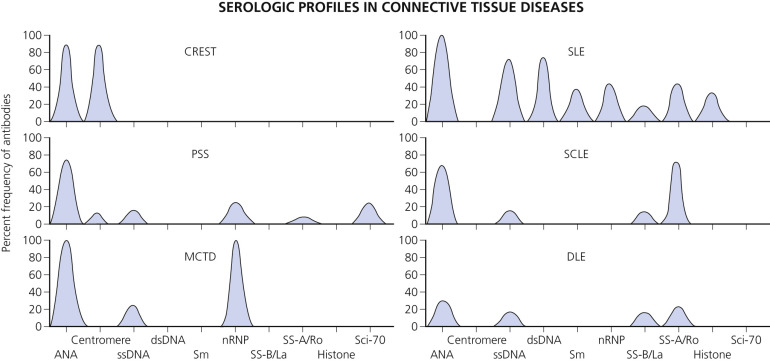
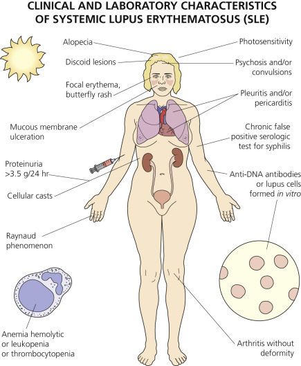
Connective tissue diseases can be more accurately described as autoimmune diseases. Many serum antibodies directed at cellular components (autoantibodies) have been found in each disease and are probably responsible for the clinical manifestations ( Fig. 17.1 ).
Diagnosis
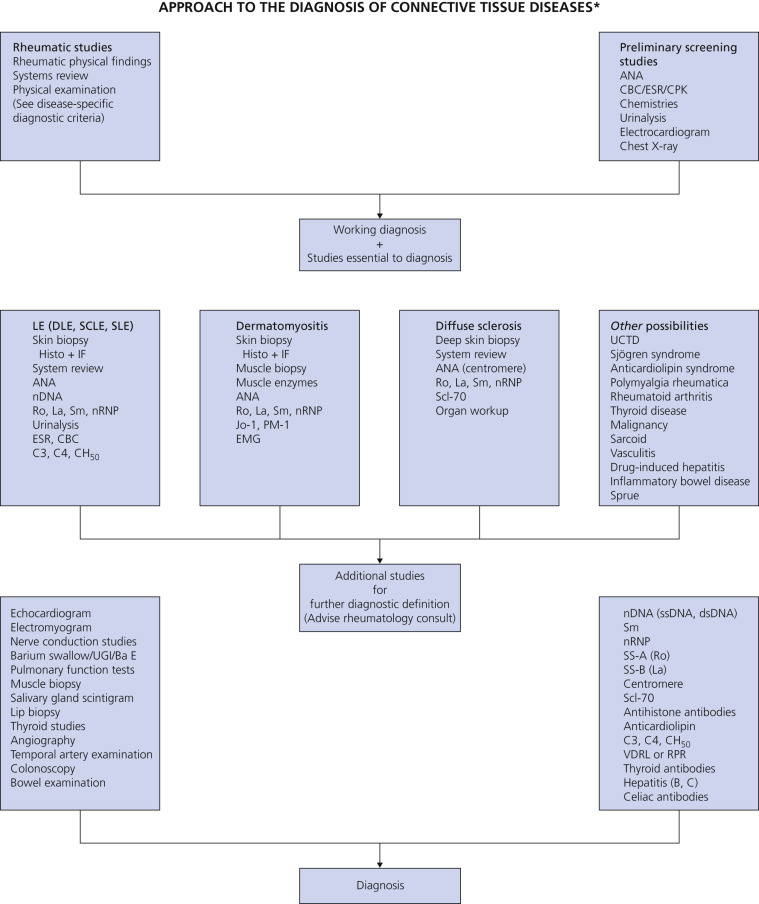
The diagnosis of connective tissue disease is made by the clinical picture. Antibodies to cell components (typically nuclear antigens) are present in these multisystem disorders. Detecting and defining them helps support the clinical diagnosis and provides information about subsets of disease and prognosis. A large number of antibodies are reported in the literature. Each systemic autoimmune disease has a characteristic antinuclear antibody (ANA) spectrum. Patients often produce multiple autoantibodies. The challenge for the clinician is determining the tests that should be ordered and then correctly interpreting the results. An approach to the diagnosis of connective tissue diseases is shown in Fig. 17.2 ![]() .
.
Antinuclear Antibody Screening
ANA is the first test to order when collagen vascular disease is suspected ( Table 17.2 ![]() ).
).
| ANA | Frequency of ANA Positivity (%) |
|---|---|
| Systemic lupus erythematosus | 95–100 |
| Drug-induced lupus erythematosus | 100 |
| Scleroderma | 60–95 |
| Sjögren syndrome | 80 |
| Polymyositis–dermatomyositis | 49–74 |
| Rheumatoid arthritis | 40–60 |
| Mixed connective tissue disease | 100 |
| Normal | <4 |
ANA tests identify antibodies present in serum that bind to autoantigens present in the nuclei of mammalian cells.
A negative result suggests that a connective tissue disease is unlikely; a positive result, especially a high titer in a patient with appropriate clinical findings, supports a diagnosis of a connective tissue disease.
False-Positive Test Results.
Positive results occur in normal blood donors and in patients with chronic liver disease, neoplasms, or active chronic infections. These patients usually have lower titers than those in patients with autoimmune diseases. Any autoantibody test must be interpreted in the context of available clinical information.
Specific Diagnostic Antibody Tests.
Specific antibody tests should be ordered. The clinical presentation and ANA pattern help to determine the tests that should be ordered ( Tables 17.3 and 17.4 ). Some laboratories offer tests for groups of antigens (i.e., ANA profiles).
| Antibody | Clinical Significance |
|---|---|
| Antinuclear antibodies | Screening for SLE and PSS |
| Centromere antibodies | Marker for CREST |
| Histone antibodies | To exclude drug-induced LE |
| ENA: Sm antibodies | Marker for SLE |
| RNP antibodies | SLE, MCTD, scleroderma |
| SS-A (Ro)/SS-B (La) antibodies | SLE, Sjögren syndrome, SCLE, and others |
| Scl-70 antibodies | Marker for scleroderma |
| Jo-1 antibodies | Marker for polymyositis |
| Ku (Ki) antibodies | Polymyositis/scleroderma overlap, SLE |
| Phospholipid antibodies (lupus anticoagulant) | Marker for SLE subset with thrombosis: frequent aborters |
| Disease | Biopsy Findings: Direct Immunofluorescence | Serum Findings | Relevance |
|---|---|---|---|
| Systemic LE | LE band (granular immune deposits, IgG, and/or IgM) IgA, C3 at DEJ in lesional and/or normal skin (over 90% in sun-exposed skin) | ANA elevated titers (about 95-99%); nDNA antibodies about 50%–75%; DNP antibodies <50%; Sm antibodies in about 20%; RNP antibodies in about 5%–30%; SS-A antibodies in about 30%–40%; SS-B antibodies in about 1%–15%; phospholipid antibodies in about 30%–50%; PCNA antibodies in about 2%–10%; Ku (Ki) antibodies in about 10% | DIF, ANA, and ENA usually diagnostic; nDNA and Sm antibodies are diagnostic markers |
| Discoid LE | LE band, mostly IgG and C in lesion ONLY | Essentially negative; ANA titers usually in normal range | LE band highly characteristic |
| Subacute, cutaneous LE | LE band in lesion | ANA positive in 70%; SS-A (Ro) antibodies positive in more than 60% | DIF and anti-SS-A (Ro) highly characteristic |
| Neonatal LE | LE band in lesion (about 50%) | ANA positive in 30%; antibodies to SS-A (Ro) in 100%; antibodies to SS-B (La) in about 60% | DIF and anti-SS-A (Ro) highly characteristic |
| Drug-induced LE | LE band in lesion (rare) | ANA positive in more than 90%; histone positive about 90%; other antibodies to nDNA and ENA negative | DIF and histone antibodies in absence of other nuclear antibodies highly characteristic |
| Mixed connective tissue disease | Nuclear IgG or LE band in normal and/or lesional epidermis | Speckled ANA antibodies in more than 95% and RNP antibodies in more than 90% | Serology and/or DIF of nuclei diagnostic for MCTD, SLE, or PSS |
| Sjögren syndrome | Negative | ANA positive in about 55%; antibodies to SS-A (Ro) in 43%–88%; SS-B (La) in 14%–60%; RF positive | Positive serum results support diagnosis |
| Progressive systemic sclerosis (scleroderma) | Nucleolar IgG in epidermis in few cases; most negative | ANA (about 85%) speckled or nucleolar; centromere antibody in CREST (70%–90%); Scl-70 antibodies in diffuse sclerosis (45%) and in acrosclerosis (15%–20%) | DIF limited value; centromere antibodies are diagnostic marker in CREST; Scl-70 antibodies are diagnostic marker in scleroderma |
| Polymyositis/ dermatomyositis | Negative | ANA usually positive (more than 80%); Jo-1 antibodies in 30% PM, 10% DM; SS-A (Ro) antibodies in 55% PM/scleroderma overlap; Ku (Ki) antibodies in 10% PM/scleroderma overlap | Limited value, but positive serum results support diagnosis |
| Rheumatoid arthritis | Negative | ANA usually negative or low titer; RF positive in about 90%; RNA positive in about 70%–90% and 95% of RF-negative cases | Positive serum results support diagnosis |
Titers.
Specimens are screened by diluting them 1 : 40 with saline. If there is no nuclear fluorescence at this dilution, the result is reported as “negative.” If there is green fluorescence, the level of ANA present is determined by repeating the test after serially diluting the specimen. ANA results are now reported by many laboratories using the international unit system (e.g., 1 international unit rather than a titer of 160).
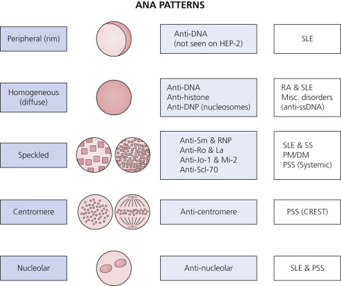
Patterns.
Antibodies to nuclear antigens attach to the various components of the nucleus. The fluorescein-labeled antihuman immunoglobulins are applied to the preparation and react with ANAs that have attached to the nucleus. The preparation is visualized with a fluorescent microscope. Diverse patterns of nuclear fluorescence (homogeneous, peripheral, speckled, or nucleolar) reflect the binding of antibodies to different nuclear components ( Fig. 17.3 ). Nuclear staining patterns were once used as criteria for subsetting, but, with the availability of direct measurements for specific autoantibodies, pattern identification has become less important. The test requires interpretation by visual inspection and consequently lacks a high degree of specificity.
Connective Tissue Laboratory Screening Tests
The signs and symptoms often associated with connective tissue disease (fatigue, arthralgias, fever, and weight loss) are not specific for autoimmune disease and occur in many other diseases. This makes early and accurate diagnosis difficult. The Mayo Medical Laboratories developed a Connective Tissue Diseases Cascade of tests for the primary care physician to evaluate patients with signs and symptoms compatible with a connective tissue disease in a setting of low disease prevalence. It provides immediate disease-specific, follow-up tests in those patients with presumptive serologic evidence of disease.
This algorithmic approach ( Fig. 17.4 ) is more efficient than a panel of tests that can result in unnecessary testing for specific autoantibodies in ANA-negative or low-titer ANA samples. Panel testing carries an increased potential to overdiagnose patients with benign autoimmunity.
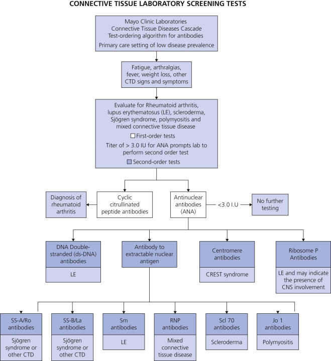
First-Order Tests.
The Connective Tissue Diseases Cascade begins with the following two tests that are done in all cases:
- 1.
Cyclic citrullinated peptide antibodies – Serum autoantibodies to cyclic citrullinated peptide (CCP) are quite specific for rheumatoid arthritis (RA), the most common connective tissue disease. Unlike the test for rheumatoid factor, which has poor specificity for RA, the test for CCP antibodies has proven to be reliable for differentiating RA from other connective tissue diseases. This test is not an ideal screening test because it is positive in fewer than 80% of patients with RA and negative test results do not conclusively exclude RA. Strongly positive tests for CCP antibodies have a very high positive predictive value for this disease.
- 2.
Antinuclear antibodies (ANAs) – A negative ANA test result is useful for excluding lupus erythematosus (LE) and scleroderma, the two most commonly encountered connective tissue diseases other than RA. Other disease-specific autoantibodies rarely occur in sera that test negative or have low levels of reactivity for ANA. Using a cutoff level of 3.0 units for ANA permits detection of >90% of sera with an identifiable specific autoantibody on follow-up testing. If the screening test for ANA is >3.0 units, second-order testing is performed for additional disease-specific autoantibodies.
Second-Order Tests.
The second level of testing includes the following four components:
- 1.
Double-stranded DNA (dsDNA) antibody, IgG, serum (anti-dsDNA) – A positive test result for anti-dsDNA antibodies is found in up to 82% of patients with active lupus erythematosus (LE). Anti-dsDNA antibodies are highly specific for LE, making this test useful for confirming the diagnosis. Low levels may account for false-positive reactivity. The levels of anti-dsDNA correlate with disease activity. Order this as a stand-alone test to monitor disease activity. dsDNA antibodies are also found in 20% to 30% of patients with Sjögren syndrome, 20% to 25% of patients with mixed connective tissue disease (MCTD), and less than 5% of patients with progressive systemic sclerosis (PSS).
- 2.
Antibodies to extractable nuclear antigens, serum (ENA) – The antibodies to the ENA group are comprised of six autoantibodies directed against small nuclear ribonucleoproteins (snRNPs) and enzymes. Autoantibodies to these individual antigens are important serologic markers of particular connective tissue diseases.
- A.
Autoantibodies to SS-A/Ro, serum (SS-A): SS-A antibodies occur with variable frequencies in several connective tissue diseases including Sjögren syndrome, LE, and RA. When present in isolation or with SS-B antibodies, the finding of this autoantibody is consistent with Sjögren syndrome. SS-A antibodies are found in approximately 60% of patients with Sjögren syndrome and 35% of patients with LE.
- B.
Autoantibodies to SS-B/La, serum (SS-B): SS-B antibodies rarely occur in isolation and are most often encountered in sera that contain SS-A antibodies. SS-B (La) antibody is seen in 50% to 60% of Sjögren syndrome cases and is specific if it is the only ENA antibody present; 15% to 25% of patients with SLE and 5% to 10% of patients with progressive systemic sclerosis also have this antibody.
- C.
Autoantibodies to Sm, serum (Sm): Sm antibodies are highly specific for LE. The test for Sm antibodies lacks sensitivity, and this autoantibody is detectable in only approximately 30% of patients with documented LE. The presence of antibodies to Smith (Sm) is often associated with renal disease.
- D.
Autoantibodies to U(1) RNP, serum (U1RNP): Antibodies to U1RNP occur in several different connective tissue diseases including MCTD and LE. The finding of U1RNP antibodies in the absence of anti-dsDNA antibodies and antibodies to other ENAs is consistent with the diagnosis of MCTD. U1RNP antibodies have been reported in 71% to 100% of patients with MCTD.
- E.
Autoantibodies to Scl-70, serum (Scl-70): Scl-70 antibodies react with the enzyme DNA topo-isomerase I and are highly specific for scleroderma. Scl-70 antibodies have been reported in approximately 40% of patients with scleroderma. The presence of Scl-70 antibodies is consistent with the diagnosis of scleroderma and indicates an increased risk for systemic involvement including pulmonary fibrosis.
- F.
Autoantibodies to Jo-1, serum (Jo-1): Jo-1 antibodies are highly specific for polymyositis PM but occur in only approximately 20% of PM patients. The presence of Jo-1 antibody is also found in patients with DM, or myositis associated with another rheumatic disease. The finding of Jo-1 antibodies indicates an increased risk for severe disease with pulmonary involvement and fibrosis.
- A.
- 3.
Centromere antibodies, IgG, serum – Centromere antibodies demonstrate a specific ANA pattern, which is present in 80% to 90% of individuals with CREST ( c alcinosis cutis, R aynaud phenomenon, e sophageal involvement, s clerodactyly, t elangiectasia) variant scleroderma. The pattern is also seen in 30% of patients with Raynaud phenomenon; 12% of patients with MCTD, diffuse scleroderma, interstitial pulmonary fibrosis, and primary biliary cirrhosis; and in a smaller percent of patients with systemic lupus erythematosus (SLE) and RA.
- 4.
Ribosome P antibodies, IgG, serum – Autoantibodies reacting with cytoplasmic ribosomes are highly specific for SLE. Ribosomal P antibodies are found in approximately 12% of patients with SLE and in 90% of patients with lupus psychosis; titers often increase more than 5-fold during and before active phases of psychosis.
Lupus Erythematosus
Clinical Classification
Systemic lupus erythematosus is a multisystem disease of unknown origin characterized by the production of numerous diverse types of autoantibodies that, through immune mechanisms (e.g., formation of immune complexes) in various tissues, cause several combinations of clinical signs, symptoms, and laboratory abnormalities ( Box 17.1 and Table 17.5 ). The natural history of SLE is characterized by episodes of relapses, flares, and remissions. The outcome is highly variable, ranging from remission to death.
* Criteria are cumulative and need not be present concurrently.
Classify a patient as having SLE if he or she satisfies four of the clinical and immunologic criteria, including at least one clinical criterion and one immunologic criterion, or if he or she has biopsy-proven nephritis compatible with SLE in the presence of ANAs or anti-dsDNA antibodies. The criteria do not need to be present concurrently.
Clinical Criteria
- 1.
Acute cutaneous lupus, including:
- •
Lupus malar rash (do not count if malar discoid)
- •
Bullous lupus
- •
Toxic epidermal necrolysis variant of SLE
- •
Maculopapular lupus rash
- •
Photosensitive lupus rash in the absence of dermatomyositis
- •
or subacute cutaneous lupus (nonindurated psoriasiform and/or annular polycyclic lesions that resolve without scarring, although occasionally with postinflammatory dyspigmentation or telangiectasias)
- •
- 2.
Chronic cutaneous lupus, including:
- •
Classic discoid rash
- •
Localized (above the neck)
- •
Generalized (above and below the neck)
- •
Hypertrophic (verrucous) lupus
- •
Lupus panniculitis (profundus)
- •
Mucosal lupus
- •
Lupus erythematosus tumidus
- •
Chilblain lupus
- •
Discoid lupus/lichen planus overlap
- •
- 3.
Oral ulcers:
- •
Palate
- •
Buccal
- •
Tongue
- •
or nasal ulcers
- •
In the absence of other causes, such as vasculitis, Behçet disease, infection (herpesvirus), inflammatory bowel disease, reactive arthritis, and acidic foods
- •
- 4.
Nonscarring alopecia (diffuse thinning or hair fragility with visible broken hairs) in the absence of other causes such as alopecia areata, drugs, iron deficiency, and androgenic alopecia
- 5.
Synovitis involving 2 or more joints, characterized by swelling or effusion
- •
or tenderness in 2 or more joints and at least 30 minutes of morning stiffness
- •
- 6.
Serositis:
- •
Typical pleurisy for more than 1 day
- •
or pleural effusions
- •
or pleural rub
- •
Typical pericardial pain (pain with recumbency improved by sitting forward) for more than 1 day
- •
or pericardial effusion
- •
or pericardial rub
- •
or pericarditis by electrocardiography
- •
in the absence of other causes, such as infection, uremia, and Dressler pericarditis
- •
- 7.
Renal:
- •
Urine protein-to-creatinine ratio (or 24-hour urine protein) representing 500 mg of protein/24 hours
- •
or red blood cell casts
- •
- 8.
Neurologic:
- •
Seizures
- •
Psychosis
- •
Mononeuritis multiplex in the absence of other known causes such as primary vasculitis
- •
Myelitis
- •
Peripheral or cranial neuropathy in the absence of other known causes such as primary vasculitis, infection, and diabetes mellitus
- •
Acute confusional state in the absence of other causes, including toxic/metabolic, uremia, drugs
- •
- 9.
Hemolytic anemia
- 10.
Leukopenia (<4000/mm 3 at least once):
- •
In the absence of other known causes such as Felty syndrome, drugs, and portal hypertension or
- •
Lymphopenia (<1000/mm 3 at least once) in the absence of other known causes such as corticosteroids, drugs, and infection
- •
- 11.
Thrombocytopenia (<100,000/mm 3 ) at least once in the absence of other known causes such as drugs, portal hypertension, and thrombotic thrombocytopenic purpura
Immunologic Criteria
- 1.
ANA level above laboratory reference range
- 2.
Anti-dsDNA antibody level above laboratory reference range (or >2-fold the reference range if tested by ELISA)
- 3.
Anti-Sm: presence of antibody to Sm nuclear antigen
- 4.
Antiphospholipid antibody positivity as determined by any of the following:
- •
Positive test result for lupus anticoagulant
- •
False-positive test result for rapid plasma reagin
- •
Medium- or high-titer anticardiolipin antibody level (IgA, IgG, or IgM)
- •
Positive test result for anti-β 2 -glycoprotein I (IgA, IgG, or IgM)
- •
- 5.
Low complement:
- •
Low C3
- •
Low C4
- •
Low CH50
- •
- 6.
Direct Coombs’ test in the absence of hemolytic anemia
ANA, antinuclear antibody; anti-dsDNA, anti-double-stranded DNA; ELISA, enzyme-linked immunosorbent assay; SLE, systemic lupus erythematosus; SLICC, Systemic Lupus International Collaborating Clinics.
| Type | Subtype | Clinical Forms | Common Sites | Clinical Features | Laboratory Features | Histologic Features |
|---|---|---|---|---|---|---|
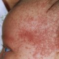
| Acute cutaneous lupus erythematosus (ACLE), 30%–50% | No subtype | Localized | Malar areas of the face | Transient ”butterfly rash” on the face. Erythema that may last for hours, days, or weeks. Indurated erythematous lesions often recur with sun exposure. Multisystem disease usually present; renal disease common | Antinuclear antibodies usually present. Anti-dsDNA antibodies present in 60%–80% of patients, often in high concentration. Hypocomplementemia common. Subepidermal immunoglobulin deposits commonly found in lesional (≥95%) and exposed nonlesional (75%) skin | Histologic features are similar among localized ACLE and generalized ACLE. Findings include interface dermatitis, apoptotic keratinocytes, hydropic changes along epidermal basal layer. Sparse mononuclear cell infiltrate and upper dermal edema |
| Generalized | Face, scalp, neck, upper chest, shoulders, extensor arms, backs of hands. Skin on the metacarpophalangeal joint and interphalangeal joints is often not affected | Erythematous maculopapular or morbilliform eruption primarily on sun-exposed skin. Vesicles or bullae possible | Same as localized | Same as localized | ||
| Toxic epidermal necrolysis–like | Can be throughout the body. Mucosal, conjunctival, buccal, and genital areas are generally unaffected | Blistering eruption. Can result in massive cleavage and shedding of epidermis | ANA and anti-dsDNA positive | Partial or full-thickness epidermal necrosis with subepidermal blistering | ||
| Subacute cutaneous lupus erythematosus (SCLE), 10%–15% | No subtype | Annular (5%) Papulosquamous (8%) | Shoulders, forearms, neck, hands, and upper torso. Though photoaggravated, the face is commonly unaffected. | Plaques combine to form polycyclic or figurative patterns. Typically nonscarring but dyspigmentation and resulting telangiectasias are common. Papulosquamous lesions can mimic psoriasis or lichen planus. Usually associated with extracutaneous disease, but severe renal or CNS disease is uncommon | Antinuclear and anticytoplasmic antibodies frequently present (60% of patients). Anti-dsDNA antibodies present in low serum concentrations (30% of patients). Hypocomplementemia rare. HLA-A1, HLA-B8, and HLA-DR3 significantly increased. Subepidermal immunoglobulin deposits only found in 50% of lesions and 30% of uninvolved skin | Moderate hyperkeratosis. Marked hydropic changes along basal cell layer, mild to moderate atrophy. Mild to moderate periappendageal mononuclear cell infiltrate only in superficial dermis. Dermal edema and possible minimal follicular plugging. Vacuolization of the basement membrane and mucin deposition in the dermis. Minimal or absent thickening of the basement membrane |
| Drug-induced | Same as annular and papulosquamous | Systemic: fever, myalgias, rash, arthralgias Typically affects sun-exposed skin with an annular or psoriasiform eruption. Macular rash on the face | Anti-dsDNA and hypocomplementemia are rare. Antinuclear antibodies present (95%–100%). Ro/SSA positive (70%–90% of patients). Procainamide- most common drug associated with drug-induced LE. Positive ANA in almost every patient on the drug for more than two years. Minocycline-A positive ANA, anti-dsDNA antibodies, and perinuclear antineutrophil cytoplasmic antibody (P-ANCA) tests have been found (83%–92%); anti-histone antibodies are uncommon (0%–13%). Hydralazine can be associated with an antineutrophil cytoplasmic antibody (ANCA)-positive vasculitis involving the kidney | Same as annular and papulosquamous | ||
| Erythrodermic | Generalized | Papulosquamous, nonscarring lesions | Positive Ro and La antibodies. Positive antinuclear antibody test with speckled pattern. Anti-dsDNA negative | Epidermal atrophy, vacuolar degeneration of the basal cell layer, perivascular inflammation | ||
| Rowell syndrome (erythema multiforme–like) | Commonly appear symmetrically on acral extensor areas. Face, neck, palms, soles, trunk, and/or flexural surfaces of extremities can be affected | “Target” lesions with a dusky or blistered, dark red center, surrounded by an erythematous halo. Edema present. Usually asymptomatic, but itching or burning can be present | Positive antinuclear antibody with a speckled pattern | Destructive interface dermatitis; extensive necrosis of epithelial cells is observed | ||
| Poikilodermatous | Sun-exposed areas. Face, chest, and neck are commonly affected | Hyper and/or hypopigmentation, telangiectasia, and atrophy | Elevated antinuclear antibody titer. Positive Ro and La antibodies | Orthokeratosis, basal cell vacuolization, and melanin inconsistency. Civatte body formation is also observed | ||
| Vesiculobullous annular | Photoexposed trunk extensor surfaces of limbs Involvement of the vermillion border and mucosal surfaces are possible | Tense vesicles and blisters addition, red or pink macules without overlying vesicles or bullae can be present. Lesions are typically nonscarring, but dyspigmentation may take years to resolve. No formation of milia | Elevated antinuclear antibody titer. Anti-collagen VII antibodies detected by ELISA or immunoblotting | Vacuolar degeneration of the epidermal basal cell layer. DIF demonstrates IgG, IgM, and IgA linear or granular deposition at the basement membrane | ||
| Chronic cutaneous lupus erythematosus (CCLE) | Discoid lupus erythematosus (DLE) (15%–30%) | Localized | Limited to sites above the neck | Indurated, erythematous, plaques with well-formed adherent scale. Scarring, atrophy, telangiectasias, and dyspigmentation is common | Usually no extracutaneous disease. Antinuclear antibodies occasionally present in low titer and anticytoplasmic antibodies not present. Anti-dsDNA antibodies rarely present. Simultaneous occurrence of severe systemic lupus erythematosus with nephritis is rare. Subepidermal immunoglobulin commonly found in lesions (75%), but rarely present in uninvolved skin | Follicular plugging. Slowly expanding plaques with active inflammation at the periphery. Vacuolar changes at the basal layer. Mononuclear cell (majority T cells) infiltrate near the dermoepidermal junction, dermal blood vessels, and appendages. Thickened basement membrane and dermal mucinosis is observed |
| Generalized | Occurs above and below the neck | Same as localized | Same as localized | Same as localized | ||
| Hypertrophic | Can be generalized or localized | Indicated by the development of prominent hyperkeratotic, verrucous plaques | Same as localized | Same as localized | ||
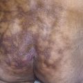
| Lupus erythematosus tumidus (LE tumidus) | No clinical variants | Face, dorsal hands and forearms, posterior and lateral neck (photodistributed) | Chronic, pink to violaceous, urticarial or edematous plaques or nodules. Annular plaques may occur, but scaling and scarring are absent | Estimated 10% of cases are ANA-positive | Moderate to dense, superficial and deep, perivascular lymphocytic infiltrate, consisting of predominately CD3 + /CD4 + lymphocytes. Mucin deposition is observed in the papillary and reticular dermis. Interface changes at the dermoepidermal junction are uncommon | |
| Lupus profundus (lupus panniculitis) | No clinical variants | Scalp, face, upper arms, chest, breasts, lower back, flank, upper thighs, or buttocks | Painful or tender indurated plaques or nodules. Overlying cutaneous changes and ulceration or calcifications at sites of involvement possible. May leave depressed areas of lipoatrophy | Typically demonstrates linear deposition of IgM and C3 along the dermoepidermal junction with immunofluorescence. Diagnosis is supported with a positive lupus band test. ANA present in 50% of cases | Panniculitis is present, as well as perivascular infiltrates of mononuclear cells manifested as hyaline fat necrosis with mononuclear cell infiltration and lymphocytic vasculitis. Presence of immune deposits in the dermoepidermal junction on direct immunofluorescence is supportive for diagnosis | |
| Chilblain lupus erythematosis (chilblain LE) | No clinical variants | Toes, fingers, nose, or ears | Tender, bright red to reddish-blue papules, nodules, or plaques with exposure to cold | Positive rheumatoid factor and speckled antinuclear antibody pattern. Cryoglobulins and cryoprecipitates are negative | Suggested by vacuolization of the basal layer of the epidermis. Immunofluorescence of lesional skin may reveal a linear deposition of immunoglobulins or complement at the dermoepidermal junction which is supportive for diagnosis, along with typical findings of pernio. | |
| Lichenoid cutaneous lupus erythematosus–lichen planus overlap syndrome (LE-LP overlap syndrome) | No clinical variants | Acral portions of the extremities, particularly the palms, soles, and nails. Nail may be absent | Persistent atrophic blue-red to violaceous plaques or patches. Not commonly associated with photosensitivity and/or pruritus | Positive lupus band test and anti-dsDNA antibodies. High antinuclear antibody titers | May share features with lichen planus, such as hyperkeratosis, hypergranulosis, irregular acanthosis, and pigment incontinence. Features similar to lichen planus, such as cytoid bodies staining for IgM and fibrin in a fibrillar pattern, can be shown by DIF. Linear granular deposition of immunoglobulin and complement along the dermoepidermal junction is also shown by DIF. |
In the United States, there are between 161,000 and 322,000 individuals with SLE and the prevalence is highest among blacks/African Americans, Asians, Hispanics/Latinos, and Native Americans/Alaska Natives. The incidence of SLE may be up to 12 times higher in women than men. Susceptibility genes located on the X chromosome, X-inactivation, other molecular genetic factors and estrogen effects may account for the disparity between men and women. The vast majority of patients develop SLE between ages 16 and 55.
The American College of Rheumatology classification criteria for SLE were replaced in 2012 (see Box 17.1 ) by the Systemic Lupus International Collaborating Clinics (SLICC) classification criteria for SLE. The criteria do not need to be present concurrently. The proposed classification rule is as follows: classify a patient as having SLE if he or she satisfies four of the clinical and immunologic criteria used in the SLICC classification criteria, including at least one clinical criterion and one immunologic criterion, or if he or she has biopsy-proven nephritis compatible with SLE in the presence of ANAs or anti-dsDNA antibodies. Those criteria in a modified form are illustrated in Fig. 17.5 .
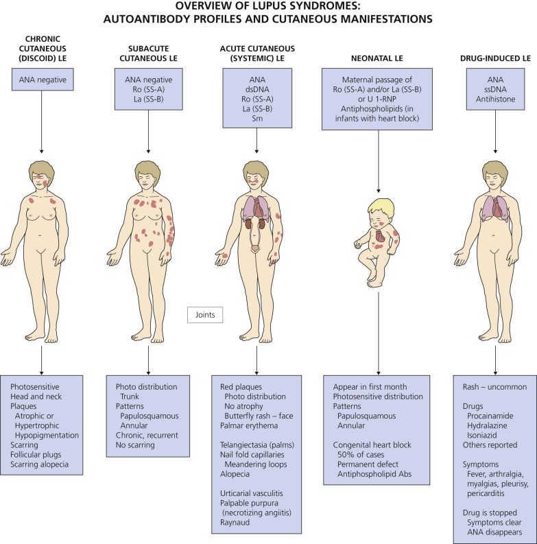
There are several forms (subsets) of cutaneous erythematosus (see Table 17.5 ).
Subsets of Cutaneous Lupus Erythematosus
Attempts have been made to group patients into subsets to define more homogeneous groups with a predictable course or response to treatment. Subsets of LE have been defined by cutaneous manifestations present in some form in most patients with lupus. The classification in Table 17.5 divides cutaneous LE into three types on the basis of the clinical appearance of the skin lesion: chronic cutaneous LE (scarring, discoid LE [DLE]), subacute cutaneous LE (SCLE), and acute cutaneous LE (ALE). A comparison of laboratory findings in these subsets is shown in Table 17.6 .
| Finding | DLE (%) | SCLE (%) | ALE (%) |
|---|---|---|---|
| ANA titer (≥1 : 160) | 4 | 63 | 98 |
| Anti-dsDNA | Rare | 30 | 60–80 |
| ESR greater than 30 | Few | 59 | 90 |
| LE cell preparation | 2 | 55 | 80 |
| Low C3 or CH50 | Rare | Rare | 90 |
| WBC count less than 4000/mm 3 | 7 | 19 | 17 |
| Rheumatoid factor latex test positive | 15 | 19 | 37 |
| Low hemoglobin level | Few | 15 | 50 |
| VDRL biologic false-positive | Few | 7 | 22 |
| DIRECT IMMUNOFLUORESCENCE AND LUPUS BAND TEST | |||
| Lesion | 90 | 60 | 95 |
| Normal sun-exposed | 0 | 46 | 75 |
| Normal nonexposed | 0 | 26 | 50 |
An overview of the lupus syndromes appears in Fig. 17.6 .
Chronic Cutaneous Lupus Erythematosus (Discoid Lupus Erythematosus)
Patients with DLE have a low incidence of systemic disease. The disease is more common in females, and it has a peak incidence in the fourth decade. Less than 2% of patients with DLE develop the disease before 10 years of age. Trauma and ultraviolet B (UVB) light exposure may initiate and exacerbate lesions. There are several clinical variations (see Table 17.5 ).
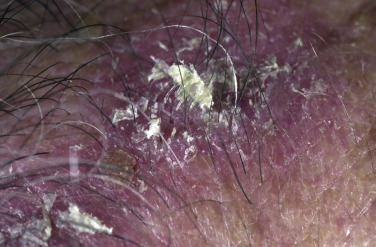
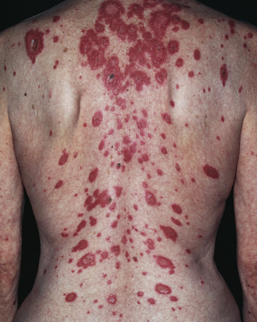
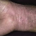
The most common manifestation is the DLE lesion. Lesions are sharply demarcated and can be round, thus giving rise to the term discoid (or disc-like) ( Figs. 17.7 to 17.15 ). The face and scalp are the most commonly affected areas, but lesions may occur on any body surface. Lesions are usually asymmetrically distributed and begin as asymptomatic, well-defined, elevated, red-to-violaceous, 1- to 2-cm, flat-topped plaques with firmly adherent scale. The scale penetrates into the orifices of the hair follicle. Peeling the scale reveals an undersurface that has the appearance of a carpet penetrated by several carpet tacks; it is called carpet tack scale. Carpet tack scale is most apparent on the face and scalp where the follicular orifices are larger.
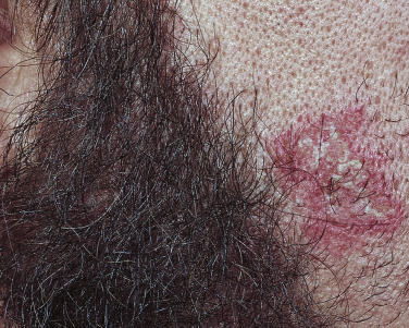
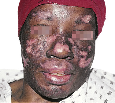
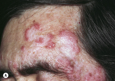
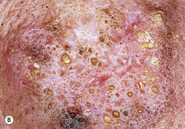
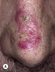
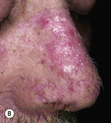
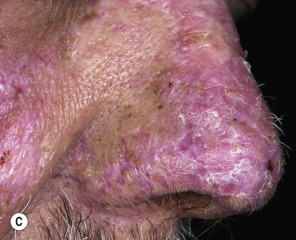
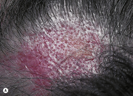
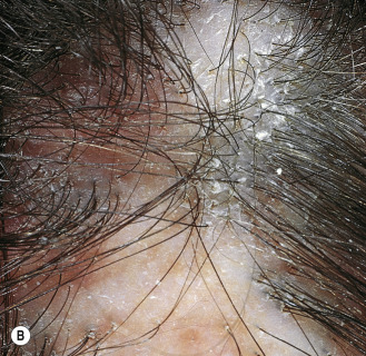
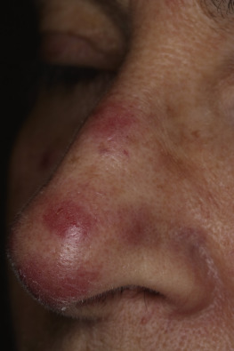
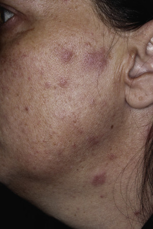
Atrophy occurs in both the epidermis and the dermis. Epidermal atrophy occurs early and gives the surface either a smooth white or a wrinkled appearance. Hypopigmentation is particularly disfiguring for black people. Follicular plugs may be prominent. These lesions endure for months and either resolve spontaneously or progress with further atrophy, ultimately forming smooth white or hyperpigmented depressed scars with telangiectasia and scarring alopecia. Occasionally plaques become thick (hypertrophic DLE). DLE can cover wide areas of the face, causing disfigurement. The laboratory and histologic features are outlined in Tables 17.5 and 17.6 . The presence of anti-single-stranded DNA (ssDNA) occurs with widespread active disease.
Impact of Smoking.
Smokers with chronic cutaneous lupus have more severe disease, worse quality of life, and are more refractory to treatment with the combination of immunomodulator and antimalarial drugs than nonsmokers.
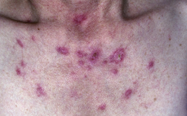
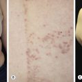
Subacute Cutaneous Lupus Erythematosus
Subacute cutaneous lupus erythematosus (SCLE) encompasses the clinical spectrum of cutaneous LE between the chronic, destructive DLE and the erythema of ALE. However, SCLE can be associated with the full spectrum of LE-associated phenomena ( Table 17.5 ). Like DLE, the individual lesions of SCLE may last for months; in contrast to DLE, they heal without scarring. Most patients with SCLE are white females. SCLE may be induced by a variety of drugs, most notably hydrochlorothiazide, calcium channel blockers, angiotensin-converting enzyme inhibitors, and terbinafine. The mean time to clinical disease after initiation of the offending drug is 27.9 weeks (median 6 weeks), whereas the mean time to improvement is 7.3 weeks after discontinuation (median 4 weeks).
Two morphologic varieties are a papulosquamous pattern ( Fig. 17.16 ) and an annular–polycyclic pattern ( Fig. 17.17 ). Both occur most often on the trunk; one predominates. The lesions spare the knuckles, the inner aspects of the arms, the axillae, and the lateral part of the trunk. They are rarely seen below the waist. A subtle gray hypopigmentation and telangiectasia are frequently seen in the center of annular lesions, bordered by erythema and a superficial scale. Follicular plugging, adherent hyperkeratosis, scarring, and dermal atrophy that are characteristic of DLE are not prominent features of SCLE. Hypopigmentation and telangiectasia become more evident as individual lesions resolve. The hypopigmentation fades after several months, but the telangiectasia may persist. The disease tends to be chronic and recurrent, lasting for years. SCLE and antibodies to Ro/SS-A have been associated with hydrochlorothiazide therapy.
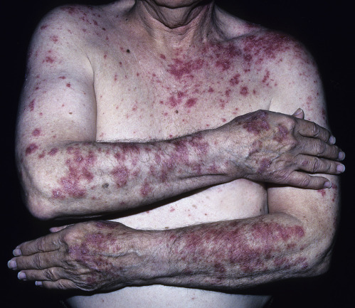
Other dermatologic manifestations are photosensitivity (52% to 85%), periungual telangiectasia (22% to 51%), DLE (19% to 35%), and vasculitis (12%). Systemic manifestations (arthritis/arthralgia [43% to 74%], renal disease [11% to 19%], serositis [12%], and central nervous system [CNS] symptoms [6% to 19%]) are not severe and follow a benign course.
The laboratory and histologic features of SCLE are outlined in Tables 17.5 and 17.6 . Antibodies to Ro/SS-A are present in 29% of patients.
Systemic Lupus Erythematosus
Systemic lupus erythematosus is a syndrome characterized by clinical diversity, by changes in the disease activity over time, and by aberrant immunologic findings and especially the presence of antinuclear antibodies.
Clinical Presentation.
The clinical presentation varies between different patients, and in a single patient the disease activity varies over time. General symptoms such as fatigue and fever are common. A vast majority of the patients have arthralgia, mostly of the hands. About half of the patients have cutaneous features, such as butterfly rash and discoid lupus as well as photosensitivity, while one third of the patients have oral ulcerations. Approximately 50% of the patients have nephropathy, which varies from mild proteinuria and microscopic hematuria to end-stage renal failure. About 20% to 40% of the patients have pleurisy. Acute pneumonitis and chronic fibrosing alveolitis are relatively rare.
Pericarditis is less common than pleuritis. T-wave changes in the electrocardiogram (ECG) are usual. Depression and headache are the most common of the neuropsychiatric symptoms. Generalized tonic–clonic seizures and organic psychoses are rare. Peripheral neuropathy is observed in about 10% of the patients, and approximately 10% of patients have a thromboembolic or hemorrhagic complication of the brain. The lymph nodes may enlarge, especially when the disease is active. There is a risk of first- and second-trimester fetal losses and of premature birth.
Laboratory Findings.
Laboratory findings are diverse and may include the following:
- •
Erythrocyte sedimentation rate (ESR) is usually elevated; the C-reactive protein (CRP) value is often normal.
- •
Mild or moderate anemia is common. A clear-cut hemolytic anemia is seen in less than 10% of patients.
- •
Leukocytopenia (lymphocytopenia) is usually present.
- •
Mild thrombocytopenia is typically present.
- •
Antinuclear antibodies are found in more than 90% of patients.
- •
Anti–deoxyribonucleic acid (anti-DNA) antibodies are found in 50% to 90% of patients.
- •
Polyclonal hypergammaglobulinemia is usually present.
- •
Decreased complement values (C3 and C4) are found.
- •
Antiphospholipid antibodies are present.
- •
Proteinuria, microscopic hematuria, and decreased creatinine clearance are present.
Diagnosis.
There is no single symptom or finding that is intrinsically sufficient for making the diagnosis. When SLE is suspected the basic laboratory investigations are as follows:
- •
Blood count
- •
Platelet count
- •
Erythrocyte sedimentation rate
- •
Measurement of antinuclear antibodies
- •
Dipstick test of the urine and urinalysis
Treatment.
The treatment is always individual and depends on the manifestations and activity of the disease. There is no need for treatment solely on the basis of the immunologic findings. Patients should be encouraged to refrain from sunbathing, use sunscreens, eat a balanced diet, exercise, and stop smoking.
The most important drugs used for treatment of SLE are the following:
- •
Nonsteroidal antiinflammatory drugs
- •
Hydroxychloroquine
- •
Corticosteroids
- •
Belimumab
- •
Immunosuppressive drugs (e.g., azathioprine, cyclophosphamide, methotrexate [MTX], mycophenolate)
Hydroxychloroquine.
Hydroxychloroquine and nonsteroidal antiinflammatory drugs are used in the treatment of mild symptoms such as cutaneous manifestations and arthralgia. When the response is insufficient or when the patient has fatigue or fever, a low dose of corticosteroids (prednisolone 5 to 7.5 mg/day) can be added. In the treatment of pleuritis or pericarditis, larger amounts of corticosteroids (about 30 mg of prednisolone per day) are used. For the treatment of severe central nervous system (CNS) symptoms and severe glomerulonephritis, thrombocytopenia, and hemolytic anemia, large corticosteroid doses and other immunosuppressive drugs are used.
Belimumab.
Belimumab is a fully humanized monoclonal antibody that binds and inactivates B-lymphocyte stimulator (a cytokine expressed by B-cell lineage cells that activates B cells and stimulates their proliferation and differentiation), inhibiting B-lymphocyte proliferation and differentiation. For adult patients with seropositive active SLE (especially musculoskeletal and cutaneous disease), belimumab represents a treatment option. Trials show a modest but statistically significant benefit for the addition of belimumab to standard care for the treatment of SLE. The inhibition of B-lymphocyte stimulator with belimumab may prevent morbidity and mortality in patients with SLE. The drug is administered by intravenous infusion and is costly.
When monitoring patients with systemic lupus, distinguishing a lupus flare from infection is critical because the former requires immunosuppressant medications and the latter antibiotics.
If there are signs of renal manifestations, the patient should be referred to a nephrologist for a renal biopsy. SLE patients are often allergic to a variety of antibiotics, especially sulfonamides.
The main causes of morbidity and mortality and the main immunologic parameters in SLE were analyzed for 1000 patients (female to male ratio, 10 : 1) in a 5-year multicenter study. Table 17.7 ![]() shows the frequencies of the main SLE clinical manifestations during the 5-year study.
shows the frequencies of the main SLE clinical manifestations during the 5-year study.
| SLE Manifestation | Percent |
|---|---|
| Arthritis | 41.3 |
| Malar rash | 26.4 |
| Nephropathy | 22.2 |
| Photosensitivity | 18.7 |
| Fever | 13.9 |
| Neurologic involvement | 13.6 |
| Raynaud phenomenon | 13.2 |
| Serositis | 12.9 |
| Thrombocytopenia | 9.5 |
| Oral ulcers | 8.9 |
| Thrombosis | 7.2 |
| Livedo reticularis | 5.5 |
| Discoid lesions | 5.4 |
| Subacute cutaneous lesions | 4.6 |
| Myositis | 4 |
| Hemolytic anemia | 3.3 |
Related posts:


Stay updated, free articles. Join our Telegram channel

Full access? Get Clinical Tree


