Fig. 36.1
Photograph of the hemiface/calvarium flap during preperation of the graft before detachment from the donor (LBN). Please note that orbita was exentrated and dura mater left under the graft. Arrow shows the vascular pedicles consisting of external jugular vein and common carotid artery. Asterisk shows the calvarial bone. In the lower photo it is possible to see the graft transferred and reperfused. Please note the vascular appearence of inner calvarium including dura mater. The enastomoses were performed to common carotid end-to-side, external jugular to end-to-end. After barium injection via common carotid artery showing vasculature of the composite hemiface/calvarium flap. Note the middle temporal artery supplying the temporalis muscle
1.
Dissection of external carotid artery branches through midline incision after excision of submandiblar gland lateral horn of hyoid and omohyoideus muscle.
2.
Facial artery, posterior auricular artery and superficial temporal artery were spared and the rest ligated and common carotid artery and external jugular vein was prepared as pedicle.
3.
After zygomatic arch was reached the ramus of mandible was excised and the orbita was exentrated, Zygomatic arch was excised, temporal muscle was dissected as a carrier for calvarium.
4.
Hemifacial graft skin edges incised and dissected subperiosteally.
5.
Coronoid process was cut using fine tipped scissors. Calvarium graft was delineated and osteotomized by using fine tipped scissors. Dura mater left intact under the flap (Fıg. 36.1).
6.
Careful dissection was conducted around infratemporal fossa and timpanic bullae and jugular bulb left intact in order not to encounter lethal bleding.
Following the preperation of the flap, pedicles divided, donor sacrificed and the graft was transfered orthotopically to the excized face of the recipient rat after arterial end-to-side anastomosis to common carotid artery and end-to-end anastomosis to external jugular vein. Mean ischemia time was 1 h per graft.
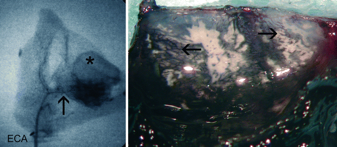
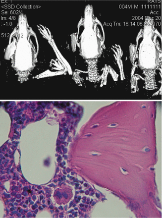
Hemiface/calvarium flap recipients received cyclosporine A intraperitoneal monotherapy. Tapered from 16 mg/kg/day by 50 % per week to 2 mg/kg/day after 4 weeks and continued that way. Indian ink and Barium angiography were used for demonstration of vascularity (Fıg. 36.2). Daily inspection and H&E sections were used for histological evaluation after sacrifice as well as CT scans to show if there is any bony resorption (Fıg. 36.3).


Fig. 36.2
Barium angiography (left) and Indian ink angiography after glycerol clearence in the right photo. In the left angiography asterisk denotes calvarial bone. Under the bone the dark fluffy area is temporalis muscle which conveys periosteal vascular supply to our graft. Arrow shows middle temporal artery which is an end branch of external carotid system. Dissection of this pedicle was made possible by preserving all soft tissue under zyomatic arch. Photo on the right shows microperfusion of the bone. Indian ink was arterially injected via common carotid pedicle and the specimen left for clearing in 50 % glycerol. Arrow to right shows sinusoidal perfusion of the bone by indian ink. Arrow to the left shows periosteal network filled by the ink

Fig. 36.3
Upper CT image shows intact non-resorbed bone component of the hemifac/calvarial allograft onlay over zygomatic arch of the recipient at days 7, 30 and 90 posotperatively. Lower photo: 200× H&E microphoto of the graft on day 105 postoperatively showing healthy bone marrow and cortical bone structures
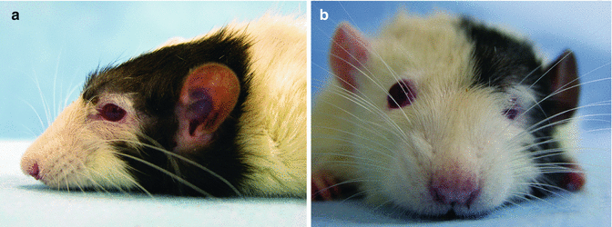
Average, 154 days post-transplant, all allografts were healthy and the longest survival was up to 300 days without any rejection or flap loss (Fıg. 36.4). At 7, 30, 63, 100 days posttransplant, bone biopsies revealed healthy bone structure showing that the graft was well vascularized. Bone marrows were also viable without any sign of rejection (Fıg. 36.3). Ct scans showed no significant resorption of bony edges secondary to vascular insufficiency.


Fig..36.4
(a) The recipient rat 100 days after transplantation from left side (a) and from anterior views (b) Please note the supraorbital fullness of the onlay calvarial bone
Stay updated, free articles. Join our Telegram channel

Full access? Get Clinical Tree


